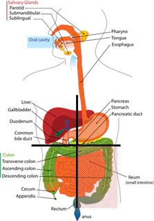
Related Research Articles

The abdominal cavity is a large body cavity in humans and many other animals that contains many organs. It is a part of the abdominopelvic cavity. It is located below the thoracic cavity, and above the pelvic cavity. Its dome-shaped roof is the thoracic diaphragm, a thin sheet of muscle under the lungs, and its floor is the pelvic inlet, opening into the pelvis.

Peritonitis is inflammation of the localized or generalized peritoneum, the lining of the inner wall of the abdomen and cover of the abdominal organs. Symptoms may include severe pain, swelling of the abdomen, fever, or weight loss. One part or the entire abdomen may be tender. Complications may include shock and acute respiratory distress syndrome.

Ascites is the abnormal build-up of fluid in the abdomen. Technically, it is more than 25 ml of fluid in the peritoneal cavity, although volumes greater than one liter may occur. Symptoms may include increased abdominal size, increased weight, abdominal discomfort, and shortness of breath. Complications can include spontaneous bacterial peritonitis.
Rovsing's sign, named after the Danish surgeon Niels Thorkild Rovsing (1862–1927), is a sign of appendicitis. If palpation of the left lower quadrant of a person's abdomen increases the pain felt in the right lower quadrant, the patient is said to have a positive Rovsing's sign and may have appendicitis. The phenomenon was first described by Swedish surgeon Emil Samuel Perman (1856-1945) writing in the journal Hygiea in 1904.

Conductive hearing loss (CHL) occurs when there is a problem transferring sound waves anywhere along the pathway through the outer ear, tympanic membrane (eardrum), or middle ear (ossicles). If a conductive hearing loss occurs in conjunction with a sensorineural hearing loss, it is referred to as a mixed hearing loss. Depending upon the severity and nature of the conductive loss, this type of hearing impairment can often be treated with surgical intervention or pharmaceuticals to partially or, in some cases, fully restore hearing acuity to within normal range. However, cases of permanent or chronic conductive hearing loss may require other treatment modalities such as hearing aid devices to improve detection of sound and speech perception.
Colic in horses is defined as abdominal pain, but it is a clinical symptom rather than a diagnosis. The term colic can encompass all forms of gastrointestinal conditions which cause pain as well as other causes of abdominal pain not involving the gastrointestinal tract. The most common forms of colic are gastrointestinal in nature and are most often related to colonic disturbance. There are a variety of different causes of colic, some of which can prove fatal without surgical intervention. Colic surgery is usually an expensive procedure as it is major abdominal surgery, often with intensive aftercare. Among domesticated horses, colic is the leading cause of premature death. The incidence of colic in the general horse population has been estimated between 4 and 10 percent over the course of the average lifespan. Clinical signs of colic generally require treatment by a veterinarian. The conditions that cause colic can become life-threatening in a short period of time.

Paracentesis is a form of body fluid sampling procedure, generally referring to peritoneocentesis in which the peritoneal cavity is punctured by a needle to sample peritoneal fluid.
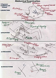
An abdominal examination is a portion of the physical examination which a physician or nurse uses to clinically observe the abdomen of a patient for signs of disease. The physical examination typically occurs after a thorough medical history is taken, that is, after the physician asks the patient the course of their symptoms. The abdominal examination is conventionally split into four different stages: first, inspection of the patient and the visible characteristics of their abdomen. Auscultation (listening) of the abdomen with a stethoscope. Palpation of the patient's abdomen. Finally, percussion (tapping) of the patient's abdomen and abdominal organs. Depending on the need to test for specific diseases such as ascites, special tests may be performed as a part of the physical examination. An abdominal examination may be performed because the physician suspects a disease of the organs inside the abdominal cavity, or simply as a part of a complete physical examination for other conditions. In a complete physical examination, the abdominal exam classically follows the respiratory examination and cardiovascular examination.
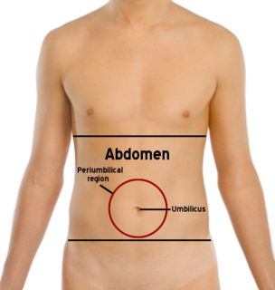
The abdomen is the part of the body between the thorax (chest) and pelvis, in humans and in other vertebrates. The abdomen is the front part of the abdominal segment of the torso. The area occupied by the abdomen is called the abdominal cavity. In arthropods it is the posterior tagma of the body; it follows the thorax or cephalothorax.
Abdominal distension occurs when substances, such as air (gas) or fluid, accumulate in the abdomen causing its expansion. It is typically a symptom of an underlying disease or dysfunction in the body, rather than an illness in its own right. People suffering from this condition often describe it as "feeling bloated". Sufferers often experience a sensation of fullness, abdominal pressure, and sometimes nausea, pain, or cramping. In the most extreme cases, upward pressure on the diaphragm and lungs can also cause shortness of breath. Through a variety of causes, bloating is most commonly due to buildup of gas in the stomach, small intestine, or colon. The pressure sensation is often relieved, or at least lessened, by belching or flatulence. Medications that settle gas in the stomach and intestines are also commonly used to treat the discomfort and lessen the abdominal distension.
In medicine, shifting dullness refers to a sign elicited on physical examination for ascites.
An acute abdomen refers to a sudden, severe abdominal pain. It is in many cases a medical emergency, requiring urgent and specific diagnosis. Several causes need immediate surgical treatment.

A trocar is a medical or veterinary device that is made up of an awl, a cannula, and a seal. Trocars are placed through the abdomen during laparoscopic surgery. The trocar functions as a portal for the subsequent placement of other instruments, such as graspers, scissors, staplers, etc. Trocars also allow the escape of gas or fluid from organs within the body.
In gastroenterology, the puddle sign is a physical examination maneuver that can be used to detect the presence of ascites. It is useful for detecting small amounts of ascites—as small as 120 mL; shifting dullness and bulging flanks typically require 500 mL.
The hepatorenal recess is the subhepatic space that separates the liver from the right kidney. As a potential space, the recess is not normally filled with fluid. However, fluid can collect here in circumstances where the abdomen fills with fluid, such as hemoperitoneum. This fluid may be seen on ultrasound or computed tomography.
Peritoneal fluid is a serous fluid made by the peritoneum in the abdominal cavity which lubricates the surface of tissue that lines the abdominal wall and pelvic cavity. It covers most of the organs in the abdomen. An increased volume of peritoneal fluid is called ascites.
Ballottement is a medical sign which indicates increased fluid in the suprapatellar pouch over the patella at the knee joint. To test ballottement the examiner would apply downward pressure towards the foot with one hand, while pushing the patella backwards against the femur with one finger of the opposite hand. A "milking" motion is used with the downward pressure. If a bogginess around the joint occurs, then the test is positive for ballottement.

A Bogota bag is a sterile plastic bag used for closure of abdominal wounds. It is generally a sterilized 3-liter genitourinary irrigation bag that is sewn to the skin or fascia of the anterior abdominal wall. Its use was first described by Oswaldo Borraez while he was a resident in Bogotá, Colombia.

In electrocardiography, left axis deviation (LAD) is a condition wherein the mean electrical axis of ventricular contraction of the heart lies in a frontal plane direction between −30° and −90°. This is reflected by a QRS complex positive in lead I and negative in leads aVF and II.
The cardiovascular examination is a portion of the physical examination that involves evaluation of the cardiovascular system. The exact contents of the examination will vary depending on the presenting complaint but a complete examination will involve the heart, lungs, belly and the blood vessels.
References
- Bickley & Szilagyi. Bate's Guide to physical examination and history taking. 2003.
| | This medical diagnostic article is a stub. You can help Wikipedia by expanding it. |