Frontal lobe injury

Last updated
This image shows the four lobes of the human brain: the frontal lobe (red), the parietal lobe (orange), the temporal lobe (green), and the occipital lobe (yellow). Also shown are the insular cortex (purple), the brain stem (black), and the cerebellum (blue). Four lobes animation small2.gif
This image shows the four lobes of the human brain: the frontal lobe (red), the parietal lobe (orange), the temporal lobe (green), and the occipital lobe (yellow). Also shown are the insular cortex (purple), the brain stem (black), and the cerebellum (blue).

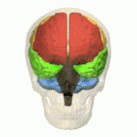
The frontal lobe of the human brain is both relatively large in mass and less restricted in movement than the posterior portion of the brain. [1] It is a component of the cerebral system, which supports goal directed behavior. [2] This lobe is often cited as the part of the brain responsible for the ability to decide between good and bad choices, as well as recognize the consequences of different actions. Because of its location in the anterior part of the head, the frontal lobe is arguably more susceptible to injuries. Following a frontal lobe injury, an individual's abilities to make good choices and recognize consequences are often impaired. Memory impairment is another common effect associated with frontal lobe injuries, but this effect is less documented and may or may not be the result of flawed testing. [3] Damage to the frontal lobe can cause increased irritability, which may include a change in mood and an inability to regulate behavior. [1] Particularly, an injury of the frontal lobe could lead to deficits in executive function, such as anticipation, goal selection, planning, initiation, sequencing, monitoring (detecting errors), and self-correction (initiating novel responses). [4] A widely reported case of frontal lobe injury was that of Phineas Gage, a railroad worker whose left frontal lobe was damaged by a large iron rod in 1848 (though Gage's subsequent personality changes are almost always grossly exaggerated).

Contents

Neuropsychological effects

Midsagittal cross-section of a human brain. The portion in color is the left frontal lobe, damaged in Phineas Gage's accident. Frontal lobe animation.gif
Midsagittal cross-section of a human brain. The portion in color is the left frontal lobe, damaged in Phineas Gage's accident.

Memory and attention

Patients with damaged frontal lobes often complain of minimal to substantial memory loss. Because of this, frontal lobe injuries have long been associated with memory problems, despite little evidence actually showing this relation to be true. In fact, when patients with such injuries are tested using standard memory tests, they often score within normal. Close relatives of these same patients, however, may describe substantial memory problems. The disparity occurs because it is not the memory system itself that is affected, but the functions of the frontal lobe that facilitate working memory. [3] Working memory is closely involved with the ability to hold attention. [5] Working memory is not simply how much information you can maintain in a brief period of time; this describes primary memory, and a small part of working memory relates to it. The important part of working memory is secondary memory, in which an individual retrieves information. Those with high working memory are able to perform this retrieval even when distracted by another task. Patients with damaged frontal lobes show lower working memory and, therefore, a lessened ability to retrieve information from their secondary memory. [6]

Risk taking

An increase in impulsivity, risk taking or both is often seen in individuals following frontal lobe damage. The two related terms differ in that impulsivity is a response disinhibition, while risk taking is related to the reward-based aspects of decision-making. [7] Put more simply, an impulsive person will make a decision quickly, without considering the consequences, leading ultimately to a lack of self-control. Contrarily, risk takers will look at the consequences but not weigh them; they will jump at the opportunity of a reward even if the likelihood of receiving that reward is slim. The increase of risk taking amongst damaged frontal lobe patients can be directly observed during gambling, and gambling tasks have been developed to measure such behavior.

Diagnosis

A screenshot from a computerized version of the Iowa gambling task IowaGamblingTask.gif
A screenshot from a computerized version of the Iowa gambling task
A screenshot from a computerized version of the Wisconsin Card Sorting Test. WisconsinCardSort.png
A screenshot from a computerized version of the Wisconsin Card Sorting Test.
In both forms of the saccade test, every eye movement is tracked, even small movements like the ones shown in this animation. Optokinetic nystagmus.gif
In both forms of the saccade test, every eye movement is tracked, even small movements like the ones shown in this animation.

Types of tests

Before more advanced technology came about, scientists tested individual behavior using more low-tech means. As technology progressed, so did the tests scientists administer to evaluate a person's cognitive function. [8]
In testing the behavioral effects of a frontal lobe injury, many of the tests are still very simple and do not involve greatly advanced technology.

Gambling task

This test has an inverse relationship between the probability of obtaining a reward and the value of the reward itself; that is to say, a low probability but a high reward or a high probability but a low reward. [7] Thus, actual gambling skills are not being tested, but simply the preference for high reward despite the risks. In one of the ways to carry this out, a set of cards will be presented face down to the individual being tested; one of the cards would be the winning card, and all the others, losers. Cards will then be continuously removed from the pile and added back in randomly, during which time the winning card could be anywhere. Subjects being tested are told they can stop the process at any time and have the cards flipped over; if the winning card is present within the pile they win points. The catch, however, is that more points are awarded when fewer cards are present; but when fewer cards are present, the probability of the winning card being within the pile is lesser. Risk takers are those that go for the higher reward (more points), even though they are less likely to actually receive that reward. They choose a higher, less likely award, over a lower, more probable reward. Subjects that have experienced a frontal lobe injury show just such behavior when tested.

Wisconsin Card Sorting Test (WCST)

The Wisconsin Card Sorting Test (WCST) can be used in conjunction with other tests to speculate to possible dysfunction of the prefrontal cortex, the front-most area of the frontal lobe, that plays an important role in executive functioning. However, since the age of modern medicine and brain imaging, the WCST has been purported to be inaccurate and inconclusive in diagnosing frontal lobe damage.[ citation needed ]

The WCST test is supposed to specifically measure an individual's competence in abstract reasoning, and the ability to change problem-solving strategies when needed. [9]

Saccade

A saccade is a fast movement of the eyes in a certain direction. In the most simplistic form, there are two types of saccade tests administered in which the only requirement is movement of the eye: the prosaccade and the antisaccade. [10] In the prosaccade, participants are required to quickly look toward a point in response to some attention-catching cue, such as a flashing light. Because there are very powerful evolutionary forces that work to automatically focus attention toward prepotent (greater in power) stimuli, this type of test does not call upon an individual's executive control; therefore, the prosaccade is not relevant when testing the effects of frontal lobe damage on executive cognitive control and working memory. [10] Conversely, the antisaccade test requires not only ignoring the flashing cue, but looking in the opposite direction. This task calls for inhibition of a prepotent response as well as planning and executing an eye movement that contradicts instinct. In the anti-saccade test, an individual has to set the goal of ignoring these instincts and continue to maintain this goal. Those with frontal lobe injuries show lower working memory, and therefore typically do not test well in the antisaccade test.

Flaws in Testing

While impulsivity and risk-taking behavior are both commonly observed following a frontal lobe injury, such traits are hard to evaluate and quantify without some degree of subjectivity. [7] The definitions of these traits are themselves not completely straightforward, nor are they always agreed upon. As a result, the methods to measure such behaviors often differ, and this should be taken into consideration when comparing data/results from different sources. Because of this, caution should be taken in how to interpret different results.

It is also important to remember that a single test, such as the WCST, cannot be used to measure the effects of a frontal lobe injury, or the aspects of cognitive function it may affect, such as working memory; variety of tests must be used. A subject may be good at one task but show dysfunction in executive function overall. Similarly, test results can be made misleading after testing the same individual over a long period of time. The subject may get better at a task, but not because of an improvement in executive cognitive function. He/she may have simply learned some strategies for doing this particular task that made it no longer a good measurement tool.[ citation needed ]

Patients with damaged frontal lobes often complain of minimal to substantial memory loss, even though when such patients are tested using standard memory tests, they often score within normal. The disparity could be the result of the limits of these standardized tests. [3] Just as likely, the scientific community may not be comparing the right groups of people. Little is understood about frontal lobe functions facilitating memory, but what is clear is that more in-depth research of brain injury patients is needed. Because most research compares those with brain injuries (whether frontal lobe or not) and those without, the scientific community is unsure whether certain memory impairment is specific to frontal lobe injuries, or just traumatic brain injuries in general. There are many factors to consider when examining the effects of a traumatic brain injury, such as the nature of the injury as well as its cause; but the severity of the injury seems to be most important in affecting memory impairment specific to frontal lobe damage. Those patients with a mild traumatic brain injury with frontal lobe damage seem to be only slightly affected, if affected at all.

Frontal lobe injuries have been shown to cause decreased ability in combining events that are temporally separated (separated by time), as well as recalling information in its correct context. However, standardized testing may mask or exaggerate a possible impairment because the patients are strictly regulated, as are their discretionary behaviors. Many times, these are behaviors thought to be directly related to disorders of the frontal lobe, when in fact, these patients have a different problem entirely, such as paying attention, anxiety, sleep disorder, etc.

The main conclusion that can be agreed upon is that tests should continuously be scrutinized; as society progresses, better tests should be designed. Without the proper tests to assess traumatic brain injury patients with frontal lobe damage in particular, we may be misrepresenting the functions of the frontal lobe, specifically the role it plays in memory. [3]

See also

Related Research Articles

Source amnesia is the inability to remember where, when or how previously learned information has been acquired, while retaining the factual knowledge. This branch of amnesia is associated with the malfunctioning of one's explicit memory. It is likely that the disconnect between having the knowledge and remembering the context in which the knowledge was acquired is due to a dissociation between semantic and episodic memory – an individual retains the semantic knowledge, but lacks the episodic knowledge to indicate the context in which the knowledge was gained.

<span class="mw-page-title-main">Brain damage</span> Destruction or degeneration of brain cells

Neurotrauma, brain damage or brain injury (BI) is the destruction or degeneration of brain cells. Brain injuries occur due to a wide range of internal and external factors. In general, brain damage refers to significant, undiscriminating trauma-induced damage.

<span class="mw-page-title-main">Frontal lobe</span> Part of the brain

The frontal lobe is the largest of the four major lobes of the brain in mammals, and is located at the front of each cerebral hemisphere. It is parted from the parietal lobe by a groove between tissues called the central sulcus and from the temporal lobe by a deeper groove called the lateral sulcus. The most anterior rounded part of the frontal lobe is known as the frontal pole, one of the three poles of the cerebrum.

<span class="mw-page-title-main">Wisconsin Card Sorting Test</span> Neuropsychological test

The Wisconsin Card Sorting Test (WCST) is a neuropsychological test of set-shifting, which is the capability to show flexibility when exposed to changes in reinforcement. The WCST was written by David A. Grant and Esta A. Berg. The Professional Manual for the WCST was written by Robert K. Heaton, Gordon J. Chelune, Jack L. Talley, Gary G. Kay, and Glenn Curtiss.

Disinhibition, also referred to as behavioral disinhibition, is medically recognized as an orientation towards immediate gratification, leading to impulsive behaviour driven by current thoughts, feelings, and external stimuli, without regard for past learning or consideration for future consequences. It is one of five pathological personality trait domains in certain psychiatric disorders. In psychology, it is defined as a lack of restraint manifested in disregard of social conventions, impulsivity, and poor risk assessment. Hypersexuality, hyperphagia, and aggressive outbursts are indicative of disinhibited instinctual drives.

<span class="mw-page-title-main">Somatic marker hypothesis</span> Hypothesis that emotional processes guide or bias decision-making

The somatic marker hypothesis, formulated by Antonio Damasio and associated researchers, proposes that emotional processes guide behavior, particularly decision-making.

Memory disorders are the result of damage to neuroanatomical structures that hinders the storage, retention and recollection of memories. Memory disorders can be progressive, including Alzheimer's disease, or they can be immediate including disorders resulting from head injury.

<span class="mw-page-title-main">Orbitofrontal cortex</span> Region of the prefrontal cortex of the brain

The orbitofrontal cortex (OFC) is a prefrontal cortex region in the frontal lobes of the brain which is involved in the cognitive process of decision-making. In non-human primates it consists of the association cortex areas Brodmann area 11, 12 and 13; in humans it consists of Brodmann area 10, 11 and 47.

<span class="mw-page-title-main">Frontal lobe disorder</span> Brain disorder

Frontal lobe disorder, also frontal lobe syndrome, is an impairment of the frontal lobe of the brain due to disease or frontal lobe injury. The frontal lobe plays a key role in executive functions such as motivation, planning, social behaviour, and speech production. Frontal lobe syndrome can be caused by a range of conditions including head trauma, tumours, neurodegenerative diseases, neurodevelopmental disorders, neurosurgery and cerebrovascular disease. Frontal lobe impairment can be detected by recognition of typical signs and symptoms, use of simple screening tests, and specialist neurological testing.

<span class="mw-page-title-main">Dorsolateral prefrontal cortex</span> Area of the prefrontal cortex of primates

The dorsolateral prefrontal cortex is an area in the prefrontal cortex of the primate brain. It is one of the most recently derived parts of the human brain. It undergoes a prolonged period of maturation which lasts into adulthood. The DLPFC is not an anatomical structure, but rather a functional one. It lies in the middle frontal gyrus of humans. In macaque monkeys, it is around the principal sulcus. Other sources consider that DLPFC is attributed anatomically to BA 9 and 46 and BA 8, 9 and 10.

Post-traumatic amnesia (PTA) is a state of confusion that occurs immediately following a traumatic brain injury (TBI) in which the injured person is disoriented and unable to remember events that occur after the injury. The person may be unable to state their name, where they are, and what time it is. When continuous memory returns, PTA is considered to have resolved. While PTA lasts, new events cannot be stored in the memory. About a third of patients with mild head injury are reported to have "islands of memory", in which the patient can recall only some events. During PTA, the patient's consciousness is "clouded". Because PTA involves confusion in addition to the memory loss typical of amnesia, the term "post-traumatic confusional state" has been proposed as an alternative.

Dysexecutive syndrome (DES) consists of a group of symptoms, usually resulting from brain damage, that fall into cognitive, behavioural and emotional categories and tend to occur together. The term was introduced by Alan Baddeley to describe a common pattern of dysfunction in executive functions, such as planning, abstract thinking, flexibility and behavioural control. It is thought to be Baddeley's hypothesized working memory system and the central executive that are the hypothetical systems impaired in DES. The syndrome was once known as frontal lobe syndrome; however 'dysexecutive syndrome' is preferred because it emphasizes the functional pattern of deficits over the location of the syndrome in the frontal lobe, which is often not the only area affected.

Traumatic brain injury can cause a variety of complications, health effects that are not TBI themselves but that result from it. The risk of complications increases with the severity of the trauma; however even mild traumatic brain injury can result in disabilities that interfere with social interactions, employment, and everyday living. TBI can cause a variety of problems including physical, cognitive, emotional, and behavioral complications.

Retrospective memory is the memory of people, words, and events encountered or experienced in the past. It includes all other types of memory including episodic, semantic and procedural. It can be either implicit or explicit. In contrast, prospective memory involves remembering something or remembering to do something after a delay, such as buying groceries on the way home from work. However, it is very closely linked to retrospective memory, since certain aspects of retrospective memory are required for prospective memory.

In psychology and neuroscience, executive dysfunction, or executive function deficit, is a disruption to the efficacy of the executive functions, which is a group of cognitive processes that regulate, control, and manage other cognitive processes. Executive dysfunction can refer to both neurocognitive deficits and behavioural symptoms. It is implicated in numerous psychopathologies and mental disorders, as well as short-term and long-term changes in non-clinical executive control.

In psychology, confabulation is a memory error consisting of the production of fabricated, distorted, or misinterpreted memories about oneself or the world. It is generally associated with certain types of brain damage or a specific subset of dementias. While still an area of ongoing research, the basal forebrain is implicated in the phenomenon of confabulation. People who confabulate present with incorrect memories ranging from subtle inaccuracies to surreal fabrications, and may include confusion or distortion in the temporal framing of memories. In general, they are very confident about their recollections, even when challenged with contradictory evidence.

Cerebellar cognitive affective syndrome (CCAS), also called Schmahmann's syndrome is a condition that follows from lesions (damage) to the cerebellum of the brain. It refers to a constellation of deficits in the cognitive domains of executive function, spatial cognition, language, and affect resulting from damage to the cerebellum. Impairments of executive function include problems with planning, set-shifting, abstract reasoning, verbal fluency, and working memory, and there is often perseveration, distractibility and inattention. Language problems include dysprosodia, agrammatism and mild anomia. Deficits in spatial cognition produce visual–spatial disorganization and impaired visual–spatial memory. Personality changes manifest as blunting of affect or disinhibited and inappropriate behavior. These cognitive impairments result in an overall lowering of intellectual function. CCAS challenges the traditional view of the cerebellum being responsible solely for regulation of motor functions. It is now thought that the cerebellum is responsible for monitoring both motor and nonmotor functions. The nonmotor deficits described in CCAS are believed to be caused by dysfunction in cerebellar connections to the cerebral cortex and limbic system.

Executive functions are a cognitive apparatus that controls and manages cognitive processes. Norman and Shallice (1980) proposed a model on executive functioning of attentional control that specifies how thought and action schemata become activated or suppressed for routine and non-routine circumstances. Schemas, or scripts, specify an individual's series of actions or thoughts under the influence of environmental conditions. Every stimulus condition turns on the activation of a response or schema. The initiation of appropriate schema under routine, well-learned situations is monitored by contention scheduling which laterally inhibits competing schemas for the control of cognitive apparatus. Under unique, non-routine procedures controls schema activation. The SAS is an executive monitoring system that oversees and controls contention scheduling by influencing schema activation probabilities and allowing for general strategies to be applied to novel problems or situations during automatic attentional processes.

The anti-saccade (AS) task is a gross estimation of injury or dysfunction of the frontal lobe, by assessing the brain’s ability to inhibit the reflexive saccade. Saccadic eye movement is primarily controlled by the frontal cortex.

Alcohol-related brain damage alters both the structure and function of the brain as a result of the direct neurotoxic effects of alcohol intoxication or acute alcohol withdrawal. Increased alcohol intake is associated with damage to brain regions including the frontal lobe, limbic system, and cerebellum, with widespread cerebral atrophy, or brain shrinkage caused by neuron degeneration. This damage can be seen on neuroimaging scans.

References

  1. 1 2 Lux WE (2007). "A neuropsychiatric perspective on traumatic brain injury". Journal of Rehabilitation Research & Development . 44 (7): 951–962. doi: 10.1682/jrrd.2007.01.0009 . PMID   18075952.
  2. Badre D, D'Esposito M (2009). "Is the rostro-caudal axis of the frontal lobe hierarchical?" (PDF). Nature Reviews Neuroscience. 10 (9): 659–669. doi:10.1038/nrn2667. PMC   3258028 . PMID   19672274. Archived from the original (PDF) on 2012-01-24. Retrieved 2011-12-31.
  3. 1 2 3 4 Kim J.S.; Kim O.L.; Seo W.S.; Koo B.H.; Joo Y.; Bai D.S. (2009). "Memory Dysfunctions after Mild and Moderate Traumatic Brain Injury : Comparison between Patients with and without Frontal Lobe Injury". Journal of Korean Neurosurgical Society . 46 (5): 459–467. doi:10.3340/jkns.2009.46.5.459. PMC   2796352 . PMID   20041056.
  4. Lezak M.D. (1989). Assessment of psychosocial dysfunction resulting from head trauma. In: Lezak MD, editor. Assessment of the behavioral consequences of head trauma. New York (NY): A. R. Liss; 113–43.
  5. Broadway, J.M., Redick, T.S., & Engle, R.W. (2010). "Working memory capacity: Self-control is (in) the goal." In R. Hassin, K. N. Ochsner, & Y. Trope (Eds.), Oxford University Press: New York, NY. Self-control in society, mind, and brain; 163-173. Retrieved from http://psychology.gatech.edu/renglelab/Publications/2010/Broadway%20Redick%20Engle%202010.pdf Archived 2012-04-25 at the Wayback Machine
  6. Kane, M.J. & Engle, R.W. (2002). "The role of prefrontal cortex in working memory capacity, executive attention, and general fluid intelligence: An individual-differences perspective." Psychonomic Bulletin & Review 9(4), 637-671. Retrieved from "Archived copy" (PDF). Archived from the original (PDF) on 2011-12-27. Retrieved 2011-12-31.{{cite web}}: CS1 maint: archived copy as title (link)
  7. 1 2 3 Floden, D., Alexander, M.P., Kubu, C.S., Katz, D., & Stuss, D.T. (2008). "Impulsivity and risk-taking behavior in focal frontal lobe lesions." Neuropsychologia 46, 213-223. Retrieved from http://www.psych.yorku.ca/joelab/jc/journalclub/gambling/2008Floden%20et%20al_RaymondMar_GamblingTask%202008.pdf
  8. Purves, D. (2008). Neuroscience (4th ed.). Sunderland, MA: Sinauer Associates, Inc.
  9. Biederam J, Faraone S, Monutaeux M, et al. (2000). "Neuropsychological functioning in nonreferred siblings of children with attention deficit/hyperactivity disorder". Journal of Abnormal Psychology. 109 (2): 252–65. doi:10.1037/0021-843X.109.2.252. PMID   10895563.
  10. 1 2 Unsworth, N., Engle, R.W., & Schrock, J.C. (2004). "Working Memory Capacity and the Antisaccade Task: Individual Differences in Voluntary Saccade Control." Journal of Experimental Psychology 30(6), 1302-1321. Retrieved from http://www.interactivemetronome.com/impublic/Research/Temporal%20Processing/MemoryWorkingMemory/Research_Working%20Memory_Unsworth2004.pdf Archived 2010-12-05 at the Wayback Machine