Ghosting (medical imaging)

Last updated

Ghosting is a visual artifact that occurs in magnetic resonance imaging(MRI) scans. This artifact can be a consequence of environmental factors or the human body (such as blood flow, implants etc.). Ghosting is a multidimensional artifact that occurs in the MRI in the phase encoded direction (short axis of the image) after applying the Fourier transform.

Contents

When the phase of the magnetic resonance signal is being encoded into the 2-D or 3-D Fourier image, a mild deviation from the actual phase and amplitude may occur. This incompatibility of parameters causes ghosting. The reasons this occurs are often physical factors, such as temperature or humidity of the environment, or movement caused by the patient or in the patient’s body. There are two types of movement that could result in ghosting:

These movements, when reflected over the anatomy may result in improper diagnosis of the disease and hence have to be identified and suppressed.

Problem statement and basics

Phase encoding

In MRI, phase encoding is the process of acquiring data by altering the phase of the spin of the atom by applying magnetic pulses before acquiring the actual data.

Ghosting in k-space

k-space is a graphic matrix of an MR that represents the Fourier transform domain of an image before undergoing the Inverse Fourier transformation.The phase deviations that occur in the k-space of an MR image decide the characteristics of the ghosts that appear in the resulting image. Even though most of the ghosting is due to the phase deviation in the phase encoded direction, they also appear in other directions of the k-space.

Echo-planar imaging

The basic principle of ghosting can be explained with the help of Echo-Planar Imaging (EPI). Echo-planar imaging is an MRI technique that reduces the time of data acquisition to reduce capture of patient movement. An image in the EPI can be captured in between 20-100 milliseconds. Multiple lines of data are created by transmitting RF pulse sequences with a gradient difference of 90° and 180°. After the 180° pulse, the frequency encoding gradient rapidly changes to a negative amplitude and the resulting echos are encoded in the phase encoded axis. The pulses that are used to excite can be classified into 2 types, namely 'single shot' and 'multi-shot' pulse sequences. The multi-shot echo planar images tend to capture more data than that of the single shot EPI.

De-ghosting

The process of removing the movement of images in an MRI scan is known as de-ghosting.

Existing approaches

Several algorithms have been proposed to remove ghosting in the medical images.

Iterative inverse problem solving

The iterative problem solving method [1] is a ghost correction algorithm that removes ghosting that occurs due to the physical movement of the patient. This is a post-processing technique which uses the simple motion models (such as translational motion, rotational motion or linear motion) to remove the ghosts that occur in the MR images. This algorithm uses an iterative approach to correct the distorted image by using the motion models.

In a standard rectangular-grid acquisition system, each row of data is acquired by applying a gradient in the direction of the y-axis with a fixed time T before the data is acquired. The process of acquiring data in a row is known as view. This data is scanned by encoding in the frequency encoding and phase encoding directions. This is followed by taking the Inverse Fourier transform for reconstruction of the image, which can contain ghosting artifacts. The iterative method is then applied to reduce the ghosting artifacts.

As this is a post-processing technique and requires the pre-defined model of the motion, the inter-view motion detected signal is compared with the existing models. The theoretically generated magnetization of the image is calculated. This magnetization should match with the magnetization that is observed.

Advantage

Iterative inverse problem solving is faster than Cardiac gating [2] and doesn't involve the patient too much.

Disadvantage

The choice of the motion model is critical, as it should be sufficiently close to the actual model.

Reference-free EPI ghost correction algorithm

Flow of ALOHA Algorithm Alohaalgorithm.gif
Flow of ALOHA Algorithm

Reference free EPI ghost correction algorithm [3] uses a method called ALOHA (Annihilating filter-based low rank Hankel structured matrix completion approach). The data of the k-space matrix is numbered consecutively and is split into odd and even data based on the samples of the index. This method was developed based on the fact that the difference between the odd and even virtual k-space data is the Fourier transform of the underlying sparse image. It is based on the principle that Ghost Nyquists are produced due to the inconsistencies between the odd and even echos of the MR images. The occurrence of the Ghosts in the images is converted into missing k-space data and is recovered with the help of the ALOHA matrix.

There are 2 types of approaches that are used to remove the Nyquist Ghost artifacts:

  1. Navigator based approach [4] - uses reference images and are obtained with phase encoding blips [5]
  2. Navigator free approach [6] - does not use reference images and uses pulse sequences for ghost correction

The odd and the even samples of data are taken from the k-space by means of interpolation. A high-performance interpolation method would be able to find the missing data from the actual even and odd sequences and remove the ghosting artifacts from the images. ALOHA converts the sparse recovery problem into a k-space interpolation problem using a low-rank interpolator.

Advantage
  1. Removes the ghost artifact without pre-scan data or the modification of the pulse sequence.
  2. Other artifacts can also be avoided by observing the existing pre-scan approach.
  3. This method can be applied to both single and multi-coil acquisitions.
  4. Faster when compared to the reference based algorithms.
Disadvantage

Reference free algorithms are prone to errors and have low performance when compared to the reference based algorithms.

Method of generalized projections

The method of generalized projections (MGP) is based on the fact that discontinuities in the phase form alternating patterns in the k-space, which leads to ghosting. Interleaved EPI gives a train of ghosts in the phase encoded direction. This EPI ghosting can be corrected from the phase using 2 or more reference scans.

According to MGP, the region of support (ROS) is defined around the parent image (the object in the image that is ghosted) of the ghosted image. The pixels outside the ROS is masked to zero. The Fourier transform of the masked image is taken. The phase of the original image is changed to the model image (the image that is modeled as ghosting arising from the phase offsets) but the magnitude is kept constant. The assumption in the MGP method is that the model k-space phase of each line is more precise than that of the original k-space image. From the MGP algorithm of ghost correction, it has been observed that it works best only when there is low degree of freedom. This method can be applied only when no more than 8 parameters are required to be found.

Advantage
  1. This method is an image based technique and does not require additional reference scan.
  2. Allows de-ghosting up to 2-interleaves even if there is phase variation between them.
  3. Relaxation parameter improves convergence
Disadvantage

The projections on the slices have their own magnetization values. These different values of magnetization will induce a different phase response. Hence a new measurement of the phase response has to be taken.

Related Research Articles

Digital signal processing (DSP) is the use of digital processing, such as by computers or more specialized digital signal processors, to perform a wide variety of signal processing operations. The digital signals processed in this manner are a sequence of numbers that represent samples of a continuous variable in a domain such as time, space, or frequency. In digital electronics, a digital signal is represented as a pulse train, which is typically generated by the switching of a transistor.

<span class="mw-page-title-main">Image compression</span> Reduction of image size to save storage and transmission costs

Image compression is a type of data compression applied to digital images, to reduce their cost for storage or transmission. Algorithms may take advantage of visual perception and the statistical properties of image data to provide superior results compared with generic data compression methods which are used for other digital data.

<span class="mw-page-title-main">Motion compensation</span> Video compression technique, used to efficiently predict and generate video frames

Motion compensation in computing is an algorithmic technique used to predict a frame in a video given the previous and/or future frames by accounting for motion of the camera and/or objects in the video. It is employed in the encoding of video data for video compression, for example in the generation of MPEG-2 files. Motion compensation describes a picture in terms of the transformation of a reference picture to the current picture. The reference picture may be previous in time or even from the future. When images can be accurately synthesized from previously transmitted/stored images, the compression efficiency can be improved.

<span class="mw-page-title-main">Deconvolution</span> Reconstruction of a filtered signal

In mathematics, deconvolution is the operation inverse to convolution. Both operations are used in signal processing and image processing. For example, it may be possible to recover the original signal after a filter (convolution) by using a deconvolution method with a certain degree of accuracy. Due to the measurement error of the recorded signal or image, it can be demonstrated that the worse the signal-to-noise ratio (SNR), the worse the reversing of a filter will be; hence, inverting a filter is not always a good solution as the error amplifies. Deconvolution offers a solution to this problem.

<span class="mw-page-title-main">Synthetic-aperture radar</span> Form of radar used to create images of landscapes

Synthetic-aperture radar (SAR) is a form of radar that is used to create two-dimensional images or three-dimensional reconstructions of objects, such as landscapes. SAR uses the motion of the radar antenna over a target region to provide finer spatial resolution than conventional stationary beam-scanning radars. SAR is typically mounted on a moving platform, such as an aircraft or spacecraft, and has its origins in an advanced form of side looking airborne radar (SLAR). The distance the SAR device travels over a target during the period when the target scene is illuminated creates the large synthetic antenna aperture. Typically, the larger the aperture, the higher the image resolution will be, regardless of whether the aperture is physical or synthetic – this allows SAR to create high-resolution images with comparatively small physical antennas. For a fixed antenna size and orientation, objects which are further away remain illuminated longer – therefore SAR has the property of creating larger synthetic apertures for more distant objects, which results in a consistent spatial resolution over a range of viewing distances.

<span class="mw-page-title-main">Tomographic reconstruction</span> Estimate object properties from a finite number of projections

Tomographic reconstruction is a type of multidimensional inverse problem where the challenge is to yield an estimate of a specific system from a finite number of projections. The mathematical basis for tomographic imaging was laid down by Johann Radon. A notable example of applications is the reconstruction of computed tomography (CT) where cross-sectional images of patients are obtained in non-invasive manner. Recent developments have seen the Radon transform and its inverse used for tasks related to realistic object insertion required for testing and evaluating computed tomography use in airport security.

Phase correlation is an approach to estimate the relative translative offset between two similar images or other data sets. It is commonly used in image registration and relies on a frequency-domain representation of the data, usually calculated by fast Fourier transforms. The term is applied particularly to a subset of cross-correlation techniques that isolate the phase information from the Fourier-space representation of the cross-correlogram.

In a mixed-signal system, a reconstruction filter, sometimes called an anti-imaging filter, is used to construct a smooth analog signal from a digital input, as in the case of a digital to analog converter (DAC) or other sampled data output device.

<span class="mw-page-title-main">Analysis of Functional NeuroImages</span>

Analysis of Functional NeuroImages (AFNI) is an open-source environment for processing and displaying functional MRI data—a technique for mapping human brain activity.

<span class="mw-page-title-main">Image scaling</span> Changing the resolution of a digital image

In computer graphics and digital imaging, imagescaling refers to the resizing of a digital image. In video technology, the magnification of digital material is known as upscaling or resolution enhancement.

Television standards conversion is the process of changing a television transmission or recording from one video system to another. Converting video between different numbers of lines, frame rates, and color models in video pictures is a complex technical problem. However, the international exchange of television programming makes standards conversion necessary so that video may be viewed in another nation with a differing standard. Typically video is fed into video standards converter which produces a copy according to a different video standard. One of the most common conversions is between the NTSC and PAL standards.

<i>k</i>-space in magnetic resonance imaging

In magnetic resonance imaging (MRI), the k-space or reciprocal space is obtained as the 2D or 3D Fourier transform of the image measured. It was introduced in 1979 by Likes and in 1983 by Ljunggren and Twieg.

Compressed sensing is a signal processing technique for efficiently acquiring and reconstructing a signal, by finding solutions to underdetermined linear systems. This is based on the principle that, through optimization, the sparsity of a signal can be exploited to recover it from far fewer samples than required by the Nyquist–Shannon sampling theorem. There are two conditions under which recovery is possible. The first one is sparsity, which requires the signal to be sparse in some domain. The second one is incoherence, which is applied through the isometric property, which is sufficient for sparse signals.

<span class="mw-page-title-main">Physics of magnetic resonance imaging</span> Overview article

The physics of magnetic resonance imaging (MRI) concerns fundamental physical considerations of MRI techniques and technological aspects of MRI devices. MRI is a medical imaging technique mostly used in radiology and nuclear medicine in order to investigate the anatomy and physiology of the body, and to detect pathologies including tumors, inflammation, neurological conditions such as stroke, disorders of muscles and joints, and abnormalities in the heart and blood vessels among others. Contrast agents may be injected intravenously or into a joint to enhance the image and facilitate diagnosis. Unlike CT and X-ray, MRI uses no ionizing radiation and is, therefore, a safe procedure suitable for diagnosis in children and repeated runs. Patients with specific non-ferromagnetic metal implants, cochlear implants, and cardiac pacemakers nowadays may also have an MRI in spite of effects of the strong magnetic fields. This does not apply on older devices, and details for medical professionals are provided by the device's manufacturer.

<span class="mw-page-title-main">Real-time MRI</span> Type of MRI

Real-time magnetic resonance imaging (RT-MRI) refers to the continuous monitoring ("filming") of moving objects in real time. Because MRI is based on time-consuming scanning of k-space, real-time MRI was possible only with low image quality or low temporal resolution. Using an iterative reconstruction algorithm these limitations have recently been removed: a new method for real-time MRI achieves a temporal resolution of 20 to 30 milliseconds for images with an in-plane resolution of 1.5 to 2.0 mm. Real-time MRI promises to add important information about diseases of the joints and the heart. In many cases MRI examinations may become easier and more comfortable for patients.

<span class="mw-page-title-main">Philip Batchelor</span>

Philip Batchelor, was a Swiss-British academic in the fields of mathematics and medical imaging.

Harmonic phase (HARP) algorithm is a medical image analysis technique capable of extracting and processing motion information from tagged magnetic resonance image (MRI) sequences. It was initially developed by N. F. Osman and J. L. Prince at the Image Analysis and Communications Laboratory at Johns Hopkins University. The method uses spectral peaks in the Fourier domain of tagged MRI, calculating the phase images of their inverse Fourier transforms, which are called harmonic phase (HARP) images. The motion of material points through time is then tracked, under the assumption that the HARP value of a fixed material point is time-invariant. The method is fast and accurate, and has been accepted as one of the most popular tagged MRI analysis methods in medical image processing.

An MRI artifact is a visual artifact in magnetic resonance imaging (MRI). It is a feature appearing in an image that is not present in the original object. Many different artifacts can occur during MRI, some affecting the diagnostic quality, while others may be confused with pathology. Artifacts can be classified as patient-related, signal processing-dependent and hardware (machine)-related.

Deep learning in photoacoustic imaging

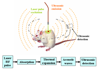
Deep learning in photoacoustic imaging combines the hybrid imaging modality of photoacoustic imaging (PA) with the rapidly evolving field of deep learning. Photoacoustic imaging is based on the photoacoustic effect, in which optical absorption causes a rise in temperature, which causes a subsequent rise in pressure via thermo-elastic expansion. This pressure rise propagates through the tissue and is sensed via ultrasonic transducers. Due to the proportionality between the optical absorption, the rise in temperature, and the rise in pressure, the ultrasound pressure wave signal can be used to quantify the original optical energy deposition within the tissue.

References

[7] [8] [9] [10] [11] [12] [13]

  1. Motion artifact correction of MRI via iterative inverse problem solving Yen-Hao Tseng; Jenq-Neng Hwang; Chun Yuan
  2. Cardiac gating technique for arterial oxygen saturation measurement Rasoul Yousefi; Mehrdad Nourani 2014 IEEE Biomedical Circuits and Systems Conference (BioCAS) Proceedings
  3. Reference-free EPI Nyquist ghost correction using annihilating filter-based low rank hankel matrix for K-space interpolation. Juyoung Lee; Kyong Hwan Jin; Jong Chul Ye 2016 IEEE 13th International Symposium on Biomedical Imaging (ISBI)
  4. Reference-free EPI Nyquist ghost correction using annihilating filter-based low rank hankel matrix for K-space interpolation. Juyoung Lee; Kyong Hwan Jin; Jong Chul Ye 2016 IEEE 13th International Symposium on Biomedical Imaging (ISBI)
  5. Reference-free EPI Nyquist ghost correction using annihilating filter-based low rank hankel matrix for K-space interpolation. Juyoung Lee; Kyong Hwan Jin; Jong Chul Ye 2016 IEEE 13th International Symposium on Biomedical Imaging (ISBI)
  6. Reference-free EPI Nyquist ghost correction using annihilating filter-based low rank hankel matrix for K-space interpolation. Juyoung Lee; Kyong Hwan Jin; Jong Chul Ye 2016 IEEE 13th International Symposium on Biomedical Imaging (ISBI)
  7. Reference-free EPI Nyquist ghost correction using annihilating filter-based low rank hankel matrix for K-space interpolation. Juyoung Lee; Kyong Hwan Jin; Jong Chul Ye 2016 IEEE 13th International Symposium on Biomedical Imaging (ISBI)
  8. A method of generalized projections (MGP) ghost correction algorithm for interleaved EPI K. J. Lee; N. G. Papadakis; D. C. Barber; I. D. Wilkinson; P. D. Griffiths; M. N. J. Paley, IEEE Transactions on Medical Imaging vol. 23, no. 7,July 2004
  9. Motion artifact correction of MRI via iterative inverse problem solving Yen-Hao Tseng; Jenq-Neng Hwang; Chun Yuan 06 August 2002
  10. Handbook of medical imaging: physics and psychologists Jacob Beutel, Harold L. Kundel, Richard L. Van Metter published by SPIE - published 2000- International society for optical engineering
  11. Imaging Systems for Medical Diagnostics: Fundamentals, Technical Solutions and Applications for Systems Applying Ionizing Radiation, Nuclear Magnetic Resonance and Ultrasound, 2nd Edition February 2006
  12. Medical Imaging systems technology, Cornelius T. Leondez, published 2005
  13. Cardiac gating technique for arterial oxygen saturation measurement Rasoul Yousefi; Mehrdad Nourani 2014 IEEE Biomedical Circuits and Systems Conference (BioCAS) Proceedings