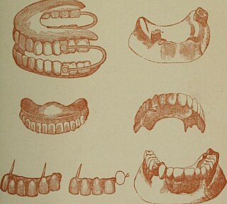
History of dental treatments

Last updated

The history of dental treatments dates back to thousands of years. [1] [2] The scope of this article is limited to the pre-1981 history.

Contents

The earliest known example of dental caries manipulation is found in a Paleolithic man, dated between 14,160 and 13,820 BP. [3] The earliest known use of a filling after removal of decayed or infected pulp is found in a Paleolithic who lived near modern-day Tuscany, Italy, from 13,000 to 12,740 BP. [4] Although inconclusive, researchers have suggested that rudimentary dental procedures have been performed as far back as 130,000 years ago by Neanderthals. [5] Regarding implants, one of the milestone progress is osseointegration which was termed in 1981 by Tomas Albrektsson. [6]

Dental implants

Greenfield's basket: one of the earliest examples of a successful endosseous implant was Greenfield's 1913 implant system Greenfield implant.jpg
Greenfield's basket: one of the earliest examples of a successful endosseous implant was Greenfield's 1913 implant system
While studying bone cells in a rabbit tibia using a titanium chamber, Branemark was unable to remove it from bone. His realization that bone would adhere to titanium led to the concept of osseointegration and the development of modern dental implants. The original x-ray film of the chamber embedded in the rabbit tibia is shown (made available by Branemark). Branemark's initial radiograph.jpg
While studying bone cells in a rabbit tibia using a titanium chamber, Branemark was unable to remove it from bone. His realization that bone would adhere to titanium led to the concept of osseointegration and the development of modern dental implants. The original x-ray film of the chamber embedded in the rabbit tibia is shown (made available by Branemark).
Panoramic radiograph of historic dental implants, taken 1978 Panoramic radiograph of historic dental implants.jpg
Panoramic radiograph of historic dental implants, taken 1978

There is archeological evidence that humans have attempted to replace missing teeth with root form implants for thousands of years. Remains from ancient China (dating 4000 years ago) have carved bamboo pegs, tapped into the bone, to replace lost teeth, and 2000-year-old remains from ancient Egypt have similarly shaped pegs made of precious metals. Some Egyptian mummies were found to have transplanted human teeth, and in other instances, teeth made of ivory. [1] :26 [2] [7] Wilson Popenoe and his wife in 1931, at a site in Honduras dating back to 600 AD, found the lower mandible of a young Mayan woman, with three missing incisors replaced by pieces of sea shells, shaped to resemble teeth. [8] Bone growth around two of the implants, and the formation of calculus, indicates that they were functional as well as esthetic. The fragment is currently part of the Osteological Collection of the Peabody Museum of Archaeology and Ethnology at Harvard University. [1] [2]

In modern times, a tooth replica implant was reported as early as 1969, but the polymethacrylate tooth analogue was encapsulated by soft tissue rather than osseointegrated. [9]

The early part of the 20th century saw a number of implants made of a variety of materials. One of the earliest successful implants was the Greenfield implant system of 1913 (also known as the Greenfield crib or basket). [10] Greenfield's implant, an iridioplatinum implant attached to a gold crown, showed evidence of osseointegration and lasted for a number of years. [10] The first use of titanium as an implantable material was by Bothe, Beaton and Davenport in 1940, who observed how close the bone grew to titanium screws, and the difficulty they had in extracting them. [11] Bothe et al. were the first researchers to describe what would later be called osseointegration (a name that would be marketed later on by Per-Ingvar Brånemark). In 1951, Gottlieb Leventhal implanted titanium rods in rabbits. [12] Leventhal's positive results led him to believe that titanium represented the ideal metal for surgery. [12]

In the 1950s research was being conducted at Cambridge University in England on blood flow in living organisms. These workers devised a method of constructing a chamber of titanium which was then embedded into the soft tissue of the ears of rabbits. In 1952 the Swedish orthopaedic surgeon, Per-Ingvar Brånemark, was interested in studying bone healing and regeneration. During his research time at Lund University he adopted the Cambridge designed "rabbit ear chamber" for use in the rabbit femur. Following the study, he attempted to retrieve these expensive chambers from the rabbits and found that he was unable to remove them. Brånemark observed that bone had grown into such close proximity with the titanium that it effectively adhered to the metal. Brånemark carried out further studies into this phenomenon, using both animal and human subjects, which all confirmed this unique property of titanium.[ citation needed ] Leonard Linkow, in the 1950s, was one of the first to insert titanium and other metal implants into the bones of the jaw. Artificial teeth were then attached to these pieces of metal. [13] In 1965 Brånemark placed his first titanium dental implant into a human volunteer. He began working in the mouth as it was more accessible for continued observations and there was a high rate of missing teeth in the general population offered more subjects for widespread study. He termed the clinically observed adherence of bone with titanium as "osseointegration". [14] :626 Since then implants have evolved into three basic types:

  1. Root form implants; the most common type of implant indicated for all uses. Within the root form type of implant, there are roughly 18 variants, all made of titanium but with different shapes and surface textures. There is limited evidence showing that implants with relatively smooth surfaces are less prone to peri-implantitis than implants with rougher surfaces and no evidence showing that any particular type of dental implant has superior long-term success. [15]
  2. Zygoma Implant; a long implant that can anchor to the cheek bone by passing through the maxillary sinus to retain a complete upper denture when bone is absent. While zygomatic implants offer a novel approach to severe bone loss in the upper jaw, it has not been shown to offer any advantage over bone grafting functionally although it may offer a less invasive option, depending on the size of the reconstruction required. [16]
  3. Small diameter implants are implants of low diameter with one piece construction (implant and abutment) that are sometimes used for denture retention or orthodontic anchorage. [17]

Ceramic implants made from alumina were introduced between 1960s and 1970s but were eventually withdrawn from the market in the early 1990s because they presented some biomechanical problems (like low fracture toughness) and were replaced by zirconia implants. [18]

Robot-assisted dental surgery, including for dental implants, [19] has also been developed in the 2000s. [20]

Dentures

Carved ivory dentures from the 18th century. Left is lower/mandibular; upper/maxillary is at right. Carved ivory upper and lower denture Wellcome L0003910.jpg
Carved ivory dentures from the 18th century. Left is lower/mandibular; upper/maxillary is at right.
Pierre Fauchard described the construction of dentures using a metal frame, animal bone teeth, and leaf springs in 1728. Pierre Fauchard - Le Chirurgien Dentiste - dentures.png
Pierre Fauchard described the construction of dentures using a metal frame, animal bone teeth, and leaf springs in 1728.

As early as the 7th century BC, Etruscans in northern Italy made partial dentures out of human or other animal teeth fastened together with gold bands. [22] The Romans had likely borrowed this technique by the 5th century BC. [22] [23]

Wooden full dentures were invented in Japan around the early 16th century. [21] Softened bees wax was inserted into the patient's mouth to create an impression, which was then filled with harder bees wax. Wooden dentures were then meticulously carved based on that model. The earliest of these dentures were entirely wooden, but later versions used natural human teeth or sculpted pagodite, ivory, or animal horn for the teeth. These dentures were built with a broad base, exploiting the principles of adhesion to stay in place. This was an advanced technique for the era; it would not be replicated in the West until the late 18th century. Wooden dentures continued to be used in Japan until the Opening of Japan to the West in the 19th century. [21]

In 1579 gold and silver dentures were created by the barber-dentist Ambroise Paré for the French King Charles IX. [23] In 1728, Pierre Fauchard described the construction of dentures using a metal frame and teeth sculpted from animal bone. [21] The first porcelain dentures were made around 1770 by Alexis Duchâteau. In 1791, the first British patent was granted to Nicholas Dubois De Chemant, previous assistant to Duchateau, for 'De Chemant's Specification':

[...] a composition for the purpose of making of artificial teeth either single double or in rows or in complete sets, and also springs for fastening or affixing the same in a more easy and effectual manner than any hitherto discovered which said teeth may be made of any shade or colour, which they will retain for any length of time and will consequently more perfectly resemble the natural teeth. [24]

He began selling his wares in 1792, with most of his porcelain paste supplied by Wedgwood. [25] [26]

17th century London's Peter de la Roche is believed to be one of the first 'operators for the teeth', men who advertised themselves as specialists in dental work. They were often professional goldsmiths, ivory turners or students of barber-surgeons. [27]

In 1820, Samuel Stockton, a goldsmith by trade, began manufacturing high-quality porcelain dentures mounted on 18-carat gold plates. Later dentures from the 1850s on were made of Vulcanite, a form of hardened rubber into which porcelain teeth were set. In the 20th century, acrylic resin and other plastics were used. [28] In Britain, sequential Adult Dental Health Surveys revealed that in 1968 79% of those aged 65–74 had no natural teeth; by 1998, this proportion had fallen to 36%. [29]

George Washington (1732–1799) had problems with his teeth throughout his life, and historians have tracked his experiences in great detail. [30] He lost his first adult tooth when he was twenty-two and had only one left by the time he became president. [31] John Adams says he lost them because he used them to crack Brazil nuts but modern historians suggest the mercury oxide, which he was given to treat illnesses such as smallpox and malaria, probably contributed to the loss. He had several sets of false teeth made, four of them by a dentist named John Greenwood. None of the sets, contrary to popular belief, were made from wood or contained any wood. [32] The set made when he became president were carved from hippopotamus and elephant ivory, held together with gold springs. [33] Prior to these, he had a set made with real human teeth, [34] likely ones he purchased from "several unnamed Negroes, presumably Mount Vernon slaves" in 1784. [35] Washington's dental problems left him in constant pain, for which he took laudanum. [36] This distress may be apparent in many of the portraits painted while he was still in office, [36] including the one still used on the $1 bill. [37] [lower-alpha 1]

Charles Darwin (1809-1882) had dental problems from an early age, resulting from the repeated vomiting that was a feature of his life-long illness and the seasickness he suffered when aboard HMS Beagle. Like patients today with repeated vomiting the regurgitation of gastric acid would have caused tooth erosions. When the Beagle was in Hobart (1836) he visited a local dental practitioner, Henry Jeanneret (1802-1886) and earlier in the voyage (1833) he recorded that he needed a denture to be repaired. [38] Later in life (1852) he had five 'grinders' (molars) removed under chloroform, and his is one of the first records of this substance being used as an anaesthetic in dentistry.

Advancements in 3D printing and applications in dentistry

3D printing

There have been significant advances in the field of dentistry within the past 50 years due to the use and development of 3D printing and imaging. These innovations include Stereolithography and Digital Light Processing. [39]

Stereolithography

The Stereolithography (SLA) technique was developed by Charles Hull and patented in 1986. The technology was originally developed for manufacturing use, and has been applied to a multitude of fields. [39] Common applications for this technology in dentistry include but are not limited to modeling, temporary prosthetics, retainers, aligners, and orthodontic models.

SLA technology and printing technique

An Ultraviolet laser is used to cure a liquid polymer resin into layers. This liquid polymer is then held in a container as the laser polymerizes the layers of resin. This process is repeated layer by layer, and the finished product is refined and hardened in an UV oven or a solvent bath. [40]

Digital Light Processing

Digital Light Processing (DLP) was created shortly after SLA technology in 1987, at Texas Instruments Company. The SLA and DLP methods are very similar, but the DLP method has a couple key process characteristics that differ. Applications of this technology include custom dental molds and models, as well as custom fit medical equipment. [41]

DLP technology and printing technique

DLP utilizes 3D printing machines that accept CAD files and .STL files. The process starts with the build platform being lowered into a vat of liquid resin. A digital light source is then used to harden each layer. The light source cures the resin, and the layers build on top of each other until they are eventually removed from the printer. The resin that is typically used is thermosetting resin, which is cured when exposed to a light source. DLP printers are efficient in that each coordinate in a layer is cured at the simultaneously. [41]

Dental applications of 3D printing technology

Toothbuds have been 3D printed that resemble real teeth. [42] Other applications are as follows: dental modeling, digital orthodontics, medical modeling, drilling and cutting guides, and partial denture frameworks. [43]

Notes

  1. The Smithsonian Institution states in "The Portrait—George Washington: A National Treasure" that:
    Stuart admired the sculpture of Washington by French artist Jean-Antoine Houdon, probably because it was based on a life mask and therefore extremely accurate. Stuart explained, "When I painted him, he had just had a set of false teeth inserted, which accounts for the constrained expression so noticeable about the mouth and lower part of the face. Houdon's bust does not suffer from this defect. I wanted him as he looked at that time." Stuart preferred the Athenaeum pose and, except for the gaze, used the same pose for the Lansdowne painting. [36]

Related Research Articles

<span class="mw-page-title-main">Dentures</span> Prosthetic devices constructed to replace missing teeth

Dentures are prosthetic devices constructed to replace missing teeth, supported by the surrounding soft and hard tissues of the oral cavity. Conventional dentures are removable. However, there are many denture designs, some of which rely on bonding or clasping onto teeth or dental implants. There are two main categories of dentures, the distinction being whether they fit onto the mandibular arch or on the maxillary arch.

Cosmetic dentistry is generally used to refer to any dental work that improves the appearance of teeth, gums and/or bite. It primarily focuses on improvement in dental aesthetics in color, position, shape, size, alignment and overall smile appearance. Many dentists refer to themselves as "cosmetic dentists" regardless of their specific education, specialty, training, and experience in this field. This has been considered unethical with a predominant objective of marketing to patients. The American Dental Association does not recognize cosmetic dentistry as a formal specialty area of dentistry. However, there are still dentists that promote themselves as cosmetic dentists.

<span class="mw-page-title-main">Dental surgery</span> Surgery of the teeth and jaw bones

Dental surgery is any of a number of medical procedures that involve artificially modifying dentition; in other words, surgery of the teeth, gums and jaw bones.

Dental restoration, dental fillings, or simply fillings are treatments used to restore the function, integrity, and morphology of missing tooth structure resulting from caries or external trauma as well as to the replacement of such structure supported by dental implants. They are of two broad types—direct and indirect—and are further classified by location and size. A root canal filling, for example, is a restorative technique used to fill the space where the dental pulp normally resides.

<span class="mw-page-title-main">Dental technician</span> Technician working on dental appliances

A dental technician is a member of the dental team who, upon prescription from a dental clinician, constructs custom-made restorative and dental appliances.

<span class="mw-page-title-main">Dental implant</span> Surgical component that interfaces with the bone of the jaw

A dental implant is a prosthesis that interfaces with the bone of the jaw or skull to support a dental prosthesis such as a crown, bridge, denture, or facial prosthesis or to act as an orthodontic anchor. The basis for modern dental implants is a biological process called osseointegration, in which materials such as titanium or zirconia form an intimate bond to the bone. The implant fixture is first placed so that it is likely to osseointegrate, then a dental prosthetic is added. A variable amount of healing time is required for osseointegration before either the dental prosthetic is attached to the implant or an abutment is placed which will hold a dental prosthetic/crown.

Osseointegration is the direct structural and functional connection between living bone and the surface of a load-bearing artificial implant. A more recent definition defines osseointegration as "functional ankylosis ", where new bone is laid down directly on the implant surface and the implant exhibits mechanical stability. Osseointegration has enhanced the science of medical bone and joint replacement techniques as well as dental implants and improving prosthetics for amputees.

Anodontia is a rare genetic disorder characterized by the congenital absence of all primary or permanent teeth. It is divided into two subsections, complete absence of teeth or only some absence of teeth. It is associated with the group of skin and nerve syndromes called the ectodermal dysplasias. Anodontia is usually part of a syndrome and seldom occurs as an isolated entity. There is usually no exact cause for anodontia. The defect results in the dental lamina obstruction during embryogenesis due to local, systemic and genetic factors.

<span class="mw-page-title-main">Toothlessness</span> Lacking teeth

Toothlessness, or edentulism, is the condition of having no teeth. In organisms that naturally have teeth, it is the result of tooth loss.

<span class="mw-page-title-main">Nobel Biocare</span> Manufacturer of dental implants

Nobel Biocare is a Swiss company originally founded in Sweden for manufacturing dental implants. It is now headquartered in Kloten, Switzerland near the Zürich Airport. Nobel Biocare in its current form was founded in 2002. It originates from a partnership formed in 1978 between Swedish medical researcher Professor Per-Ingvar Brånemark and Bofors, a Swedish company, to industrialize Brånemark's discovery of osseointegration. Beside dental implants, it also presently develops and commercializes CAD/CAM-based prosthetics equiments to scan teeth and to fabricate individualized dental prosthesis using digital technologies in place of silicone molding.

<span class="mw-page-title-main">Denturist</span>

A denturist in the United States and Canada, clinical dental technologist in the United Kingdom and Ireland, dental prosthetist in Australia, or a clinical dental technician in New Zealand is a member of the oral health care team and role as primary oral health care provider who provides an oral health examination, planning treatment, takes impressions of the surrounding oral tissues, constructs and delivers removable oral prosthesis treatment directly to the patient.

<span class="mw-page-title-main">CAD/CAM dentistry</span> Computer-aided design and manufacturing of dental prostheses

CAD/CAM dentistry is a field of dentistry and prosthodontics using CAD/CAM to improve the design and creation of dental restorations, especially dental prostheses, including crowns, crown lays, veneers, inlays and onlays, fixed dental prostheses (bridges), dental implant supported restorations, dentures, and orthodontic appliances. CAD/CAM technology allows the delivery of a well-fitting, aesthetic, and a durable prostheses for the patient. CAD/CAM complements earlier technologies used for these purposes by any combination of increasing the speed of design and creation; increasing the convenience or simplicity of the design, creation, and insertion processes; and making possible restorations and appliances that otherwise would have been infeasible. Other goals include reducing unit cost and making affordable restorations and appliances that otherwise would have been prohibitively expensive. However, to date, chairside CAD/CAM often involves extra time on the part of the dentist, and the fee is often at least two times higher than for conventional restorative treatments using lab services.

<span class="mw-page-title-main">Per-Ingvar Brånemark</span>

Per-Ingvar Brånemark was a Swedish physician and research professor, acknowledged as the "father of modern dental implantology". The Brånemark Osseointegration Center (BOC), named after its founder, was founded in 1989 in Gothenburg, Sweden.

In dentistry, an abutment is a connecting element. This is used in the context of a fixed bridge, partial removable dentures and in implants. The implant fixture is the screw-like component that is osseointegrated.

<span class="mw-page-title-main">All-on-4</span>

The term All-on-4, also known as All‐on‐Four and All‐in‐Four, refers to 'all' teeth being supported 'on four' dental implants, a prosthodontics procedure for total rehabilitation of the edentulous (toothless) patient, or for patients with badly broken down teeth, decayed teeth, or compromised teeth due to gum disease. It consists of the rehabilitation of either edentulous or dentate maxilla and / or mandible with fixed prosthesis by placing four implants in the anterior maxilla, where bone density is higher. The four implants support a fixed prosthesis with 10 to 14 teeth, and it is placed immediately, typically within 24 hours of surgery.

Zygoma implants are different from conventional dental implants in that they anchor in to the zygomatic bone rather than the maxilla. They may be used when maxillary bone quality or quantity is inadequate for the placement of regular dental implants. Inadequate maxillary bone volume may be due to bone resorption as well as to pneumatization of the maxillary sinus or to a combination of both. The minimal bone height for a standard implant placement in the posterior region of the upper jaw should be about 10 mm to ensure acceptable implant survival. When there is inadequate bone available, bone grafting procedures and sinus lift procedures may be carried out to increase the volume of bone. Bone grafting procedures in the jaws have the disadvantage of prolonged treatment time, restriction of denture wear, morbidity of the donor surgical site and graft rejection.

<span class="mw-page-title-main">Daniel Y. Sullivan</span>

Daniel Y. Sullivan was an American dentist, prosthodontist, author, and teacher credited with helping to bring the practice of osseointegrated dental implants, or the fusion of bone and titanium inside the mouth, to the United States. He worked alongside Swedish pioneer Per-Ingvar Brånemark to insert the U.S.’s first osseointegrated dental implants in 1982. In later years, he taught the technique to thousands of dentists, served as president of two prestigious dental membership organizations, and co-wrote one of the first textbooks in the United States on the subject of esthetic dental implants.

A complete denture is a removable appliance used when all teeth within a jaw have been lost and need to be prosthetically replaced. In contrast to a partial denture, a complete denture is constructed when there are no more teeth left in an arch, hence it is an exclusively tissue-supported prosthesis. A complete denture can be opposed by natural dentition, a partial or complete denture, fixed appliances or, sometimes, soft tissues.

<span class="mw-page-title-main">Overdenture</span> Removable dental prosthesis

Overdenture is any removable dental prosthesis that covers and rests on one or more remaining natural teeth, the roots of natural teeth, and/or dental implants. It is one of the most practical measures used in preventive dentistry. Overdentures can be either tooth supported or implant supported. It is found to help in the preservation of alveolar bone and delay the process of complete edentulism.

A root-analog dental implant (RAI) – also known as a truly anatomic dental implant, or an anatomical/custom implant – is a medical device to replace one or more roots of a single tooth immediately after extraction. In contrast to common titanium screw type implants, these implants are custom-made to exactly match the extraction socket of the specific patient. Thus there is usually no need for surgery.

References

  1. 1 2 3 Misch, Carl E (2007). Contemporary Implant Dentistry. St. Louis, Missouri: Mosby Elsevier.
  2. 1 2 3 Balaji, S. M. (2007). Textbook of Oral and Maxillofacial Surgery. New Delhi: Elsevier India. pp. 301–302. ISBN   9788131203002.
  3. Oxilia, Gregorio; Peresani, Marco; Romandini, Matteo; Matteucci, Chiara; Spiteri, Cynthianne Debono; Henry, Amanda G.; Schulz, Dieter; Archer, Will; Crezzini, Jacopo; Boschin, Francesco; Boscato, Paolo (2015-07-16). "Earliest evidence of dental caries manipulation in the Late Upper Palaeolithic". Scientific Reports. 5 (1): 12150. Bibcode:2015NatSR...512150O. doi: 10.1038/srep12150 . ISSN   2045-2322. PMC   4504065 . PMID   26179739.
  4. Oxilia, Gregorio; Fiorillo, Flavia; Boschin, Francesco; Boaretto, Elisabetta; Apicella, Salvatore A.; Matteucci, Chiara; Panetta, Daniele; Pistocchi, Rossella; Guerrini, Franca; Margherita, Cristiana; Andretta, Massimo (July 2017). "The dawn of dentistry in the late upper Paleolithic: An early case of pathological intervention at Riparo Fredian". American Journal of Physical Anthropology. 163 (3): 446–461. doi:10.1002/ajpa.23216. hdl: 11585/600517 . ISSN   0002-9483. PMID   28345756.
  5. "KU Discoveries of Neanderthal teeth marks uncovers evidence of prehistoric dentistry". The University of Kansas. 2017-06-23. Retrieved 2021-03-07.
  6. Albrektsson, T; Branemark, PI; Hansson, HA; Lindstrom, J (1981). "Osseointegrated titanium implants. Requirements for ensuring a long-lasting, direct bone-to- implant anchorage in man". Acta Orthop Scand. 52 (2): 155–170. doi:10.3109/17453678108991776. PMID   7246093.
  7. Anusavice, Kenneth J. (2003). Phillips' Science of Dental Materials. St. Louis, Missouri: Saunders Elsevier. p. 6. ISBN   978-0-7020-2903-5.
  8. Misch, Carl E (2015). "Chapter 2: Generic Root Form Component Terminology". Dental Implant Prosthetics (2nd ed.). Mosby. pp. 26–45. ISBN   9780323078450.
  9. Hodosh, M; Shklar, G; Povar, M (1974). "The porous vitreous carbon/polymethacrylate tooth implant: Preliminary studies". J. Prosthet. Dent. 32 (3): 326–334. doi:10.1016/0022-3913(74)90037-7. PMID   4612143.
  10. 1 2 Greenfield, E.J. (1913). "Implantation of artificial crown and bridge abutments". Dental Cosmos. 55: 364–369.
  11. Bothe, R.T.; Beaton, K.E.; Davenport, H.A. (1940). "Reaction of bone to multiple metallic implants". Surg Gynecol Obstet. 71: 598–602.
  12. 1 2 Leventhal, Gottlieb S. (1951). "Titanium, a metal for surgery". J Bone Joint Surg Am. 33-A (2): 473–474. doi:10.2106/00004623-195133020-00021. PMID   14824196.
  13. Fraunhofer, J.A. von (2013). Dental materials at a glance (Second ed.). John Wiley & Sons. p. 115. ISBN   9781118646649.
  14. Newman, Michael; Takei, Henry; Klokkevold, Perry, eds. (2012). Carranza's Clinical Periodontology (in English). St. Louis, Missouri: Elsevier Saunders. ISBN   9781437704167.
  15. Esposito, Marco; Ardebili, Yasmin; Worthington, Helen V. (2014-07-22). "Interventions for replacing missing teeth: different types of dental implants". The Cochrane Database of Systematic Reviews (7): CD003815. doi:10.1002/14651858.CD003815.pub4. ISSN   1469-493X. PMID   25048469.
  16. Esposito, M.; Worthington, H. V. (2013). "Interventions for replacing missing teeth: dental implants in zygomatic bone for the rehabilitation of the severely deficient edentulous maxilla". Cochrane Database of Systematic Reviews. 9 (Cochrane Database of Systematic Reviews): CD004151. doi:10.1002/14651858.CD004151.pub3. PMC   7197366 . PMID   24009079.
  17. Chen, Y.; Kyung, H. M.; Zhao, W. T.; Yu, W. J. (2009). "Critical factors for the success of orthodontic mini-implants: A systematic review". American Journal of Orthodontics and Dentofacial Orthopedics. 135 (3): 284–291. doi:10.1016/j.ajodo.2007.08.017. PMID   19268825.
  18. Cionca, Norbert; Hashim, Dena; Mombelli, Andrea (2017). "Zirconia dental implants: where are we now, and where are we heading?". Periodontology 2000. 73 (1): 241–258. doi: 10.1111/prd.12180 . ISSN   1600-0757. PMID   28000266.
  19. "Implant Surgical Guides: State of the Art". HammasOskari. Retrieved 2019-11-06.
  20. Lorsakul A, Suthakorn J. Vol. 21. International Conference on Robotics and Biomimetics; 2009. Toward robot-assisted dental surgery: Path generation and navigation system using optical tracking. Proceedings of the 2008 IEEE; pp. 1212–6.
  21. 1 2 3 4 Moriyama, N. & Hasegawa, M. (1987). "The history of the characteristic Japanese wooden denture". Bulletin of the History of Dentistry. 35 (1): 9–16. PMID   3552092.
  22. 1 2 Donaldson, J. A. (1980). "The use of gold in dentistry" (PDF). Gold Bulletin. 13 (3): 117–124. doi: 10.1007/BF03216551 . PMID   11614516. S2CID   137571298.
  23. 1 2 Malik, Ursman. "History of Dentures from Beginning to Early 19th Century". Exhibits. Retrieved 2023-05-03.
  24. British Journal of Dental Science, Volume 4. Vol. IV (62):p.208. London: John Churchill. August 1861. Retrieved March 15, 2017.
  25. "Dental art: A French dentist showing his artificial teeth". British Dental Association. 30 June 2010. Archived from the original on 6 November 2011.
  26. "The Henry J. McKellops Collection in Dental Medicine".
  27. John Woodforde, The Strange Story of False Teeth, London: Routledge & Kegan Paul, 1968
  28. Eden S. E.; Kerr W. J. S.; Brown J. (2002). "A clinical trial of light cure acrylic resin for orthodontic use". Journal of Orthodontics. 29 (1): 51–55. doi:10.1093/ortho/29.1.51. PMID   11907310.
  29. Murray JJ (Nov 2011). "Adult dental health surveys: 40 years on". Br Dent J. 211 (9): 407–8. doi: 10.1038/sj.bdj.2011.903 . PMID   22075880.
  30. Van Horn, Jennifer (2016). "George Washington's Dentures: Disability, Deception, and the Republican Body". Early American Studies. 14: 2–47. doi:10.1353/eam.2016.0000. S2CID   147542950.
  31. Lloyd, John; Mitchinson, John (2006). The Book of General Ignorance . New York: Harmony Books. p.  97. ISBN   978-0-307-39491-0 . Retrieved July 3, 2011.
  32. Earls, Stephanie (22 February 2014). "Re-enactor brings George Washington to life". The Washington Times. Retrieved 21 March 2014.
  33. Glover, Barbara (Summer–Fall 1998). "George Washington—A Dental Victim". The Riversdale Letter. Retrieved June 30, 2006.
  34. Dentures, 1790–1799 Archived 2014-04-13 at the Wayback Machine , George Washington's Mount Vernon Estate, Museum and Gardens
  35. Mary V. Thompson, "The Private Life of George Washington's Slaves", Frontline, PBS
  36. 1 2 3 "The Portrait—George Washington:A National Treasure". Smithsonian National Portrait Gallery. Retrieved January 21, 2011.
  37. Stuart, Gilbert. "George Washington (the Athenaeum portrait)". National Portrait Gallery. Archived from the original on December 5, 2011. Retrieved December 18, 2011.
  38. Hayman, John; van Wyhe, John (2018). [PMID: 30184385 "Charles Darwin and the Dentists"]. J Hist Dent. 66 (1): 25–35.{{cite journal}}: Check |url= value (help)
  39. 1 2 Tian, Yueyi; Chen, ChunXu; Xu, Xiaotong; Wang, Jiayin; Hou, Xingyu; Li, Kelun; Lu, Xinyue; Shi, HaoYu; Lee, Eui-Seok; Jiang, Heng Bo (2021). "A Review of 3D Printing in Dentistry: Technologies, Affecting Factors, and Applications". Scanning. 2021: 9950131. doi: 10.1155/2021/9950131 . ISSN   1932-8745. PMC   8313360 . PMID   34367410.
  40. Turkyilmaz, Ilser; Wilkins, Gregory Neil (July 2021). "3D printing in dentistry - Exploring the new horizons". Journal of Dental Sciences. 16 (3): 1037–1038. doi:10.1016/j.jds.2021.04.004. ISSN   2213-8862. PMC   8189864 . PMID   34141126.
  41. 1 2 Cavallo, Christian (March 2, 2023). "All About Digital Light Processing 3D Printing". Thomasnet.com.
  42. Nayyar, Namrata; Ojcius, David M.; Dugoni, Arthur A. (March 2020). "The Role of Medicine and Technology in Shaping the Future of Oral Health". Journal of the California Dental Association. 48 (3): 127–130. ISSN   1043-2256. PMC   7080193 . PMID   32189957.
  43. Dawood, A.; Marti, B. Marti; Sauret-Jackson, V.; Darwood, A. (2015-12-11). "3D printing in dentistry". British Dental Journal. 219 (11): 521–529. doi:10.1038/sj.bdj.2015.914. ISSN   0007-0610.