Related Research Articles

The lungs are the primary organs of the respiratory system in humans and many other animals including a few fish and some snails. In mammals and most other vertebrates, two lungs are located near the backbone on either side of the heart. Their function in the respiratory system is to extract oxygen from the atmosphere and transfer it into the bloodstream, and to release carbon dioxide from the bloodstream into the atmosphere, in a process of gas exchange. Respiration is driven by different muscular systems in different species. Mammals, reptiles and birds use their different muscles to support and foster breathing. In early tetrapods, air was driven into the lungs by the pharyngeal muscles via buccal pumping, a mechanism still seen in amphibians. In humans, the main muscle of respiration that drives breathing is the diaphragm. The lungs also provide airflow that makes vocal sounds including human speech possible.
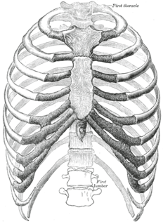
The rib cage is the arrangement of ribs attached to the vertebral column and sternum in the thorax of most vertebrates, that encloses and protects the heart and lungs. In humans, the rib cage, also known as the thoracic cage, is a bony and cartilaginous structure which surrounds the thoracic cavity and supports the shoulder girdle to form the core part of the human skeleton. A typical human rib cage consists of 24 ribs in 12 pairs, the sternum and xiphoid process, the costal cartilages, and the 12 thoracic vertebrae.

In humans, the respiratory tract is the part of the anatomy of the respiratory system involved with the process of respiration. The respiratory tract is lined with respiratory mucosa or respiratory epithelium. Air is breathed in through the nose or the mouth. In the nasal cavity, a layer of nasal mucosa acts as a filter and traps pollutants and other harmful substances found in the air. Next, air moves into the pharynx, a passage that contains the intersection between the oesophagus and the larynx. The opening of the larynx has a special flap of cartilage, the epiglottis, that opens to allow air to pass through but closes to prevent food from moving into the airway.

The thoracic diaphragm, or simply the diaphragm, is a sheet of internal skeletal muscle in humans and other mammals that extends across the bottom of the thoracic cavity. The diaphragm separates the thoracic cavity, containing the heart and lungs, from the abdominal cavity and performs an important function in respiration: as the diaphragm contracts, the volume of the thoracic cavity increases, creating a negative pressure there, which draws air into the lungs.
Exhalation is the flow of the breath out of an organism. In animals, it is the movement of air from the lungs out of the airways, to the external environment during breathing.
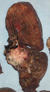
A pneumonectomy is a surgical procedure to remove a lung. Removal of just one lobe of the lung is specifically referred to as a lobectomy, and that of a segment of the lung as a wedge resection.

A respiratory examination, or lung examination, is performed as part of a physical examination, in response to respiratory symptoms such as shortness of breath, cough, or chest pain, and is often carried out with a cardiac examination.
Flail chest is a life-threatening medical condition that occurs when a segment of the rib cage breaks due to trauma and becomes detached from the rest of the chest wall. Two of the symptoms of flail chest are chest pain and shortness of breath.

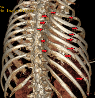
A rib fracture is a break in a rib bone. This typically results in chest pain that is worse with breathing in. Bruising may occur at the site of the break. When several ribs are broken in several places a flail chest results. Potential complications include a pneumothorax, pulmonary contusion, and pneumonia.
The muscles of respiration are those muscles that contribute to inhalation and exhalation, by aiding in the expansion and contraction of the thoracic cavity. The diaphragm and, to a lesser extent, the intercostal muscles drive respiration during quiet breathing. Additional 'accessory muscles of respiration' are typically only used under conditions of high metabolic demand or respiratory dysfunction. However, in instances where these accessory muscles become stiff and hard, expansion of the rib cage can be restricted. Maintenance of the elasticity of these muscles is crucial to the health of the respiratory system and to maximize its functional capabilities.
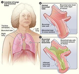
Bronchitis is inflammation of the bronchi in the lungs that causes coughing. Symptoms include coughing up sputum, wheezing, shortness of breath, and chest pain. Bronchitis can be acute or chronic.
Critical illness polyneuropathy (CIP) and critical illness myopathy (CIM) are overlapping syndromes of diffuse, symmetric, flaccid muscle weakness occurring in critically ill patients and involving all extremities and the diaphragm with relative sparing of the cranial nerves. CIP and CIM have similar symptoms and presentations and are often distinguished largely on the basis of specialized electrophysiologic testing or muscle and nerve biopsy. The causes of CIP and CIM are unknown, though they are thought to be a possible neurological manifestation of systemic inflammatory response syndrome. Corticosteroids and neuromuscular blocking agents, which are widely used in intensive care, may contribute to the development of CIP and CIM, as may elevations in blood sugar, which frequently occur in critically ill patients.

Diaphragmatic rupture is a tear of the diaphragm, the muscle across the bottom of the ribcage that plays a crucial role in respiration. Most commonly, acquired diaphragmatic tears result from physical trauma. Diaphragmatic rupture can result from blunt or penetrating trauma and occurs in about 5% of cases of severe blunt trauma to the trunk.

Shoulder impingement syndrome is a syndrome involving tendonitis of the rotator cuff muscles as they pass through the subacromial space, the passage beneath the acromion. It is particularly associated with tendonitis of the supraspinatus muscle. This can result in pain, weakness, and loss of movement at the shoulder.
Hoover’s sign of leg paresis is one of two signs named for Charles Franklin Hoover. It is a maneuver aimed to separate organic from non-organic paresis of the leg. The sign relies on the principle of synergistic contraction.
Charles Franklin Hoover (1865–1927) was an American physician born in Cleveland, Ohio, who read medicine at Harvard. He worked in Vienna under Neusser, and in Strasbourg with F Kraus before returning to Cleveland. He was appointed Professor of Medicine in 1907. His main interests were in diseases of the diaphragm, lungs, and liver.
Dahl's sign is a clinical sign in which areas of darkened (hyperpigmentation) and thickened (hyperkeratotic) skin are seen on the lower thighs and elbows. It occurs in patients with longstanding severe chronic obstructive pulmonary disease.
Chronic obstructive pulmonary disease (COPD) is a type of obstructive lung disease characterized by long-term breathing problems and poor airflow. The main symptoms include shortness of breath and cough with sputum production. COPD is a progressive disease, meaning it typically worsens over time. Eventually, everyday activities such as walking or getting dressed become difficult. Chronic bronchitis and emphysema are older terms used for different types of COPD. The term "chronic bronchitis" is still used to define a productive cough that is present for at least three months each year for two years. Those with such a cough are at a greater risk of developing COPD. The term "emphysema" is also used for the abnormal presence of air or other gas within tissues.
Litten's sign, also known as the diaphragm phenomenon, is a paralyzed hemidiaphragm, the portion of the diaphragm in contact with the parietal pleura during respiration in the base of the pleural cavity.

Pneumatosis, also known as emphysema, is the abnormal presence of air or other gas within tissues.
References
- ↑ "George Crile, Charles Hoover and John Phillips". Archived from the original on 2016-03-03.
- ↑ Binazzi B, Bianchi R, Romagnoli I, et al. (February 2008). "Chest wall kinematics and Hoover's sign". Respir Physiol Neurobiol. 160 (3): 325–33. doi:10.1016/j.resp.2007.10.019. PMID 18088571.
- ↑ Garcia-Pachon E, Padilla-Navas I (May 2006). "Frequency of Hoover's sign in stable patients with chronic obstructive pulmonary disease". Int. J. Clin. Pract. 60 (5): 514–7. doi:10.1111/j.1368-5031.2006.00850.x. PMID 16700846.