The neck is the part of the body on many vertebrates that connects the head with the torso. The neck supports the weight of the head and protects the nerves that carry sensory and motor information from the brain down to the rest of the body. In addition, the neck is highly flexible and allows the head to turn and flex in all directions. The structures of the human neck are anatomically grouped into four compartments: vertebral, visceral and two vascular compartments. Within these compartments, the neck houses the cervical vertebrae and cervical part of the spinal cord, upper parts of the respiratory and digestive tracts, endocrine glands, nerves, arteries and veins. Muscles of the neck are described separately from the compartments. They bound the neck triangles.

In anatomy, the thigh is the area between the hip (pelvis) and the knee. Anatomically, it is part of the lower limb.

In human anatomy, the thoracic duct is the larger of the two lymph ducts of the lymphatic system. The thoracic duct usually begins from the upper aspect of the cisterna chyli, passing out of the abdomen through the aortic hiatus into first the posterior mediastinum and then the superior mediastinum, extending as high up as the root of the neck before descending to drain into the systemic (blood) circulation at the venous angle.

The external jugular vein receives the greater part of the blood from the exterior of the cranium and the deep parts of the face, being formed by the junction of the posterior division of the retromandibular vein with the posterior auricular vein.

In human anatomy, the cephalic vein is a superficial vein in the arm. It originates from the radial end of the dorsal venous network of hand, and ascends along the radial (lateral) side of the arm before emptying into the axillary vein. At the elbow, it communicates with the basilic vein via the median cubital vein.

The cavernous sinus within the human head is one of the dural venous sinuses creating a cavity called the lateral sellar compartment bordered by the temporal bone of the skull and the sphenoid bone, lateral to the sella turcica.
The straight sinus, also known as tentorial sinus or the sinus rectus, is an area within the skull beneath the brain. It receives blood from the inferior sagittal sinus and the great cerebral vein, and drains into the confluence of sinuses.
The superior sagittal sinus, within the human head, is an unpaired area along the attached margin of the falx cerebri. It allows blood to drain from the lateral aspects of anterior cerebral hemispheres to the confluence of sinuses. Cerebrospinal fluid drains through arachnoid granulations into the superior sagittal sinus and is returned to venous circulation.
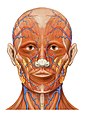
The facial vein is a relatively large vein in the human face. It commences at the side of the root of the nose and is a direct continuation of the angular vein where it also receives a small nasal branch. It lies behind the facial artery and follows a less tortuous course. It receives blood from the external palatine vein before it either joins the anterior branch of the retromandibular vein to form the common facial vein, or drains directly into the internal jugular vein.

The inferior thyroid veins appear two, frequently three or four, in number, and arise in the venous plexus on the thyroid gland, communicating with the middle and superior thyroid veins. While the superior and middle thyroid veins serve as direct tributaries to the internal jugular vein, the inferior thyroid veins drain directly to the brachiocephalic veins.
The maxillary vein or internal maxillary vein is a vein of the head. It is a short trunk which accompanies the maxillary artery. It is formed by a confluence of the veins of the pterygoid plexus. It and passes posterior-ward between the sphenomandibular ligament and the neck of the mandible to enter the parotid gland where unites with the superficial temporal vein to form the retromandibular vein.
The supraorbital vein is a vein of the forehead. It communicates with the frontal branch of the superficial temporal vein. It passes through the supraorbital notch, and merges with the angular vein to form the superior ophthalmic vein. The supraorbital vein helps to drain blood from the forehead, eyebrow, and upper eyelid.
The superficial temporal vein is a vein of the side of the head which collects venous blood from the region of the temple. It arises from an anastomosing venous plexus on the side and vertex of the head. The superficial temporal vein terminates within the substance of the parotid gland by uniting with the maxillary vein to form the retromandibular vein.
The angular vein is a vein of the face. It is the upper part of the facial vein, above its junction with the superior labial vein. It is formed by the junction of the supratrochlear vein and supraorbital vein, and joins with the superior labial vein. It drains the medial canthus, and parts of the nose and the upper lip. It can be a route of spread of infection from the danger triangle of the face to the cavernous sinus.
The retromandibular vein is a major vein of the face. It is formed within the parotid gland by the confluence of the maxillary vein, and superficial temporal vein. It descends in the gland and splits into two branches upon emerging from the gland. Its anterior branch then joins the (anterior) facial vein forming the common facial vein, while its posterior branch joins the posterior auricular vein forming the external jugular vein.
The posterior auricular vein is a vein of the head. It begins from a plexus with the occipital vein and the superficial temporal vein, descends behind the auricle, and drains into the external jugular vein.

The intercostal arteries are a group of arteries passing within an intercostal space. There are 9 anterior and 11 posterior intercostal arteries on each side of the body. The anterior intercostal arteries are branches of the internal thoracic artery and its terminal branch - the musculophrenic artery. The posterior intercostal arteries are branches of the supreme intercostal artery and thoracic aorta.
The superior labial vein is the vein receiving blood from the upper lip.

The following outline is provided as an overview of and topical guide to human anatomy:

In anatomy, the venae cavae are two large veins that return deoxygenated blood from the body into the heart. In humans they are the superior vena cava and the inferior vena cava, and both empty into the right atrium. They are located slightly off-center, toward the right side of the body.