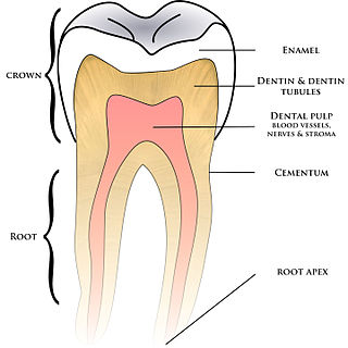
Cementum is a specialized calcified substance covering the root of a tooth. The cementum is the part of the periodontium that attaches the teeth to the alveolar bone by anchoring the periodontal ligament.

Tooth enamel is one of the four major tissues that make up the tooth in humans and many animals, including some species of fish. It makes up the normally visible part of the tooth, covering the crown. The other major tissues are dentin, cementum, and dental pulp. It is a very hard, white to off-white, highly mineralised substance that acts as a barrier to protect the tooth but can become susceptible to degradation, especially by acids from food and drink. In rare circumstances enamel fails to form, leaving the underlying dentin exposed on the surface.

Ameloblasts are cells present only during tooth development that deposit tooth enamel, which is the hard outermost layer of the tooth forming the surface of the crown.
Tomes's processes are a histologic landmark identified on an ameloblast, cells involved in the production of tooth enamel. During the synthesis of enamel, the ameloblast moves away from the enamel, forming a projection surrounded by the developing enamel. Tomes's processes are those projections and give the ameloblast a "picket-fence" appearance under a microscope.
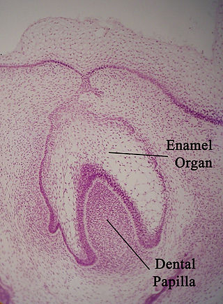
The enamel organ, also known as the dental organ, is a cellular aggregation seen in a developing tooth and it lies above the dental papilla. The enamel organ which is differentiated from the primitive oral epithelium lining the stomodeum. The enamel organ is responsible for the formation of enamel, initiation of dentine formation, establishment of the shape of a tooth's crown, and establishment of the dentoenamel junction.

Tooth development or odontogenesis is the complex process by which teeth form from embryonic cells, grow, and erupt into the mouth. For human teeth to have a healthy oral environment, all parts of the tooth must develop during appropriate stages of fetal development. Primary (baby) teeth start to form between the sixth and eighth week of prenatal development, and permanent teeth begin to form in the twentieth week. If teeth do not start to develop at or near these times, they will not develop at all, resulting in hypodontia or anodontia.
Amelogenesis is the formation of enamel on teeth and begins when the crown is forming during the advanced bell stage of tooth development after dentinogenesis forms a first layer of dentin. Dentin must be present for enamel to be formed. Ameloblasts must also be present for dentinogenesis to continue.

In embryology and prenatal development, the dental papilla is a condensation of ectomesenchymal cells called odontoblasts, seen in histologic sections of a developing tooth. It lies below a cellular aggregation known as the enamel organ. The dental papilla appears after 8–10 weeks of intra uteral life. The dental papilla gives rise to the dentin and pulp of a tooth.
In vertebrates, an odontoblast is a cell of neural crest origin that is part of the outer surface of the dental pulp, and whose biological function is dentinogenesis, which is the formation of dentin, the substance beneath the tooth enamel on the crown and the cementum on the root.
In animal tooth development, dentinogenesis is the formation of dentin, a substance that forms the majority of teeth. Dentinogenesis is performed by odontoblasts, which are a special type of biological cell on the outer wall of dental pulps, and it begins at the late bell stage of a tooth development. The different stages of dentin formation after differentiation of the cell result in different types of dentin: mantle dentin, primary dentin, secondary dentin, and tertiary dentin.

The dental follicle, also known as dental sac, is made up of mesenchymal cells and fibres surrounding the enamel organ and dental papilla of a developing tooth. It is a vascular fibrous sac containing the developing tooth and its odontogenic organ. The dental follicle (DF) differentiates into the periodontal ligament. In addition, it may be the precursor of other cells of the periodontium, including osteoblasts, cementoblasts and fibroblasts. They develop into the alveolar bone, the cementum with Sharpey's fibers and the periodontal ligament fibers respectively. Similar to dental papilla, the dental follicle provides nutrition to the enamel organ and dental papilla and also have an extremely rich blood supply.

The outer enamel epithelium, also known as the external enamel epithelium, is a layer of cuboidal cells located on the periphery of the enamel organ in a developing tooth. This layer is first seen during the bell stage.
The cervical loop is the location on an enamel organ in a developing tooth where the outer enamel epithelium and the inner enamel epithelium join. The cervical loop is a histologic term indicating a specific epithelial structure at the apical side of the tooth germ, consisting of loosely aggregated stellate reticulum in the center surrounded by stratum intermedium. These tissues are enveloped by a basal layer of epithelium known on the outside of the tooth as outer enamel epithelium and on the inside as inner enamel epithelium. During root formation the inner layers of epithelium disappear and only the basal layers are left creating Hertwig's epithelial root sheath (HERS). At this point it is usually referred to as HERS instead of the cervical loop to indicate the structural difference.
In tooth development, the enamel knot is a localization of cells on an enamel organ that appear thickened in the center of the inner enamel epithelium. The enamel knot is frequently associated with an enamel cord. It is formed in the cap stage and undergoes apoptosis in the bell stage.
In animal tooth development, the stellate reticulum is a group of cells located in the center of the enamel organ of a developing tooth. These cells are star-shaped and synthesize glycosaminoglycans. As glycosaminoglycans are produced, water is drawn in between the cells, stretching them apart. As they are moved further away from one another, the stellate reticular cells maintain contact with one another through desmosomes, resulting in their unique appearance. The stellate reticulum is lost after the first layer of enamel is laid down. This brings cells in the inner enamel epithelium closer to blood vessels at the periphery.
The stratum intermedium in a developing tooth is a layer of two or three cells between the inner enamel epithelium and the newly forming cells of the stellate reticulum. It first appears during the early bell stage of tooth development, at around the 14th week of intrauterine life. These cells are closely attached by desmosomes and gap junctions .The stratum intermedium has a notably high alkaline phosphatase activity. This layer, along with the inner enamel epithelium, is responsible for the tooth enamel formation. It is a part of the dental (enamel) organ. Stratum intermedium stores glycogen. It is absent in the part of the tooth germ that outlines the root portions of the tooth which does not form enamel.

The Hertwig epithelial root sheath (HERS) or epithelial root sheath is a proliferation of epithelial cells located at the cervical loop of the enamel organ in a developing tooth. Hertwig epithelial root sheath initiates the formation of dentin in the root of a tooth by causing the differentiation of odontoblasts from the dental papilla. The root sheath eventually disintegrates with the periodontal ligament, but residual pieces that do not completely disappear are seen as epithelial cell rests of Malassez (ERM). These rests can become cystic, presenting future periodontal infections.
The reduced enamel epithelium, sometimes called reduced dental epithelium, overlies a developing tooth and is formed by two layers: a layer of ameloblast cells and the adjacent layer of cuboidal cells from the dental lamina. As the cells of the reduced enamel epithelium degenerate, the tooth is revealed progressively with its eruption into the mouth. The degeneration of reduced enamel epithelium also mediates the initial epithelial attachment to the tooth, which is called the junctional epithelium.
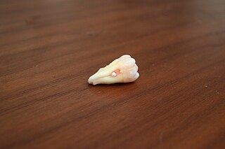
Enamel pearls are developmental variations of teeth that present as beads or nodules of enamel in places where they are not normally observed.
In dental anatomy, the junctional epithelium (JE) is that epithelium which lies at, and in health also defines, the base of the gingival sulcus. The probing depth of the gingival sulcus is measured by a calibrated periodontal probe. In a healthy-case scenario, the probe is gently inserted, slides by the sulcular epithelium (SE), and is stopped by the epithelial attachment (EA). However, the probing depth of the gingival sulcus may be considerably different from the true histological gingival sulcus depth.
