Related Research Articles

Human teeth function to mechanically break down items of food by cutting and crushing them in preparation for swallowing and digesting. As such, they are considered part of the human digestive system. Humans have four types of teeth: incisors, canines, premolars, and molars, which each have a specific function. The incisors cut the food, the canines tear the food and the molars and premolars crush the food. The roots of teeth are embedded in the maxilla or the mandible and are covered by gums. Teeth are made of multiple tissues of varying density and hardness.

Cementum is a specialized calcified substance covering the root of a tooth. The cementum is the part of the periodontium that attaches the teeth to the alveolar bone by anchoring the periodontal ligament.

Ameloblasts are cells present only during tooth development that deposit tooth enamel, which is the hard outermost layer of the tooth forming the surface of the crown.
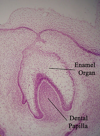
The enamel organ, also known as the dental organ, is a cellular aggregation seen in a developing tooth and it lies above the dental papilla. The enamel organ which is differentiated from the primitive oral epithelium lining the stomodeum.The enamel organ is responsible for the formation of enamel, initiation of dentine formation, establishment of the shape of a tooth's crown, and establishment of the dentoenamel junction.

Tooth development or odontogenesis is the complex process by which teeth form from embryonic cells, grow, and erupt into the mouth. For human teeth to have a healthy oral environment, all parts of the tooth must develop during appropriate stages of fetal development. Primary (baby) teeth start to form between the sixth and eighth week of prenatal development, and permanent teeth begin to form in the twentieth week. If teeth do not start to develop at or near these times, they will not develop at all, resulting in hypodontia or anodontia.
Amelogenesis is the formation of enamel on teeth and begins when the crown is forming during the advanced bell stage of tooth development after dentinogenesis forms a first layer of dentin. Dentin must be present for enamel to be formed. Ameloblasts must also be present for dentinogenesis to continue.

In embryology and prenatal development, the dental papilla is a condensation of ectomesenchymal cells called odontoblasts, seen in histologic sections of a developing tooth. It lies below a cellular aggregation known as the enamel organ. The dental papilla appears after 8–10 weeks intra uteral life. The dental papilla gives rise to the dentin and pulp of a tooth.
Dentinogenesis is the formation of dentin, a substance that forms the majority of teeth. Dentinogenesis is performed by odontoblasts, which are a special type of biological cell on the outer wall of dental pulps, and it begins at the late bell stage of a tooth development. The different stages of dentin formation after differentiation of the cell result in different types of dentin: mantle dentin, primary dentin, secondary dentin, and tertiary dentin.

The dental follicle, also known as dental sac, is made up of mesenchymal cells and fibres surrounding the enamel organ and dental papilla of a developing tooth. It is a vascular fibrous sac containing the developing tooth and its odontogenic organ. The dental follicle (DF) differentiates into the periodontal ligament. In addition, it may be the precursor of other cells of the periodontium, including osteoblasts, cementoblasts and fibroblasts. They develop into the alveolar bone, the cementum with Sharpey's fibers and the periodontal ligament fibers respectively. Similar to dental papilla, the dental follicle provides nutrition to the enamel organ and dental papilla and also have an extremely rich blood supply.
The oral mucosa is the mucous membrane lining the inside of the mouth. It comprises stratified squamous epithelium, termed "oral epithelium", and an underlying connective tissue termed lamina propria. The oral cavity has sometimes been described as a mirror that reflects the health of the individual. Changes indicative of disease are seen as alterations in the oral mucosa lining the mouth, which can reveal systemic conditions, such as diabetes or vitamin deficiency, or the local effects of chronic tobacco or alcohol use. The oral mucosa tends to heal faster and with less scar formation compared to the skin. The underlying mechanism remains unknown, but research suggests that extracellular vesicles might be involved.

The outer enamel epithelium, also known as the external enamel epithelium, is a layer of cuboidal cells located on the periphery of the enamel organ in a developing tooth. This layer is first seen during the bell stage.
The inner enamel epithelium, also known as the internal enamel epithelium, is a layer of columnar cells located on the rim nearest the dental papilla of the enamel organ in a developing tooth. This layer is first seen during the cap stage, in which these inner enamel epithelium cells are pre-ameloblast cells. These will differentiate into Ameloblasts which are responsible for secretion of enamel during tooth development.
The cervical loop is the location on an enamel organ in a developing tooth where the outer enamel epithelium and the inner enamel epithelium join. The cervical loop is a histologic term indicating a specific epithelial structure at the apical side of the tooth germ, consisting of loosely aggregated stellate reticulum in the center surrounded by stratum intermedium. These tissues are enveloped by a basal layer of epithelium known on the outside of the tooth as outer enamel epithelium and on the inside as inner enamel epithelium. During root formation the inner layers of epithelium disappear and only the basal layers are left creating Hertwig's epithelial root sheath (HERS). At this point it is usually referred to as HERS instead of the cervical loop to indicate the structural difference.
In tooth development, the enamel knot is a localization of cells on an enamel organ that appear thickened in the center of the inner enamel epithelium. The enamel knot is frequently associated with an enamel cord. It is formed in the cap stage and undergoes apoptosis in the bell stage.
The stellate reticulum is a group of cells located in the center of the enamel organ of a developing tooth. These cells are star-shaped and synthesize glycosaminoglycans. As glycosaminoglycans are produced, water is drawn in between the cells, stretching them apart. As they are moved further away from one another, the stellate reticular cells maintain contact with one another through desmosomes, resulting in their unique appearance. The stellate reticulum is lost after the first layer of enamel is laid down. This brings cells in the inner enamel epithelium closer to blood vessels at the periphery.

The Hertwig epithelial root sheath (HERS) or epithelial root sheath is a proliferation of epithelial cells located at the cervical loop of the enamel organ in a developing tooth. Hertwig epithelial root sheath initiates the formation of dentin in the root of a tooth by causing the differentiation of odontoblasts from the dental papilla. The root sheath eventually disintegrates with the periodontal ligament, but residual pieces that do not completely disappear are seen as epithelial cell rests of Malassez (ERM). These rests can become cystic, presenting future periodontal infections.
The reduced enamel epithelium, sometimes called reduced dental epithelium, overlies a developing tooth and is formed by two layers: a layer of ameloblast cells and the adjacent layer of cuboidal cells from the dental lamina. As the cells of the reduced enamel epithelium degenerate, the tooth is revealed progressively with its eruption into the mouth. The degeneration of reduced enamel epithelium also mediates the initial epithelial attachment to the tooth, which is called the junctional epithelium.

This article describes the anatomy of the head and neck of the human body, including the brain, bones, muscles, blood vessels, nerves, glands, nose, mouth, teeth, tongue, and throat.
Dental anatomy is a field of anatomy dedicated to the study of human tooth structures. The development, appearance, and classification of teeth fall within its purview. Tooth formation begins before birth, and the teeth's eventual morphology is dictated during this time. Dental anatomy is also a taxonomical science: it is concerned with the naming of teeth and the structures of which they are made, this information serving a practical purpose in dental treatment.
The junctional epithelium (JE) is that epithelium which lies at, and in health also defines, the base of the gingival sulcus. The probing depth of the gingival sulcus is measured by a calibrated periodontal probe. In a healthy-case scenario, the probe is gently inserted, slides by the sulcular epithelium (SE), and is stopped by the epithelial attachment (EA). However, the probing depth of the gingival sulcus may be considerably different from the true histological gingival sulcus depth.
References
- Cate, A.R. Ten. Oral Histology: development, structure, and function. 5th ed. 1998. ISBN 0-8151-2952-1.
- BKB Berkovitz, GR Holland, BJMoxham. Oral Anatomy Histology and Embryology. 3rd edition. Mosby ISBN 0-7234-3181-7
| | This dentistry article is a stub. You can help Wikipedia by expanding it. |