Interleukin 33

Last updated
IL33
2KLL.pdb.png
Available structures
PDB Ortholog search: PDBe RCSB
Identifiers
Aliases IL33 , C9orf26, DVS27, IL1F11, NF-HEV, NFEHEV, Interleukin 33, IL-33, IL-1F11
External IDs OMIM: 608678 MGI: 1924375 HomoloGene: 14126 GeneCards: IL33
Orthologs
SpeciesHumanMouse
Entrez
Ensembl
UniProt
RefSeq (mRNA)

NM_001164724
NM_133775
NM_001360725

RefSeq (protein)

NP_001158196
NP_598536
NP_001347654

Location (UCSC) Chr 9: 6.22 – 6.26 Mb Chr 19: 29.9 – 29.94 Mb
PubMed search [3] [4]
Wikidata
View/Edit Human View/Edit Mouse

Interleukin 33 (IL-33) is a protein that in humans is encoded by the IL33 gene. [5]

Contents

Interleukin 33 is a member of the IL-1 family that potently drives production of T helper-2 (Th2)-associated cytokines (e.g., IL-4). IL33 is a ligand for ST2 (IL1RL1), an IL-1 family receptor that is highly expressed on Th2 cells, mast cells and group 2 innate lymphocytes. [6]

IL-33 is expressed by a wide variety of cell types, including fibroblasts, mast cells, dendritic cells, macrophages, osteoblasts, endothelial cells, and epithelial cells. [7]

Structure

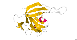
IL-33 is a member of the IL-1 superfamily of cytokines, a determination based in part on the molecules β-trefoil structure, a conserved structure type described in other IL-1 cytokines, including IL-1α, IL-1β, IL-1Ra and IL-18. In this structure, the 12 β-strands of the β-trefoil are arranged in three pseudorepeats of four β-strand units, of which the first and last β-strands are antiparallel staves in a six-stranded β-barrel, while the second and third β-strands of each repeat form a β-hairpin sitting atop the β-barrel. IL-33 is a ligand that binds to a high-affinity receptor family member ST2. The complex of these two molecules with IL-1RAcP indicates a ternary complex formation. The binding area appears to be a mix of polar and non-polar regions that create a specific binding between ligand and receptor. The interface between the molecules has been shown to be extensive. Structural data on the IL-33 molecule was determined by solution NMR and small angle X-ray scattering. [8]

Function

Interleukin 33 (IL-33) is a cytokine belonging to the IL-1 superfamily. IL-33 induces helper T cells, mast cells, eosinophils and basophils to produce type 2 cytokines. This cytokine was previously named NF-HEV 'nuclear factor (NF) in high endothelial venules' (HEVs) since it was originally identified in these specialized cells. [9] IL-33 acts intracellularly as a nuclear factor and extracellularly as a cytokine.

Nuclear role

IL-33 is constitutively located in the nucleus of structural cells of humans and mice [10] and has a helix-turn-helix domain [9] presumably allowing it to bind to DNA. There is a paucity of research into the nuclear role of IL-33 but amino acids 40-58 in human IL-33 are sufficient for nuclear localisation and histone binding. [11] IL-33 also interacts with the histone methyltransferase SUV39H1 [12] and murine appears to IL-33 interact to NF-κB. [13]

Cytokine role

As a cytokine, IL-33 interacts with the receptors ST2 (also known as IL1RL1) and IL-1 Receptor Accessory Protein (IL1RAP), activating intracellular molecules in the NF-κB and MAP kinase signaling pathways that drive production of type 2 cytokines (e.g. IL-5 and IL-13) from polarized Th2 cells. The induction of type 2 cytokines by IL-33 in vivo is believed to induce the severe pathological changes observed in mucosal organs following administration of IL-33. [14] [15] IL-33 is also effective in reversing Alzheimer-like symptoms in APP/PS1 mice, by reversing the buildup and preventing the new formation of amyloid plaques. [16]

Regulation

Extracellularly, IL-33 is rapidly oxidised. The oxidation process results in the formation of two disulphide bridges and a change in the conformation of the molecule, which prevents it from binding to its receptor, ST2. This is believed to limit the range and duration of the action of IL-33. [17]

Clinical significance

IL-33 has been associated with several disease states through Genome Wide Association Studies: asthma, [18] allergy, [19] endometriosis, [20] and hay fever. [21] In particular, a single-nucleotide polymorphism rs928413 (A/G), is located in the 5′ upstream region of IL33 gene, and its minor “G” allele was identified as a susceptible variant for early childhood asthma [22] and atopic asthma [23] development. The rs928413(G) allele creates a binding site for the cAMP responsive element-binding protein 1 transcription factor that may explain the negative effect of the rs928413 minor “G” allele on asthma development. [24] “T” allele of the polymorphism rs4742170 located in the second intron of IL33 gene was linked to specific wheezing phenotype (intermediate-onset wheeze). [25] Risk “T” rs4742170 allele disrupts binding of GR transcription factor to IL33 putative enhancer that may explain the negative effect of the rs4742170 (T) risk allele on the development of wheezing phenotype that strongly correlates with allergic sensitization in childhood. [26]

This protein is one of many that acts as a cytokine and signals inflammation in the body by acting upon macrophages, neutrophils, B cells, Th2 cells, eosinophils, basophils and mast cells. [27] This protein is also thought to cause the itching that is associated with dermatitis. The IL-33 protein resides in keratinocytes of the skin and when subjected to irritation or allergic conditions will communicate with nearby sensory neurons and initiate an itchy feeling. [28] In IL-33 knockout mice, it was discovered that nuclear IL-33 is associated with wound healing as mice without the protein healed significantly slower than mice with the IL-33 protein. [29] Elevated levels of IL-33 are associated with asthma. [30]

In mice, IL-33 was found to effect the production of methionine-enkephalin peptides in group 2 innate lymphocytes, in turn promoting the emergence of beige adipocytes, which leads to increased energy expenditure and decreased adiposity. [31]

Elevated levels of IL-33 have been reported in some patients with nonsmall cell lung carcinomas. The source of elevated serum levels of IL-33 during the early stages could be bronchial and vascular epithelium. [32] IL-33 knockdown showed lower growth of nonsmall cell lung carcinomas, while overexpression of IL-33 resulted in increased growth. Blocking of IL-33 reduced the growth of human nonsmall cell lung carcinomas. I mice model blocking of IL-33 inhibited tumor growth in immunodeficient mice. [33] [34]

In the mouse colon carcinoma model, IL-33 was expressed by tumor stromal cells, while the colon carcinoma cells did not express ST2 with or without IL-33 stimulation. The IL-33 knockout model had higher tumor growth than wild type. Similarly, IFN- γ expression was increased in the IL-33 knockout model as well as the number of T regulatory cells and CD8+ T cells. [35]

Age-related macular degeneration is a retinal disease leading to neovascularization and thus impaired vision. Current treatment includes administration of anti-VEGF but is not sufficient. Retinal pigment epithelial cells can express IL-33 at both mRNA and protein levels. IL-33 expression is upregulated during inflammatory stimuli. IL-33 can inhibit fibroblasts and endothelial cells that express ST2, which can lead to reduced angiogenesis. [36]

In a mouse model of chronic asthma, anti-IL-33 administration decreased antigen-induced immune response. Similar results were found in ST2 deficient mice. IL-33 activated innate lymphoid cells 2 remained in the lymph nodes for several weeks. CD4 + Th2 cells were formed after repeated exposure to IL-33. This type of cells highly produced IL-5. [37]

Chronic inflammation is characteristic for IBD ( inflammatory bowel disease). Under normal conditions, IL-33 is present in healthy intestinal tissue, but during inflammatory conditions its expression is increased. However, IL-33 has also a protective role under inflammatory conditions and is involved in wound healing. [38]

In brain, IL-33 is expressed in oligodendrocytes and astrocytes and is implicated in the pathophysiology of intracerebral hemorrhage. [39]

Related Research Articles

<span class="mw-page-title-main">T helper cell</span> Type of immune cell

The T helper cells (Th cells), also known as CD4+ cells or CD4-positive cells, are a type of T cell that play an important role in the adaptive immune system. They aid the activity of other immune cells by releasing cytokines. They are considered essential in B cell antibody class switching, breaking cross-tolerance in dendritic cells, in the activation and growth of cytotoxic T cells, and in maximizing bactericidal activity of phagocytes such as macrophages and neutrophils. CD4+ cells are mature Th cells that express the surface protein CD4. Genetic variation in regulatory elements expressed by CD4+ cells determines susceptibility to a broad class of autoimmune diseases.

<span class="mw-page-title-main">Interleukin 12</span> Interleukin

Interleukin 12 (IL-12) is an interleukin that is naturally produced by dendritic cells, macrophages, neutrophils, and human B-lymphoblastoid cells (NC-37) in response to antigenic stimulation. IL-12 belongs to the family of interleukin-12. IL-12 family is unique in comprising the only heterodimeric cytokines, which includes IL-12, IL-23, IL-27 and IL-35. Despite sharing many structural features and molecular partners, they mediate surprisingly diverse functional effects.

<span class="mw-page-title-main">Interleukin 4</span> Mammalian protein found in Mus musculus

The interleukin 4 is a cytokine that induces differentiation of naive helper T cells (Th0 cells) to Th2 cells. Upon activation by IL-4, Th2 cells subsequently produce additional IL-4 in a positive feedback loop. IL-4 is produced primarily by mast cells, Th2 cells, eosinophils and basophils. It is closely related and has functions similar to IL-13.

<span class="mw-page-title-main">Interleukin 9</span> Protein-coding gene in the species Homo sapiens

Interleukin 9, also known as IL-9, is a pleiotropic cytokine belonging to the group of interleukins. IL-9 is produced by variety of cells like mast cells, NKT cells, Th2, Th17, Treg, ILC2, and Th9 cells in different amounts. Among them, Th9 cells are regarded as the major CD4+ T cells that produce IL-9.

<span class="mw-page-title-main">Interleukin 13</span> Protein and coding gene in humans

Interleukin 13 (IL-13) is a protein that in humans is encoded by the IL13 gene. IL-13 was first cloned in 1993 and is located on chromosome 5q31.1 with a length of 1.4kb. It has a mass of 13 kDa and folds into 4 alpha helical bundles. The secondary structural features of IL-13 are similar to that of Interleukin 4 (IL-4); however it only has 25% sequence identity to IL-4 and is capable of IL-4 independent signaling. IL-13 is a cytokine secreted by T helper type 2 (Th2) cells, CD4 cells, natural killer T cell, mast cells, basophils, eosinophils and nuocytes. Interleukin-13 is a central regulator in IgE synthesis, goblet cell hyperplasia, mucus hypersecretion, airway hyperresponsiveness, fibrosis and chitinase up-regulation. It is a mediator of allergic inflammation and different diseases including asthma.

<span class="mw-page-title-main">Interleukin 15</span> Cytokine with structural similarity to Interleukin-2

Interleukin-15 (IL-15) is a protein that in humans is encoded by the IL15 gene. IL-15 is an inflammatory cytokine with structural similarity to Interleukin-2 (IL-2). Like IL-2, IL-15 binds to and signals through a complex composed of IL-2/IL-15 receptor beta chain (CD122) and the common gamma chain. IL-15 is secreted by mononuclear phagocytes following infection by virus(es). This cytokine induces the proliferation of natural killer cells, i.e. cells of the innate immune system whose principal role is to kill virally infected cells.

<span class="mw-page-title-main">Interleukin 31</span>

Interleukin-31 (IL-31) is a protein that in humans is encoded by the IL31 gene that resides on chromosome 12. IL-31 is an inflammatory cytokine that helps trigger cell-mediated immunity against pathogens. It has also been identified as a major player in a number of chronic inflammatory diseases, including atopic dermatitis.

<span class="mw-page-title-main">Interleukin 25</span> Cytokine that belongs to the IL-17 cytokine family

Interleukin-25 (IL-25) – also known as interleukin-17E (IL-17E) – is a protein that in humans is encoded by the IL25 gene on chromosome 14. IL-25 was discovered in 2001 and is made up of 177 amino acids.

<span class="mw-page-title-main">Interleukin 17</span> Group of proteins

Interleukin 17 family is a family of pro-inflammatory cystine knot cytokines. They are produced by a group of T helper cell known as T helper 17 cell in response to their stimulation with IL-23. Originally, Th17 was identified in 1993 by Rouvier et al. who isolated IL17A transcript from a rodent T-cell hybridoma. The protein encoded by IL17A is a founding member of IL-17 family. IL17A protein exhibits a high homology with a viral IL-17-like protein encoded in the genome of T-lymphotropic rhadinovirus Herpesvirus saimiri. In rodents, IL-17A is often referred to as CTLA8.

<span class="mw-page-title-main">Interleukin 19</span> Protein-coding gene in the species Homo sapiens

Interleukin 19 (IL-19) is an immunosuppressive protein that belongs to the IL-10 cytokine subfamily.

<span class="mw-page-title-main">Tyrosine kinase 2</span> Enzyme and coding gene in humans

Non-receptor tyrosine-protein kinase TYK2 is an enzyme that in humans is encoded by the TYK2 gene.

<span class="mw-page-title-main">STAT6</span> Protein and coding gene in humans

Signal transducer and activator of transcription 6 (STAT6) is a transcription factor that belongs to the Signal Transducer and Activator of Transcription (STAT) family of proteins. The proteins of STAT family transmit signals from a receptor complex to the nucleus and activate gene expression. Similarly as other STAT family proteins, STAT6 is also activated by growth factors and cytokines. STAT6 is mainly activated by cytokines interleukin-4 and interleukin-13.

<span class="mw-page-title-main">Interleukin 37</span> Protein-coding gene in the species Homo sapiens

Interleukin 37 (IL-37), also known as Interleukin-1 family member 7 (IL-1F7), is an anti-inflammatory cytokine important for the downregulation of pro-inflammatory cytokine production as well as the suppression of tumor cell growth.

<span class="mw-page-title-main">Thymic stromal lymphopoietin</span> Protein-coding gene in the species Homo sapiens

Thymic stromal lymphopoietin (TSLP) is a protein belonging to the cytokine family. It is known to play an important role in the maturation of T cell populations through activation of antigen-presenting cells.

The interleukin-13 receptor is a type I cytokine receptor, binding Interleukin-13. It consists of two subunits, encoded by IL13RA1 and IL4R, respectively. These two genes encode the proteins IL-13Rα1 and IL-4Rα. These form a dimer with IL-13 binding to the IL-13Rα1 chain and IL-4Rα stabilises this interaction. This IL-13 receptor can also instigate IL-4 signalling. In both cases this occurs via activation of the Janus kinase (JAK)/Signal Transducer and Activator of Transcription (STAT) pathway, resulting in phosphorylation of STAT6. Phosphorylated STAT6 dimerises and acts as a transcription factor activating many genes, such as eotaxin.

<span class="mw-page-title-main">Interleukin-17A</span> Protein-coding gene in the species Homo sapiens

Interleukin-17A is a protein that in humans is encoded by the IL17A gene. In rodents, IL-17A used to be referred to as CTLA8, after the similarity with a viral gene.

<span class="mw-page-title-main">Interleukin-1 family</span> Group of cytokines playing a key role in the regulation of immune and inflammatory responses

The Interleukin-1 family is a group of 11 cytokines that plays a central role in the regulation of immune and inflammatory responses to infections or sterile insults.

<span class="mw-page-title-main">IL1RL1</span> Protein-coding gene in the species Homo sapiens

Interleukin 1 receptor-like 1, also known as IL1RL1 and ST2, is a protein that in humans is encoded by the IL1RL1 gene.

<span class="mw-page-title-main">ILC2</span>

ILC2 cells, or type 2 innate lymphoid cells are a type of innate lymphoid cell. Not to be confused with the ILC. They are derived from common lymphoid progenitor and belong to the lymphoid lineage. These cells lack antigen specific B or T cell receptor because of the lack of recombination activating gene. ILC2s produce type 2 cytokines and are involved in responses to helminths, allergens, some viruses, such as influenza virus and cancer.

In cell biology, TH9 cells are a sub-population of CD4+T cells that produce interleukin-9 (IL-9). They play a role in defense against helminth infections, in allergic responses, in autoimmunity, and tumor suppression.

References

  1. 1 2 3 GRCh38: Ensembl release 89: ENSG00000137033 - Ensembl, May 2017
  2. 1 2 3 GRCm38: Ensembl release 89: ENSMUSG00000024810 - Ensembl, May 2017
  3. "Human PubMed Reference:". National Center for Biotechnology Information, U.S. National Library of Medicine.
  4. "Mouse PubMed Reference:". National Center for Biotechnology Information, U.S. National Library of Medicine.
  5. "Entrez Gene: Interleukin 33".
  6. Yagami A, Orihara K, Morita H, Futamura K, Hashimoto N, Matsumoto K, et al. (November 2010). "IL-33 mediates inflammatory responses in human lung tissue cells". Journal of Immunology. 185 (10): 5743–50. doi: 10.4049/jimmunol.0903818 . PMID   20926795. S2CID   27317847.
  7. Mirchandani AS, Salmond RJ, Liew FY (August 2012). "Interleukin-33 and the function of innate lymphoid cells". Trends in Immunology. 33 (8): 389–96. doi:10.1016/j.it.2012.04.005. PMID   22609147.
  8. Lingel A, Weiss TM, Niebuhr M, Pan B, Appleton BA, Wiesmann C, et al. (October 2009). "Structure of IL-33 and its interaction with the ST2 and IL-1RAcP receptors--insight into heterotrimeric IL-1 signaling complexes". Structure. 17 (10): 1398–410. doi:10.1016/j.str.2009.08.009. PMC   2766095 . PMID   19836339.
  9. 1 2 Baekkevold ES, Roussigné M, Yamanaka T, Johansen FE, Jahnsen FL, Amalric F, et al. (July 2003). "Molecular characterization of NF-HEV, a nuclear factor preferentially expressed in human high endothelial venules". The American Journal of Pathology. 163 (1): 69–79. doi:10.1016/S0002-9440(10)63631-0. PMC   1868188 . PMID   12819012.
  10. Pichery M, Mirey E, Mercier P, Lefrancais E, Dujardin A, Ortega N, Girard JP (April 2012). "Endogenous IL-33 is highly expressed in mouse epithelial barrier tissues, lymphoid organs, brain, embryos, and inflamed tissues: in situ analysis using a novel Il-33-LacZ gene trap reporter strain". Journal of Immunology. 188 (7): 3488–95. doi: 10.4049/jimmunol.1101977 . PMID   22371395. S2CID   42558099.
  11. Roussel L, Erard M, Cayrol C, Girard JP (October 2008). "Molecular mimicry between IL-33 and KSHV for attachment to chromatin through the H2A-H2B acidic pocket". EMBO Reports. 9 (10): 1006–12. doi:10.1038/embor.2008.145. PMC   2572127 . PMID   18688256.
  12. Shao D, Perros F, Caramori G, Meng C, Dormuller P, Chou PC, et al. (August 2014). "Nuclear IL-33 regulates soluble ST2 receptor and IL-6 expression in primary human arterial endothelial cells and is decreased in idiopathic pulmonary arterial hypertension". Biochemical and Biophysical Research Communications. 451 (1): 8–14. doi:10.1016/j.bbrc.2014.06.111. hdl: 10044/1/32413 . PMID   25003325.
  13. Ali S, Mohs A, Thomas M, Klare J, Ross R, Schmitz ML, Martin MU (August 2011). "The dual function cytokine IL-33 interacts with the transcription factor NF-κB to dampen NF-κB-stimulated gene transcription". Journal of Immunology. 187 (4): 1609–16. doi: 10.4049/jimmunol.1003080 . PMID   21734074. S2CID   27523266.
  14. Schmitz J, Owyang A, Oldham E, Song Y, Murphy E, McClanahan TK, et al. (November 2005). "IL-33, an interleukin-1-like cytokine that signals via the IL-1 receptor-related protein ST2 and induces T helper type 2-associated cytokines". Immunity. 23 (5): 479–90. doi: 10.1016/j.immuni.2005.09.015 . PMID   16286016.
  15. Chackerian AA, Oldham ER, Murphy EE, Schmitz J, Pflanz S, Kastelein RA (August 2007). "IL-1 receptor accessory protein and ST2 comprise the IL-33 receptor complex". Journal of Immunology. 179 (4): 2551–5. doi: 10.4049/jimmunol.179.4.2551 . PMID   17675517. S2CID   9289093.
  16. Fu AK, Hung KW, Yuen MY, Zhou X, Mak DS, Chan IC, et al. (May 2016). "IL-33 ameliorates Alzheimer's disease-like pathology and cognitive decline". Proceedings of the National Academy of Sciences of the United States of America. 113 (19): E2705-13. Bibcode:2016PNAS..113E2705F. doi: 10.1073/pnas.1604032113 . PMC   4868478 . PMID   27091974.
  17. Cohen ES, Scott IC, Majithiya JB, Rapley L, Kemp BP, England E, et al. (September 2015). "Oxidation of the alarmin IL-33 regulates ST2-dependent inflammation". Nature Communications. 6: 8327. Bibcode:2015NatCo...6.8327C. doi:10.1038/ncomms9327. PMC   4579851 . PMID   26365875.
  18. Moffatt MF, Gut IG, Demenais F, Strachan DP, Bouzigon E, Heath S, et al. (September 2010). "A large-scale, consortium-based genomewide association study of asthma". The New England Journal of Medicine. 363 (13): 1211–1221. doi:10.1056/NEJMoa0906312. PMC   4260321 . PMID   20860503.
  19. Hinds DA, McMahon G, Kiefer AK, Do CB, Eriksson N, Evans DM, et al. (August 2013). "A genome-wide association meta-analysis of self-reported allergy identifies shared and allergy-specific susceptibility loci". Nature Genetics. 45 (8): 907–11. doi:10.1038/ng.2686. PMC   3753407 . PMID   23817569.
  20. Albertsen HM, Chettier R, Farrington P, Ward K (2013-01-01). "Genome-wide association study link novel loci to endometriosis". PLOS ONE. 8 (3): e58257. Bibcode:2013PLoSO...858257A. doi: 10.1371/journal.pone.0058257 . PMC   3589333 . PMID   23472165.
  21. Ferreira MA, Matheson MC, Tang CS, Granell R, Ang W, Hui J, et al. (June 2014). "Genome-wide association analysis identifies 11 risk variants associated with the asthma with hay fever phenotype". The Journal of Allergy and Clinical Immunology. 133 (6): 1564–71. doi:10.1016/j.jaci.2013.10.030. PMC   4280183 . PMID   24388013.
  22. Bønnelykke K, Sleiman P, Nielsen K, Kreiner-Møller E, Mercader JM, Belgrave D, et al. (January 2014). "A genome-wide association study identifies CDHR3 as a susceptibility locus for early childhood asthma with severe exacerbations". Nature Genetics. 46 (1): 51–5. doi:10.1038/ng.2830. PMID   24241537. S2CID   20754856.
  23. Chen J, Zhang J, Hu H, Jin Y, Xue M (December 2015). "Polymorphisms of RAD50, IL33 and IL1RL1 are associated with atopic asthma in Chinese population". Tissue Antigens. 86 (6): 443–7. doi:10.1111/tan.12688. PMID   26493291.
  24. Gorbacheva AM, Korneev KV, Kuprash DV, Mitkin NA (September 2018). "IL33 Promoter in Lung Epithelial Cells". International Journal of Molecular Sciences. 19 (10): E2911. doi: 10.3390/ijms19102911 . PMC   6212888 . PMID   30257479.
  25. Savenije OE, Mahachie John JM, Granell R, Kerkhof M, Dijk FN, de Jongste JC, et al. (July 2014). "Association of IL33-IL-1 receptor-like 1 (IL1RL1) pathway polymorphisms with wheezing phenotypes and asthma in childhood". The Journal of Allergy and Clinical Immunology. 134 (1): 170–7. doi:10.1016/j.jaci.2013.12.1080. PMID   24568840.
  26. Gorbacheva AM, Kuprash DV, Mitkin NA (December 2018). "IL33 Enhancer and is Disrupted by rs4742170 (T) Allele Associated with Specific Wheezing Phenotype in Early Childhood". International Journal of Molecular Sciences. 19 (12): E3956. doi: 10.3390/ijms19123956 . PMC   6321062 . PMID   30544846.
  27. Tizard I (2012). Veterinary immunology: an introduction (9th ed.). St. Louis, Mo.: Elsevier/Saunders. ISBN   978-1-4557-0362-3.
  28. Liu B, Tai Y, Achanta S, Kaelberer MM, Caceres AI, Shao X, et al. (November 2016). "IL-33/ST2 signaling excites sensory neurons and mediates itch response in a mouse model of poison ivy contact allergy". Proceedings of the National Academy of Sciences of the United States of America. 113 (47): E7572–E7579. Bibcode:2016PNAS..113E7572L. doi: 10.1073/pnas.1606608113 . PMC   5127381 . PMID   27821781.
  29. Oshio T, Komine M, Tsuda H, Tominaga SI, Saito H, Nakae S, Ohtsuki M (February 2017). "Nuclear expression of IL-33 in epidermal keratinocytes promotes wound healing in mice". Journal of Dermatological Science. 85 (2): 106–114. doi:10.1016/j.jdermsci.2016.10.008. PMID   27839630.
  30. Bahrami Mahneh S, Movahedi M, Aryan Z, Bahar MA, Rezaei A, Sadr M, Rezaei N (2015). "Serum IL-33 Is Elevated in Children with Asthma and Is Associated with Disease Severity". International Archives of Allergy and Immunology. 168 (3): 193–6. doi: 10.1159/000442413 . PMID   26797312. S2CID   40501434.
  31. Brestoff JR, Kim BS, Saenz SA, Stine RR, Monticelli LA, Sonnenberg GF, et al. (March 2015). "Group 2 innate lymphoid cells promote beiging of white adipose tissue and limit obesity". Nature. 519 (7542): 242–6. Bibcode:2015Natur.519..242B. doi:10.1038/nature14115. PMC   4447235 . PMID   25533952.
  32. Casciaro M, Cardia R, Di Salvo E, Tuccari G, Ieni A, Gangemi S (May 2019). "Interleukin-33 Involvement in Nonsmall Cell Lung Carcinomas: An Update". Biomolecules. 9 (5): 203. doi: 10.3390/biom9050203 . PMC   6572046 . PMID   31130612.
  33. Wang K, Shan S, Yang Z, Gu X, Wang Y, Wang C, Ren T (September 2017). "IL-33 blockade suppresses tumor growth of human lung cancer through direct and indirect pathways in a preclinical model". Oncotarget. 8 (40): 68571–68582. doi:10.18632/oncotarget.19786. PMC   5620278 . PMID   28978138.
  34. Wang C, Chen Z, Bu X, Han Y, Shan S, Ren T, Song W (October 2016). "IL-33 signaling fuels outgrowth and metastasis of human lung cancer". Biochemical and Biophysical Research Communications. 479 (3): 461–468. doi:10.1016/j.bbrc.2016.09.081. PMID   27644880.
  35. Xia Y, Ohno T, Nishii N, Bhingare A, Tachinami H, Kashima Y, et al. (August 2019). "+ T cell antitumor responses overcoming pro-tumor effects by regulatory T cells in a colon carcinoma model". Biochemical and Biophysical Research Communications. 518 (2): 331–336. doi:10.1016/j.bbrc.2019.08.058. PMID   31421832. S2CID   201062815.
  36. Theodoropoulou S, Copland DA, Liu J, Wu J, Gardner PJ, Ozaki E, et al. (January 2017). "Interleukin-33 regulates tissue remodelling and inhibits angiogenesis in the eye". The Journal of Pathology. 241 (1): 45–56. doi:10.1002/path.4816. PMC   5683707 . PMID   27701734.
  37. Drake LY, Kita H (July 2017). "IL-33: biological properties, functions, and roles in airway disease". Immunological Reviews. 278 (1): 173–184. doi:10.1111/imr.12552. PMC   5492954 . PMID   28658560.
  38. Chen, J.; He, Y.; Tu, L.; Duan, L. (2019). "Dual immune functions of IL-33 in inflammatory bowel disease". Histology and Histopathology. 35 (2): 137–146. doi:10.14670/HH-18-149. PMID   31294456.
  39. Zhu H, Wang Z, Yu J, Yang X, He F, Liu Z, Che F, Chen X, Ren H, Hong M, Wang J (March 2019). "Role and mechanisms of cytokines in the secondary brain injury after intracerebral hemorrhage". Prog. Neurobiol. 178: 101610. doi:10.1016/j.pneurobio.2019.03.003. PMID   30923023. S2CID   85495400.

This article incorporates text from the United States National Library of Medicine, which is in the public domain.