Ischiocavernosus muscle

Last updated
Ischiocavernosus muscle
Ischiocavernosus-male.png
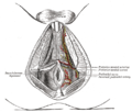
Muscles of male perineum (ischiocavernosus visible at upper left)
Ischiocavernosus-female.png
Muscles of the female perineum (ischiocavernosus visible at upper right)
Details
Origin Ischial tuberosity
Insertion Crus of penis (male) or crus of clitoris (female)
Artery Perineal artery
Nerve Pudendal nerve
Actions Maintains penile erection (male) or clitoral erection (female)
Identifiers
Latin musculus ischiocavernosus
TA98 A09.5.02.004
TA2 2417
FMA 19730
Anatomical terms of muscle

The ischiocavernosus muscle (erectores penisorerector clitoridis in older texts) is a muscle just below the surface of the perineum, present in both men and women. [1]

Contents

Structure

It arises by tendinous and fleshy fibers from the inner surface of the tuberosity of the ischium, behind the crus penis; and from the inferior pubic rami and ischium on either side of the crus.

From these points fleshy fibers succeed, and end in an aponeurosis which is inserted into the sides and under surface of the crus penis.

Function

In females, the ischiocavernosus muscle assists with clitoral erection. [2] In males, it helps to stabilize the erect penis by compressing the crus penis and retarding the return of blood through the veins.

Additional images

Related Research Articles

<span class="mw-page-title-main">Perineum</span> Region of the body between the genitals and anus

The perineum in mammals is the space between the anus and the genitals. The human perineum is between the anus and scrotum in the male or between the anus and vulva in the female. The perineum is the region of the body between the pubic symphysis and the coccyx, including the perineal body and surrounding structures. The perineal raphe is visible and pronounced to varying degrees. The perineum is an erogenous zone. This area is also known as the taint or gooch in American slang.

<span class="mw-page-title-main">Levator ani</span> Broad, thin muscle group, situated on either side of the pelvis

The levator ani is a broad, thin muscle group, situated on either side of the pelvis. It is formed from three muscle components: the pubococcygeus, the iliococcygeus, and the puborectalis.

<span class="mw-page-title-main">Bulbospongiosus muscle</span> Superficial muscle of the perineum

The bulbospongiosus muscles are a subgroup of the superficial muscles of the perineum. They have a slightly different origin, insertion and function in males and females. In males, these muscles cover the bulb of the penis, while in females, they cover the vestibular bulbs.

<span class="mw-page-title-main">Internal pudendal artery</span> Blood vessel supplying blood to the external genitalia

The internal pudendal artery is one of the three pudendal arteries. It branches off the internal iliac artery, and provides blood to the external genitalia.

<span class="mw-page-title-main">External anal sphincter</span> Flat plane of skeletal muscle fibers

The external anal sphincter is an oval tube of skeletal muscle fibers. Distally, it is adherent to the skin surrounding the margin of the anus. It exhibits a resting state of tonical contraction.

<span class="mw-page-title-main">External sphincter muscle of male urethra</span>

The external sphincter muscle of male urethra, also sphincter urethrae membranaceae, sphincter urethrae externus, surrounds the whole length of the membranous urethra, and is enclosed in the fascia of the urogenital diaphragm.

<span class="mw-page-title-main">Crus of clitoris</span> One of two erectile tissue structures of the human clitoris

The clitoral crura are two erectile tissue structures, which together form a "V" shape. Crus is a Latin word that means "leg". Each "leg" of the V converges on the clitoral body. At each divergent point is a corpus cavernosum. Together with the vestibular bulbs, they form the clitoral root. The crura are attached to the pubic arch, and are adjacent to the vestibular bulbs. The crura flank the urethra, urethral sponge, and vagina and extend back toward the pubis. Each clitoral crus connects to the rami of the pubis and the ischium.

<span class="mw-page-title-main">Ischium</span> Lower and back region of the hip bone

The ischium forms the lower and back region of the hip bone.

<span class="mw-page-title-main">Sacrotuberous ligament</span>

The sacrotuberous ligament is situated at the lower and back part of the pelvis. It is flat, and triangular in form; narrower in the middle than at the ends.

<span class="mw-page-title-main">Perineal nerve</span> Nerve of the perineum

The perineal nerve is a nerve of the pelvis. It arises from the pudendal nerve in the pudendal canal. It gives superficial branches to the skin, and a deep branch to muscles. It supplies the skin and muscles of the perineum. Its latency is tested with electrodes.

<span class="mw-page-title-main">Fascia of Colles</span>

The membranous layer of the superficial fascia of the perineum is the deeper layer of the superficial perineal fascia. It is thin, aponeurotic in structure, and of considerable strength, serving to bind down the muscles of the root of the penis. Colles' fascia emerges from the perineal membrane, which divides the base of the penis from the prostate. Colles' fascia emerges from the inferior side of the perineal membrane and continues along the ventral (inferior) penis without covering the scrotum. It separates the skin and subcutaneous fat from the superficial perineal pouch.

<span class="mw-page-title-main">Fascia of Camper</span> Layer of the anterior abdominal wall

The fascia of Camper is a thick superficial layer of the anterior abdominal wall.

<span class="mw-page-title-main">Transverse perineal muscles</span>

The transverse perineal muscles are the superficial and the deep transverse perineal muscles.

<span class="mw-page-title-main">Perineal membrane</span> Anatomical term

The perineal membrane is an anatomical term for a fibrous membrane in the perineum. The term "inferior fascia of urogenital diaphragm", used in older texts, is considered equivalent to the perineal membrane.

<span class="mw-page-title-main">Superficial perineal pouch</span>

The superficial perineal pouch is a compartment of the perineum.

<span class="mw-page-title-main">Deep perineal pouch</span> Anatomic space enclosed partly by the perineum

The deep perineal pouch is the anatomic space enclosed in part by the perineum and located superior to the perineal membrane.

<span class="mw-page-title-main">Urogenital triangle</span> Anterior part of the perineum

The urogenital triangle is the anterior part of the perineum. In female mammals, it contains the vagina and associated parts of the internal genitalia.

<span class="mw-page-title-main">Posterior labial nerves</span>

The posterior labial nerves are branches of the pudendal nerve. They supply the female labia majora.

<span class="mw-page-title-main">Muscular branches of perineal nerve</span> Nerve of the perineum

The deep branch of the perineal nerve is a nerve of the perineum. It is a branch of the perineal nerve, from the pudendal nerve. It supplies the superficial transverse perineal muscle, bulbospongiosus muscle, ischiocavernosus muscle, the bulb of penis, levator ani, and the external anal sphincter.

<span class="mw-page-title-main">Vaginal support structures</span> Structures that maintain the position of the vagina within the pelvic cavity

The vaginal support structures are those muscles, bones, ligaments, tendons, membranes and fascia, of the pelvic floor that maintain the position of the vagina within the pelvic cavity and allow the normal functioning of the vagina and other reproductive structures in the female. Defects or injuries to these support structures in the pelvic floor leads to pelvic organ prolapse. Anatomical and congenital variations of vaginal support structures can predispose a woman to further dysfunction and prolapse later in life. The urethra is part of the anterior wall of the vagina and damage to the support structures there can lead to incontinence and urinary retention.

References

PD-icon.svgThis article incorporates text in the public domain from page 428 of the 20th edition of Gray's Anatomy (1918)

  1. Maclean, Allan; Reid, Wendy (2011). "40". In Shaw, Robert (ed.). Gynaecology. Edinburgh New York: Churchill Livingstone/Elsevier. pp. 599–612. ISBN   978-0-7020-3120-5; Access provided by the University of Pittsburgh{{cite book}}: CS1 maint: postscript (link)
  2. Dean O'Loughlin, Valerie; Stouter Bidle, Theresa; McKinley, Michael P. (2022). "Muscular System: Axial and Appendicular Muscles". Anatomy and Physiology: An Integrative Approach (Fourth ed.). McGraw Hill. p. 395. ISBN   9781264265411.