The external anal sphincter (or sphincter ani externus) is an oval tube of skeletal muscle fibers.[1] Distally, it is adherent to the skin surrounding the margin of the anus.[2] It exhibits a resting state of tonic contraction[1] and also contracts during the bulbospongiosus reflex.[3][4][5][6]
The external anal sphincter is far more substantial than the internal anal sphincter. The proximal portion of external anal sphincter overlaps the internal anal sphincter (which terminates distally a little distance proximal to the anal orifice) superficially; where the two overlap, they are separated by the intervening conjoint longitudinal muscle.[1]
Structure
Historically, the sphincter was described as consisting of three parts (deep, superficial, and subcutaneous). This is not supported by current anatomical knowledge. Some sources still describe it in two layers, deep (or proximal) and superficial (or distal or subcutaneous).[1]
Some of the muscles fibres decussate at the anterior midline and posterior midline, so forming an anterior commissure and posterior commissure.[1]
Function
The external anal sphincter keeps feces retained inside the rectum and prevents them from coming out of the rectum involuntarily.
Attachments
This section needs expansion. You can help by adding to it. (July 2023)
The muscle attaches anteriorly onto the perineal body, and posteriorly onto the anococcygeal ligament.[1]
Innervation
The sphincter receives innervation from the bilaterally paired inferior anal nerve (each a branch of the pudendal nerve which is derived from ventral rami of S2-S4). It may also receive additional motor innervation from the nerve to levator ani.[1]
Histology
The sphincter consists mostly of slow twitch fibers that allow extended continuous contraction.[1]
Gallery
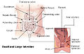
Intestines
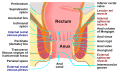
Anatomy of the human anus.
Sagittal (vertical) section of bladder, penis, and urethra.
↑ Sarica Y, Karacan I (July 1987). "Bulbocavernosus reflex to somatic and visceral nerve stimulation in normal subjects and in diabetics with erectile impotence". The Journal of Urology. 138 (1): 55–58. doi:10.1016/S0022-5347(17)42987-9. PMID3599220.
↑ Jiang XZ, Zhou CK, Guo LH, Chen J, Wang HQ, Zhang DQ, etal. (December 2009). "[Role of bulbocavernosus reflex to stimulation of prostatic urethra in pathologic mechanism of primary premature ejaculation]". Zhonghua Yi Xue Za Zhi (in Chinese). 89 (46): 3249–3252. PMID20193361.
This page is based on this Wikipedia article Text is available under the CC BY-SA 4.0 license; additional terms may apply. Images, videos and audio are available under their respective licenses.


