Transverse folds of rectum

Last updated
Transverse folds of rectum
Gray1078.png
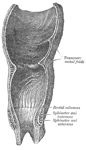
Coronal section of rectum and anal canal.
Anorectum-en.svg
Anatomy of the anus and rectum
Details
Identifiers
Latin plicae transversae recti
TA98 A05.7.04.007
TA2 3004
FMA 75657
Anatomical terminology

The transverse folds of rectum (or Houston's valves or the valves of Houston) are semi-lunar transverse folds of the rectal wall that protrude into the rectum, not the anal canal as that lies below the rectum. Their use seems to be to support the weight of fecal matter, and prevent its urging toward the anus, which would produce a strong urge to defecate. Although the term rectum means straight, these transverse folds overlap each other during the empty state of the intestine to such an extent that, as Houston remarked, they require considerable maneuvering to conduct an instrument along the canal, as often occurs in sigmoidoscopy and colonoscopy.

These folds are about 12 mm. in width and are composed of the circular muscle coat of the rectum. They are usually three in number; sometimes a fourth is found, and occasionally only two are present.

Transverse folds were first described by Irish anatomist John Houston, curator of the Royal College of Surgeons in Ireland Museum, in 1830. They appear to be peculiar to human physiology: Baur (1863) looked for Houston's valves in a number of mammals, including wolf, bear, rhinoceros, and several Old World primates, but found no evidence. They are formed very early during human development, and may be visible in embryos of as little as 55 mm in length (10 weeks of gestational age.) [1]

Related Research Articles

<span class="mw-page-title-main">Bladder</span> Organ in vertebrates that collects and stores urine from the kidneys before disposal

The bladder is a hollow organ in humans and other vertebrates that stores urine from the kidneys before disposal by urination. In humans, the bladder is a distensible organ that sits on the pelvic floor. Urine enters the bladder via the ureters and exits via the urethra. The typical adult human bladder will hold between 300 and 500 ml before the urge to empty occurs, but can hold considerably more.

<span class="mw-page-title-main">Large intestine</span> Last part of the digestive system in vertebrates

The large intestine, also known as the large bowel, is the last part of the gastrointestinal tract and of the digestive system in tetrapods. Water is absorbed here and the remaining waste material is stored in the rectum as feces before being removed by defecation. The colon is the longest portion of the large intestine, and the terms are often used interchangeably but most sources define the large intestine as the combination of the cecum, colon, rectum, and anal canal. Some other sources exclude the anal canal.

<span class="mw-page-title-main">Gastrointestinal tract</span> Organ system within humans and other animals

The gastrointestinal tract is the tract or passageway of the digestive system that leads from the mouth to the anus. The GI tract contains all the major organs of the digestive system, in humans and other animals, including the esophagus, stomach, and intestines. Food taken in through the mouth is digested to extract nutrients and absorb energy, and the waste expelled at the anus as faeces. Gastrointestinal is an adjective meaning of or pertaining to the stomach and intestines.

<span class="mw-page-title-main">Cecum</span> Pouch in the large intestine

The cecum or caecum is a pouch within the peritoneum that is considered to be the beginning of the large intestine. It is typically located on the right side of the body. The word cecum stems from the Latin caecus meaning blind.

<span class="mw-page-title-main">Amnion</span> Innermost membranous sac that surrounds and protects the developing embryo

The amnion is a membrane that closely covers the human and various other embryos when first formed. It fills with amniotic fluid, which causes the amnion to expand and become the amniotic sac that provides a protective environment for the developing embryo. The amnion, along with the chorion, the yolk sac and the allantois protect the embryo. In birds, reptiles and monotremes, the protective sac is enclosed in a shell. In marsupials and placental mammals, it is enclosed in a uterus.

<span class="mw-page-title-main">Levator ani</span> Broad, thin muscle group, situated on either side of the pelvis

The levator ani is a broad, thin muscle group, situated on either side of the pelvis. It is formed from three muscle components: the pubococcygeus, the iliococcygeus, and the puborectalis.

<span class="mw-page-title-main">Sigmoid colon</span> Section of the large intestine closest to the rectum and anus

The sigmoid colon is the part of the large intestine that is closest to the rectum and anus. It forms a loop that averages about 35–40 centimetres (14–16 in) in length. The loop is typically shaped like a Greek letter sigma (ς) or Latin letter S. This part of the colon normally lies within the pelvis, but due to its freedom of movement it is liable to be displaced into the abdominal cavity.

<span class="mw-page-title-main">Mesentery</span> Contiguous fold of tissues that supports the intestines

The mesentery is an organ that attaches the intestines to the posterior abdominal wall and is formed by the double fold of peritoneum. It helps in storing fat and allowing blood vessels, lymphatics, and nerves to supply the intestines, among other functions.

<span class="mw-page-title-main">Yolk sac</span> Membranous sac attached to an embryo

The yolk sac is a membranous sac attached to an embryo, formed by cells of the hypoblast layer of the bilaminar embryonic disc. This is alternatively called the umbilical vesicle by the Terminologia Embryologica (TE), though yolk sac is far more widely used. In humans, the yolk sac is important in early embryonic blood supply, and much of it is incorporated into the primordial gut during the fourth week of embryonic development.

<span class="mw-page-title-main">Anal canal</span> Functional segment of the large intestine

The anal canal is the part that connects the rectum to the anus, located below the level of the pelvic diaphragm. It is located within the anal triangle of the perineum, between the right and left ischioanal fossa. As the final functional segment of the bowel, it functions to regulate release of excrement by two muscular sphincter complexes. The anus is the aperture at the terminal portion of the anal canal.

<span class="mw-page-title-main">Ascending colon</span> Section of the large intestine

In the anatomy of humans and homologous primates, the ascending colon is the part of the colon located between the cecum and the transverse colon.

<span class="mw-page-title-main">Abdomen</span> Part of the body between the chest and pelvis

The abdomen is the part of the body between the thorax (chest) and pelvis, in humans and in other vertebrates. The abdomen is the front part of the abdominal segment of the torso. The area occupied by the abdomen is called the abdominal cavity. In arthropods, it is the posterior tagma of the body; it follows the thorax or cephalothorax.

<span class="mw-page-title-main">Human embryonic development</span> Development and formation of the human embryo

Human embryonic development or human embryogenesis is the development and formation of the human embryo. It is characterised by the processes of cell division and cellular differentiation of the embryo that occurs during the early stages of development. In biological terms, the development of the human body entails growth from a one-celled zygote to an adult human being. Fertilization occurs when the sperm cell successfully enters and fuses with an egg cell (ovum). The genetic material of the sperm and egg then combine to form the single cell zygote and the germinal stage of development commences. Embryonic development in the human, covers the first eight weeks of development; at the beginning of the ninth week the embryo is termed a fetus. The eight weeks has 23 stages.

The development of the reproductive system is the part of embryonic growth that results in the sex organs and contributes to sexual differentiation. Due to its large overlap with development of the urinary system, the two systems are typically described together as the genitourinary system.

<span class="mw-page-title-main">Rectum</span> Final portion of the large intestine

The rectum is the final straight portion of the large intestine in humans and some other mammals, and the gut in others. The adult human rectum is about 12 centimetres (4.7 in) long, and begins at the rectosigmoid junction at the level of the third sacral vertebra or the sacral promontory depending upon what definition is used. Its diameter is similar to that of the sigmoid colon at its commencement, but it is dilated near its termination, forming the rectal ampulla. It terminates at the level of the anorectal ring or the dentate line, again depending upon which definition is used. In humans, the rectum is followed by the anal canal which is about 4 centimetres (1.6 in) long, before the gastrointestinal tract terminates at the anal verge. The word rectum comes from the Latin rectumintestinum, meaning straight intestine.

<span class="mw-page-title-main">Human anus</span> External opening of the rectum

In humans, the anus is the external opening of the rectum located inside the intergluteal cleft. Two sphincters control the exit of feces from the body during an act of defecation, which is the primary function of the anus. These are the internal anal sphincter and the external anal sphincter, which are circular muscles that normally maintain constriction of the orifice and which relaxes as required by normal physiological functioning. The inner sphincter is involuntary and the outer is voluntary. Above the anus is the perineum, which is also located beneath the vulva or scrotum.

<span class="mw-page-title-main">Vertebral column</span> Bony structure found in vertebrates

The vertebral column, also known as the backbone or spine, is the core part of the axial skeleton in vertebrate animals. The vertebral column is the defining characteristic of vertebrate endoskeleton in which the notochord found in all chordates has been replaced by a segmented series of mineralized irregular bones called vertebrae, separated by fibrocartilaginous intervertebral discs. The dorsal portion of the vertebral column houses the spinal canal, a cavity formed by alignment of the neural arches that encloses and protects the spinal cord.

The development of the digestive system in the human embryo concerns the epithelium of the digestive system and the parenchyma of its derivatives, which originate from the endoderm. Connective tissue, muscular components, and peritoneal components originate in the mesoderm. Different regions of the gut tube such as the esophagus, stomach, duodenum, etc. are specified by a retinoic acid gradient that causes transcription factors unique to each region to be expressed. Differentiation of the gut and its derivatives depends upon reciprocal interactions between the gut endoderm and its surrounding mesoderm. Hox genes in the mesoderm are induced by a Hedgehog signaling pathway secreted by gut endoderm and regulate the craniocaudal organization of the gut and its derivatives. The gut system extends from the oropharyngeal membrane to the cloacal membrane and is divided into the foregut, midgut, and hindgut.

<span class="mw-page-title-main">Heart development</span> Prenatal development of the heart

Heart development, also known as cardiogenesis, refers to the prenatal development of the heart. This begins with the formation of two endocardial tubes which merge to form the tubular heart, also called the primitive heart tube. The heart is the first functional organ in vertebrate embryos.

<span class="mw-page-title-main">Human digestive system</span> Digestive system in humans

The human digestive system consists of the gastrointestinal tract plus the accessory organs of digestion. Digestion involves the breakdown of food into smaller and smaller components, until they can be absorbed and assimilated into the body. The process of digestion has three stages: the cephalic phase, the gastric phase, and the intestinal phase.

References

PD-icon.svgThis article incorporates text in the public domain from page 1183 of the 20th edition of Gray's Anatomy (1918)

  1. P.H.S.Silver (1955). "Observation on the Valves of Houston in the human embryo and foetus". Journal of Anatomy. 89 (2): 217–24. PMC   1244784 . PMID   14367217.