Retropharyngeal space

Last updated
Retropharyngeal space
Gray384.png
Section of the neck at about the level of the sixth cervical vertebra. Showing the arrangement of the deep cervical fascia
Gray994.png
Details
Identifiers
Latin spatium retropharyngeum
TA98 A05.3.01.118
TA2 2884
FMA 84965
Anatomical terminology

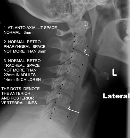
The retropharyngeal space (abbreviated as "RPS" [1] [2] ) is a potential space [2] [3] and deep compartment of the head and neck [1] situated posterior to the pharynx. [4] The RPS is bounded anteriorly by the buccopharyngeal fascia, posteriorly by the alar fascia, and laterally by the carotid sheath. It extends between the base of the skull superiorly, and the mediastinum inferiorly. [1] It contains the retropharyngeal lymph nodes. [2] Its function is to facilitate movements in the superoinferior axis of the larynx, pharynx, and esophagus in relation to the cervical spine. [3]

Contents

Sources consider the retropharyngeal space to be in principle subdivided into the so-called "true retropharyngeal space" [1] [5] or "retropharyngeal space proper" (part of the RPS situated anterior to the alar fascia), [5] [2] and the danger space (part of the RPS situated posterior to the alar fascia). [1] [2] [5] The danger space is sometimes also lumped together with the true RPS and the whole referred to as the RPS because the alar fascia is an ineffective barrier. [2] Infections from the head and neck can spread down through the danger space into the posterior mediastinum. [2]

Anatomy

Superiorly, the retropharyngeal space terminates at the base of the skull (more specifically, at the clivus [2] ). [1] [5] Inferiorly, the true RPS terminates at a variable level along the upper thoracic spine with the fusion of alar fascia and visceral fascia; [1] sources either give the inferior termination of the true RPS as occurring at approximately the vertebral level of T4 [2] or at a variable level anywhere between the T1-T6. [1] [5] The danger space component of the RPS meanwhile extends further inferiorly, entering the posterior mediastinum to reach the level of the diaphragm. [1] The retropharyngeal space drains into the superior mediastinum, whereas the danger space drains into the posterior mediastinum.

Contents

The retropharyngeal space contains the retropharyngeal lymph nodes, [2] adipose tissue, and loose connective tissue. [3] The suprahyoid portion of the RPS contains the lymph nodes as well as adipose tissue, while the infrahyoid portion contains adipose tissue only. [1]

A midline raphe is sometimes present in the RPS, subdividing it into a left half and a right half. [2]

Anatomical relations

Positions of adjacent anatomical structures in relation to the retropharyngeal space are as follows:

Clinical significance

A midline raphe may be present in this the RPS, [2] making some infections appear unilateral. However without treatment infections can easily spread from one space to the adjacent space.[ citation needed ]

If more than half of the size of the C2 vertebra, it may indicate retropharyngeal abscess. [7]

Additional images

See also

Related Research Articles

Articles related to anatomy include:

<span class="mw-page-title-main">Thoracic duct</span> Lymphatic vessel

In human anatomy, the thoracic duct is the larger of the two lymph ducts of the lymphatic system. The thoracic duct usually begins from the upper aspect of the cisterna chyli, passing out of the abdomen through the aortic hiatus into first the posterior mediastinum and then the superior mediastinum, extending as high up as the root of the neck before descending to drain into the systemic (blood) circulation at the venous angle.

<span class="mw-page-title-main">Mediastinum</span> Central part of the thoracic cavity

The mediastinum is the central compartment of the thoracic cavity. Surrounded by loose connective tissue, it is an undelineated region that contains a group of structures within the thorax, namely the heart and its vessels, the esophagus, the trachea, the phrenic and cardiac nerves, the thoracic duct, the thymus and the lymph nodes of the central chest.

<span class="mw-page-title-main">Superior thoracic aperture</span>

The superior thoracic aperture, also known as the thoracic outlet, or thoracic inlet refers to the opening at the top of the thoracic cavity. It is also clinically referred to as the thoracic outlet, in the case of thoracic outlet syndrome. A lower thoracic opening is the inferior thoracic aperture.

<span class="mw-page-title-main">Superior pharyngeal constrictor muscle</span> Muscle

The superior pharyngeal constrictor muscle is a quadrilateral muscle of the pharynx. It is the uppermost and thinnest of the three pharyngeal constrictors.

<span class="mw-page-title-main">Stylopharyngeus muscle</span> Muscle

The stylopharyngeus muscle is a muscle in the head. It originates from the temporal styloid process. Some of its fibres insert onto the thyroid cartilage, while others end by intermingling with proximal structures. It is innervated by the glossopharyngeal nerve. It acts to elevate the larynx and pharynx, and dilate the pharynx, thus facilitating swallowing.

<span class="mw-page-title-main">Carotid sheath</span>

The carotid sheath is a condensation of the deep cervical fascia enveloping multiple vital neurovascular structures of the neck, including the common and internal carotid arteries, the internal jugular vein, the vagus nerve, and ansa cervicalis. The carotid sheath helps protects the structures contained therein.

<span class="mw-page-title-main">Deep cervical fascia</span>

The deep cervical fascia lies under cover of the platysma, and invests the muscles of the neck; it also forms sheaths for the carotid vessels, and for the structures situated in front of the vertebral column. Its attachment to the hyoid bone prevents the formation of a dewlap.

<span class="mw-page-title-main">Pterygomandibular raphe</span> Ligament formed from the buccopharyngeal fascia

The pterygomandibular raphe is a thin tendinous band of buccopharyngeal fascia. It is attached superiorly to the pterygoid hamulus of the medial pterygoid plate, and inferiorly to the posterior end of the mylohyoid line of the mandible. It gives attachment to the buccinator muscle, and the superior pharyngeal constrictor muscle (behind).

<span class="mw-page-title-main">Cervical lymph nodes</span> Lymph nodes found in the neck

Cervical lymph nodes are lymph nodes found in the neck. Of the 800 lymph nodes in the human body, 300 are in the neck. Cervical lymph nodes are subject to a number of different pathological conditions including tumours, infection and inflammation.

<span class="mw-page-title-main">Prevertebral fascia</span> Layer of deep cervical fascia that surrounds the vertebral column

The prevertebral fascia is the layer of deep cervical fascia that surrounds the vertebral column. It is the deepest layer of deep cervical fascia.

<span class="mw-page-title-main">Buccopharyngeal fascia</span>

The buccopharyngeal fascia is a fascia of the pharynx. It represents the posterior portion of the pretracheal fascia. It covers the superior pharyngeal constrictor muscles, and buccinator muscle.

The pharyngobasilar fascia is a fascia of the pharynx. It is situated between the mucous and muscular layers of the pharynx. It is formed as a thickening of the pharyngeal mucosa superior to the superior pharyngeal constrictor muscle. It attaches to the basilar part of occipital bone, the petrous part of the temporal bone, the medial pterygoid plate, and the pterygomandibular raphe. It diminishes in thickness inferiorly. Posteriorly, it is reinforced by the pharyngeal raphe. It reinforces the pharyngeal wall where muscle is deficient.

<span class="mw-page-title-main">Retropharyngeal lymph nodes</span>

The retropharyngeal lymph nodes, from one to three in number, lie in the buccopharyngeal fascia, behind the upper part of the pharynx and in front of the arch of the atlas, being separated, however, from the latter by the longus capitis.

<span class="mw-page-title-main">Outline of human anatomy</span> Overview of and topical guide to human anatomy

The following outline is provided as an overview of and topical guide to human anatomy:

<span class="mw-page-title-main">Retropharyngeal abscess</span> Medical condition

Retropharyngeal abscess (RPA) is an abscess located in the tissues in the back of the throat behind the posterior pharyngeal wall. Because RPAs typically occur in deep tissue, they are difficult to diagnose by physical examination alone. RPA is a relatively uncommon illness, and therefore may not receive early diagnosis in children presenting with stiff neck, malaise, difficulty swallowing, or other symptoms listed below. Early diagnosis is key, while a delay in diagnosis and treatment may lead to death. Parapharyngeal space communicates with retropharyngeal space and an infection of retropharyngeal space can pass down behind the esophagus into the mediastinum. RPAs can also occur in adults of any age.

<span class="mw-page-title-main">Alar fascia</span>

The alar fascia a portion or prevertebral fascia that may or may not be considered a distinct anatomical structure. When acknowledged, it is described as anterior to the prevertebral fascia.

<span class="mw-page-title-main">Danger space</span> Potential space in the neck

The danger space or alar space, is a region of the neck. The common name originates from the risk that an infection in this space can spread directly to the thorax, and, due to being a space continuous on the left and right, can furthermore allow infection to spread easily to either side.

<span class="mw-page-title-main">Parapharyngeal space</span>

The parapharyngeal space, is a potential space in the head and the neck. It has clinical importance in otolaryngology due to parapharyngeal space tumours and parapharyngeal abscess developing in this area. It is also a key anatomic landmark for localizing disease processes in the surrounding spaces of the neck; the direction of its displacement indirectly reflects the site of origin for masses or infection in adjacent areas, and consequently their appropriate differential diagnosis.

<span class="mw-page-title-main">Submental space</span>

The submental space is a fascial space of the head and neck. It is a potential space located between the mylohyoid muscle superiorly, the platysma muscle inferiorly, under the chin in the midline. The space coincides with the anatomic region termed the submental triangle, part of the anterior triangle of the neck.

References

PD-icon.svgThis article incorporates text in the public domain from page 390 of the 20th edition of Gray's Anatomy (1918)

  1. 1 2 3 4 5 6 7 8 9 10 11 12 13 14 Mnatsakanian A, Minutello K, Bordoni B (2022). "Anatomy, Head and Neck, Retropharyngeal Space". StatPearls. Treasure Island (FL): StatPearls Publishing. PMID   30725729 . Retrieved 2022-07-24.
  2. 1 2 3 4 5 6 7 8 9 10 11 12 Chong VF, Fan YF (October 2000). "Radiology of the Retropharyngeal Space". Clinical Radiology. 55 (10): 740–748. doi:10.1053/crad.2000.0510. PMID   11052873.
  3. 1 2 3 Morton, David A. (2019). The Big Picture: Gross Anatomy. K. Bo Foreman, Kurt H. Albertine (2nd ed.). New York. p. 266. ISBN   978-1-259-86264-9. OCLC   1044772257.{{cite book}}: CS1 maint: location missing publisher (link)
  4. "Spatium retropharyngeum". TheFreeDictionary.com. Retrieved 2022-07-24.
  5. 1 2 3 4 5 Debnam JM, Guha-Thakurta N (December 2012). "Retropharyngeal and prevertebral spaces: anatomic imaging and diagnosis". Otolaryngologic Clinics of North America. 45 (6): 1293–1310. doi:10.1016/j.otc.2012.08.004. PMC   3994542 . PMID   23153750.
  6. 1 2 Jain H, Knorr TL, Sinha V (2022). Retropharyngeal Abscess. Treasure Island (FL): StatPearls Publishing. PMID   28722903 . Retrieved 2022-07-24.{{cite book}}: |work= ignored (help)
  7. Frank G, Shah SS, Catallozzi M, Zaoutis LB (1 June 2005). The Philadelphia guide: inpatient pediatrics. Lippincott Williams & Wilkins. pp. 181–. ISBN   978-1-4051-0428-9 . Retrieved 26 May 2010.