Duodenojejunal flexure

Last updated
Duodenojejunal flexure
Gray1041.png
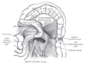
Superior and inferior duodenal fossæ.
Illu small intestine.jpg
Small intestine
Details
Identifiers
Latin flexura duodenojejunalis
TA98 A05.6.02.009
TA2 2952
FMA 15957
Anatomical terminology

The duodenojejunal flexure or duodenojejunal junction, also known as the angle of Treitz, [1] [2] is the border between the duodenum and the jejunum.

Contents

Structure

The ascending portion of the duodenum ascends on the left side of the aorta, as far as the level of the upper border of the second lumbar vertebra. At this point, it turns abruptly forward to merge with the jejunum, forming the duodenojejunal flexure. This forms the beginning of the jejunum. [3] The duodenojejunal flexure is surrounded by the suspensory muscle of the duodenum. [4] :274 It is retroperitoneal, so is less mobile than the jejunum that comes after it, helping to stabilise the jejunum. [5]

The duodenojejunal flexure lies in front of the left psoas major muscle, the left renal artery, and the left renal vein. It is covered in front, and partly at the sides, by peritoneum continuous with the left portion of the mesentery.

Clinical significance

The ligament of Treitz, a peritoneal fold, from the right crus of diaphragm, is an identification point for the duodenojejunal flexure during abdominal surgery. [6] :85

Additional images

See also

Related Research Articles

<span class="mw-page-title-main">Gastrointestinal tract</span> Organ system within humans and other animals

The gastrointestinal tract is the tract or passageway of the digestive system that leads from the mouth to the anus. The GI tract contains all the major organs of the digestive system, in humans and other animals, including the esophagus, stomach, and intestines. Food taken in through the mouth is digested to extract nutrients and absorb energy, and the waste expelled at the anus as faeces. Gastrointestinal is an adjective meaning of or pertaining to the stomach and intestines.

<span class="mw-page-title-main">Duodenum</span> First section of the small intestine

The duodenum is the first section of the small intestine in most higher vertebrates, including mammals, reptiles, and birds. In mammals it may be the principal site for iron absorption. The duodenum precedes the jejunum and ileum and is the shortest part of the small intestine.

<span class="mw-page-title-main">Ileum</span> Final section of the small intestine

The ileum is the final section of the small intestine in most higher vertebrates, including mammals, reptiles, and birds. In fish, the divisions of the small intestine are not as clear and the terms posterior intestine or distal intestine may be used instead of ileum. Its main function is to absorb vitamin B12, bile salts, and whatever products of digestion that were not absorbed by the jejunum.

<span class="mw-page-title-main">Jejunum</span> Part of the small intestine

The jejunum is the second part of the small intestine in humans and most higher vertebrates, including mammals, reptiles, and birds. Its lining is specialized for the absorption by enterocytes of small nutrient molecules which have been previously digested by enzymes in the duodenum.

<span class="mw-page-title-main">Small intestine</span> Organ in the gastrointestinal tract

The small intestine or small bowel is an organ in the gastrointestinal tract where most of the absorption of nutrients from food takes place. It lies between the stomach and large intestine, and receives bile and pancreatic juice through the pancreatic duct to aid in digestion. The small intestine is about 5.5 metres long and folds many times to fit in the abdomen. Although it is longer than the large intestine, it is called the small intestine because it is narrower in diameter.

<span class="mw-page-title-main">Suspensory muscle of duodenum</span> Muscle between the duodenum and jejunum

The suspensory muscle of duodenum is a thin muscle connecting the junction between the duodenum and jejunum, as well as the duodenojejunal flexure to connective tissue surrounding the superior mesenteric and coeliac arteries. The suspensory muscle most often connects to both the third and fourth parts of the duodenum, as well as the duodenojejunal flexure, although the attachment is quite variable.

<span class="mw-page-title-main">Superior mesenteric artery</span> Artery which supplies blood to the intestines and pancreas

In human anatomy, the superior mesenteric artery (SMA) is an artery which arises from the anterior surface of the abdominal aorta, just inferior to the origin of the celiac trunk, and supplies blood to the intestine from the lower part of the duodenum through two-thirds of the transverse colon, as well as the pancreas.

<span class="mw-page-title-main">Inferior mesenteric artery</span>

In human anatomy, the inferior mesenteric artery, often abbreviated as IMA, is the third main branch of the abdominal aorta and arises at the level of L3, supplying the large intestine from the distal transverse colon to the upper part of the anal canal. The regions supplied by the IMA are the descending colon, the sigmoid colon, and part of the rectum.

<span class="mw-page-title-main">Gastroduodenal artery</span>

In anatomy, the gastroduodenal artery is a small blood vessel in the abdomen. It supplies blood directly to the pylorus and proximal part of the duodenum. It also indirectly supplies the pancreatic head.

<span class="mw-page-title-main">Superior mesenteric vein</span> Vein which drains blood from the small intestine

In human anatomy, the superior mesenteric vein (SMV) is a blood vessel that drains blood from the small intestine. Behind the neck of the pancreas, the superior mesenteric vein combines with the splenic vein to form the portal vein that carries blood to the liver. The superior mesenteric vein lies to the right of the similarly named artery, the superior mesenteric artery, which originates from the abdominal aorta.

<span class="mw-page-title-main">Ascending colon</span> Section of the large intestine

In the anatomy of humans and homologous primates, the ascending colon is the part of the colon located between the cecum and the transverse colon.

<span class="mw-page-title-main">Right colic artery</span> Artery supplying the colon

The right colic artery is an artery of the abdomen, a branch of the superior mesenteric artery supplying the ascending colon. It divides into two terminal branches - an ascending branch and a descending branch - which form anastomoses with the middle colic artery, and ileocolic artery (respectively).

<span class="mw-page-title-main">Middle colic artery</span> Artery supplying the colon

The middle colic artery is an artery of the abdomen; a branch of the superior mesenteric artery distributed to parts of the ascending and transverse colon. It usually divides into two terminal branches - a left one and a right one - which go on to form anastomoses with the left colic artery, and right colic artery (respectively), thus participating in the formation of the marginal artery of the colon.

<span class="mw-page-title-main">Left colic artery</span> Artery

The left colic artery is a branch of the inferior mesenteric artery distributed to the descending colon, and left part of the transverse colon. It ends by dividing into an ascending branch and a descending branch; the terminal branches of the two branches go on to form anastomoses with the middle colic artery, and a sigmoid artery (respectively).

<span class="mw-page-title-main">Transverse colon</span> Longest section of the large intestine

In human anatomy, the transverse colon is the longest and most movable part of the colon.

<span class="mw-page-title-main">Greater omentum</span> Fat sheath under abdominal wall

The greater omentum is a large apron-like fold of visceral peritoneum that hangs down from the stomach. It extends from the greater curvature of the stomach, passing in front of the small intestines and doubles back to ascend to the transverse colon before reaching to the posterior abdominal wall. The greater omentum is larger than the lesser omentum, which hangs down from the liver to the lesser curvature. The common anatomical term "epiploic" derives from "epiploon", from the Greek epipleein, meaning to float or sail on, since the greater omentum appears to float on the surface of the intestines. It is the first structure observed when the abdominal cavity is opened anteriorly.

<span class="mw-page-title-main">Circular folds</span> Valvular flaps in the small intestine

The circular folds are large valvular flaps projecting into the lumen of the small intestine.

<span class="mw-page-title-main">Arterial arcades</span>

The arterial arcades are a series of anastomosing arterial arches between the arterial branches of the jejunum and ileum.

<span class="mw-page-title-main">Peritoneal recesses</span>

Peritoneal recesses are the spaces formed by peritoneum draping over viscera.

<span class="mw-page-title-main">Human digestive system</span> Digestive system in humans

The human digestive system consists of the gastrointestinal tract plus the accessory organs of digestion. Digestion involves the breakdown of food into smaller and smaller components, until they can be absorbed and assimilated into the body. The process of digestion has three stages: the cephalic phase, the gastric phase, and the intestinal phase.

References

PD-icon.svgThis article incorporates text in the public domain from page 1170 of the 20th edition of Gray's Anatomy (1918)

  1. Bracale, Umberto et.al. Laparoscopic segmental resection for tumours of the Angle of Treitz: a challenging but feasible surgical option. Results from a retrospective case-series analysis. Updates Surg. 2021; 73(1): 179–186. doi : 10.1007/s13304-020-00910-7
  2. Lissauer et.al. Neonatology at a Glance. John Wiley & Sons, 2020, p.125.
  3. Federle, Michael P.; Rosado-de-Christenson, Melissa L.; Raman, Siva P.; Carter, Brett W., eds. (2017-01-01), "Small Intestine", Imaging Anatomy: Chest, Abdomen, Pelvis (Second Edition), Elsevier, pp. 636–665, doi:10.1016/B978-0-323-47781-9.50031-3, ISBN   978-0-323-47781-9 , retrieved 2021-01-26
  4. Drake, Richard L.; Vogl, Wayne; Tibbitts, Adam W.M. Mitchell; illustrations by Richard; Richardson, Paul (2005). Gray's anatomy for students. Philadelphia: Elsevier/Churchill Livingstone. ISBN   978-0-8089-2306-0.
  5. Schneider, Armin; Feussner, Hubertus (2017-01-01), Schneider, Armin; Feussner, Hubertus (eds.), "Chapter 2 - Anatomy, Physiology, and Selected Pathologies of the Gastrointestinal Tract", Biomedical Engineering in Gastrointestinal Surgery, Academic Press, pp. 11–39, ISBN   978-0-12-803230-5 , retrieved 2021-01-26
  6. Jacob, S. (2007) Chapter 4: Abdomen; Human anatomy, A clinically-orientated approach.