Jejunum

Last updated
Jejunum
Illu small intestine.jpg
Small intestine
Gray1041.png
Superior and inferior duodenal fossæ.
Details
Pronunciation /ɪˈnəm/ [1] [2]
Precursor Midgut
Part of Small intestine
System Digestive system
Artery Jejunal arteries
Vein Jejunal veins
Nerve Celiac ganglia, vagus [3]
Identifiers
Latin jejunum
MeSH D007583
TA98 A05.6.03.001
TA2 2958
FMA 7207
Anatomical terminology

The jejunum is the second part of the small intestine in humans and most higher vertebrates, including mammals, reptiles, and birds. Its lining is specialized for the absorption by enterocytes of small nutrient molecules which have been previously digested by enzymes in the duodenum.

Contents

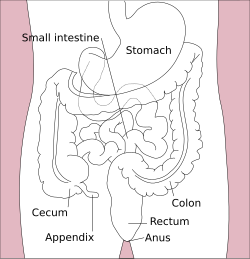
The jejunum lies between the duodenum and the ileum and is considered to start at the suspensory muscle of the duodenum, a location called the duodenojejunal flexure. [4] The division between the jejunum and ileum is not anatomically distinct. [5] In adult humans, the small intestine is usually 6–7 m (20–23 ft) long (post mortem), about two-fifths of which (about 2.5 m (8.2 ft)) is the jejunum. [4]

Structure

The interior surface of the jejunum—which is exposed to ingested food—is covered in finger–like projections of mucosa, called villi, which increase the surface area of tissue available to absorb nutrients from ingested foodstuffs. The epithelial cells which line these villi have microvilli. The transport of nutrients across epithelial cells through the jejunum and ileum includes the passive transport of sugar fructose and the active transport of amino acids, small peptides, vitamins, and most glucose. The villi in the jejunum are much longer than in the duodenum or ileum.

The pH in the jejunum is usually between 7 and 8 (neutral or slightly alkaline).

The jejunum and the ileum are suspended by mesentery which gives the bowel great mobility within the abdomen. It also contains circular and longitudinal smooth muscle which helps to move food along by a process known as peristalsis.

Histology

The jejunum contains very few Brunner's glands (found in the duodenum) or Peyer's patches (found in the ileum). However, there are a few jejunal lymph nodes suspended in its mesentery. The jejunum has many large circular folds in its submucosa called plicae circulares that increase the surface area for nutrient absorption. The plicae circulares are best developed in the jejunum.

There is no line of demarcation between the jejunum and the ileum. However, there are subtle histological differences:

Function

The lining of the jejunum is specialized for the absorption by enterocytes of small nutrient particles which have been previously digested by enzymes in the duodenum. Once absorbed, nutrients (with the exception of fat, which goes to the lymph) pass from the enterocytes into the enterohepatic circulation and enter the liver via the hepatic portal vein, where the blood is processed. [6]

Other animals

In fish, the divisions of the small intestine are not as clear and the terms middle intestine or mid-gut may be used instead of jejunum. [7]

History

Etymology

Jejunum is derived from the Latin word jējūnus (iēiūnus), meaning "fasting." It was so called because this part of the small intestine was frequently found to be void of food following death, [8] due to its intensive peristaltic activity relative to the duodenum and ileum.

The Early Modern English adjective jejune is derived from the same root. [9]

Related Research Articles

<span class="mw-page-title-main">Gastrointestinal tract</span> Organ system within humans and other animals

The gastrointestinal tract is the tract or passageway of the digestive system that leads from the mouth to the anus. The GI tract contains all the major organs of the digestive system, in humans and other animals, including the esophagus, stomach, and intestines. Food taken in through the mouth is digested to extract nutrients and absorb energy, and the waste expelled at the anus as faeces. Gastrointestinal is an adjective meaning of or pertaining to the stomach and intestines.

<span class="mw-page-title-main">Duodenum</span> First section of the small intestine

The duodenum is the first section of the small intestine in most higher vertebrates, including mammals, reptiles, and birds. In mammals it may be the principal site for iron absorption. The duodenum precedes the jejunum and ileum and is the shortest part of the small intestine.

<span class="mw-page-title-main">Ileum</span> Final section of the small intestine

The ileum is the final section of the small intestine in most higher vertebrates, including mammals, reptiles, and birds. In fish, the divisions of the small intestine are not as clear and the terms posterior intestine or distal intestine may be used instead of ileum. Its main function is to absorb vitamin B12, bile salts, and whatever products of digestion that were not absorbed by the jejunum.

<span class="mw-page-title-main">Small intestine</span> Organ in the gastrointestinal tract

The small intestine or small bowel is an organ in the gastrointestinal tract where most of the absorption of nutrients from food takes place. It lies between the stomach and large intestine, and receives bile and pancreatic juice through the pancreatic duct to aid in digestion. The small intestine is about 5.5 metres long and folds many times to fit in the abdomen. Although it is longer than the large intestine, it is called the small intestine because it is narrower in diameter.

<span class="mw-page-title-main">Intestinal villus</span> Finger-like projection of the small intestine

Intestinal villi are small, finger-like projections that extend into the lumen of the small intestine. Each villus is approximately 0.5–1.6 mm in length, and has many microvilli projecting from the enterocytes of its epithelium which collectively form the striated or brush border. Each of these microvilli are about 1 µm in length, around 1000 times shorter than a single villus. The intestinal villi are much smaller than any of the circular folds in the intestine.

<span class="mw-page-title-main">Mesentery</span> Contiguous fold of tissues that supports the intestines

The mesentery is an organ that attaches the intestines to the posterior abdominal wall and is formed by the double fold of peritoneum. It helps in storing fat and allowing blood vessels, lymphatics, and nerves to supply the intestines, among other functions.

<span class="mw-page-title-main">Enterocyte</span> Type of intestinal cell

Enterocytes, or intestinal absorptive cells, are simple columnar epithelial cells which line the inner surface of the small and large intestines. A glycocalyx surface coat contains digestive enzymes. Microvilli on the apical surface increase its surface area. This facilitates transport of numerous small molecules into the enterocyte from the intestinal lumen. These include broken down proteins, fats, and sugars, as well as water, electrolytes, vitamins, and bile salts. Enterocytes also have an endocrine role, secreting hormones such as leptin.

<span class="mw-page-title-main">Peyer's patch</span> Lymphatic tissue in the lower small intestine

Peyer's patches are organized lymphoid follicles, named after the 17th-century Swiss anatomist Johann Conrad Peyer. They are an important part of gut associated lymphoid tissue usually found in humans in the lowest portion of the small intestine, mainly in the distal jejunum and the ileum, but also could be detected in the duodenum.

<span class="mw-page-title-main">Superior mesenteric artery</span> Artery which supplies blood to the intestines and pancreas

In human anatomy, the superior mesenteric artery (SMA) is an artery which arises from the anterior surface of the abdominal aorta, just inferior to the origin of the celiac trunk, and supplies blood to the intestine from the lower part of the duodenum through two-thirds of the transverse colon, as well as the pancreas.

Gut-associated lymphoid tissue (GALT) is a component of the mucosa-associated lymphoid tissue (MALT) which works in the immune system to protect the body from invasion in the gut.

<span class="mw-page-title-main">Intestinal arteries</span>

The intestinal arteries arise from the convex side of the superior mesenteric artery. They are usually from twelve to fifteen in number, and are distributed to the jejunum and ileum.

<span class="mw-page-title-main">Duodenojejunal flexure</span> Border between the duodenum and the jejunum

The duodenojejunal flexure or duodenojejunal junction, also known as the angle of Treitz, is the border between the duodenum and the jejunum.

<span class="mw-page-title-main">Circular folds</span> Valvular flaps in the small intestine

The circular folds are large valvular flaps projecting into the lumen of the small intestine.

<span class="mw-page-title-main">Solitary lymphatic nodule</span>

The Solitary lymphatic nodules are structures found in the small intestine and large intestine.

<span class="mw-page-title-main">Intestinal atresia</span> Medical condition

Intestinal atresia is any congenital malformation of the structure of the intestine that causes bowel obstruction. The malformation can be a narrowing (stenosis), absence or malrotation of a portion of the intestine. These defects can either occur in the small or large intestine.

<span class="mw-page-title-main">Vasa recta (intestines)</span>

Vasa recta are straight arteries arising from arterial arcades in the mesentery of the jejunum and ileum that supply the jejunum and ileum.

<span class="mw-page-title-main">Superior mesenteric lymph nodes</span>

The superior mesenteric lymph nodes may be divided into three principal groups:

<span class="mw-page-title-main">Gastrointestinal wall</span> Digestive system structure

The gastrointestinal wall of the gastrointestinal tract is made up of four layers of specialised tissue. From the inner cavity of the gut outwards, these are the mucosa, the submucosa, the muscular layer and the serosa or adventitia.

<span class="mw-page-title-main">Human digestive system</span> Digestive system in humans

The human digestive system consists of the gastrointestinal tract plus the accessory organs of digestion. Digestion involves the breakdown of food into smaller and smaller components, until they can be absorbed and assimilated into the body. The process of digestion has three stages: the cephalic phase, the gastric phase, and the intestinal phase.

In poultry farming, early feeding is the ad lib availability of feed and water for day-old chicks from the moment of hatching from the egg.

References

  1. OED 2nd edition, 1989.
  2. Entry "jejunum" in Merriam-Webster Online Dictionary .
  3. Nosek, Thomas M. "Section 6/6ch2/s6ch2_30". Essentials of Human Physiology. Archived from the original on 2016-03-24.
  4. 1 2 Drake, Richard L.; Vogl, Wayne; Tibbitts, Adam W. M. Mitchell; illustrations by Richard; Richardson, Paul (2005). Gray's anatomy for students. Philadelphia: Elsevier/Churchill Livingstone. pp. 273–275. ISBN   978-0-8089-2306-0.
  5. Deakin, Barbara Young; et al. (2006). Wheater's functional histology : a text and colour atlas (5th ed.). Churchill Livingstone/Elsevier. p. 263. ISBN   978-0-443-068-508.
  6. CRANE, RK (Oct 1960). "Intestinal absorption of sugars". Physiological Reviews. 40 (4): 789–825. doi:10.1152/physrev.1960.40.4.789. PMID   13696269.
  7. Guillaume, Jean; Praxis Publishing; Sadasivam Kaushik; Pierre Bergot; Robert Metailler (2001). Nutrition and Feeding of Fish and Crustaceans. Springer. p. 31. ISBN   9781852332419 . Retrieved 2009-01-09.
  8. Harper, Douglas. "jejunum". Etymology Online. Retrieved 15 November 2014.
  9. "Oxford English Dictionary: jejune, adj".