Subserosa

Last updated
Tela subserosa
Illu stomach layers.jpg
Details
Identifiers
Latin tela subserosa
TA A12.1.08.009
FMA 77001
Anatomical terminology

The tela subserosa (or just subserosa) is a thin layer of tissue in the walls of various organs. It is a layer of connective tissue (usually of the areolar type) between the muscular layer (muscularis externa) and the serosa (serous membrane).

Tissue (biology) An ensemble of similar cells and their matrix with similar origin and function

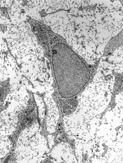
In biology, tissue is a cellular organizational level between cells and a complete organ. A tissue is an ensemble of similar cells and their extracellular matrix from the same origin that together carry out a specific function. Organs are then formed by the functional grouping together of multiple tissues.

Organ (anatomy) collection of tissues joined as a structural unit that serves a common function

Organs are collections of tissues with similar functions. Plant and animal life relies on many organs that coexist in organ systems.

Connective tissue type of biological tissue

Connective tissue (CT) is one of the four basic types of animal tissue, along with epithelial tissue, muscle tissue, and nervous tissue. It develops from the mesoderm. Connective tissue is found in between other tissues everywhere in the body, including the nervous system. In the central nervous system, the three outer membranes that envelop the brain and spinal cord are composed of connective tissue. They support and protect the body. All connective tissue consists of three main components: fibers, ground substance and cells. Not all authorities include blood or lymph as connective tissue because they lack the fiber component. All are immersed in the body water.

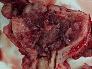
The subserosa has clinical importance particularly in cancer staging (for example, in staging stomach cancer [1] or uterine cancer).

Cancer staging is the process of determining the extent to which a cancer has developed by growing and spreading. Contemporary practice is to assign a number from I to IV to a cancer, with I being an isolated cancer and IV being a cancer which has spread to the limit of what the assessment measures. The stage generally takes into account the size of a tumor, whether it has invaded adjacent organs, how many regional (nearby) lymph nodes it has spread to, and whether it has appeared in more distant locations (metastasized).

Stomach cancer gastrointestinal system cancer that is located in the stomach

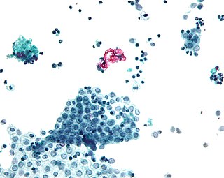
Stomach cancer, also known as gastric cancer, is a cancer which develops from the lining of the stomach. Early symptoms may include heartburn, upper abdominal pain, nausea and loss of appetite. Later signs and symptoms may include weight loss, yellowing of the skin and whites of the eyes, vomiting, difficulty swallowing and blood in the stool among others. The cancer may spread from the stomach to other parts of the body, particularly the liver, lungs, bones, lining of the abdomen and lymph nodes.

Uterine cancer female reproductive organ cancer that is located in the uterus

Uterine cancer, also known as womb cancer, are two types of cancer that develops from the tissues of the uterus. Endometrial cancer forms from the lining of the uterus and uterine sarcoma forms from the muscles or support tissue of the uterus. Symptoms of endometrial cancer include unusual vaginal bleeding or pain in the pelvis. Symptoms of uterine sarcoma include unusual vaginal bleeding or a mass in the vagina.

The subserosa ( sub- + serosa) is to a serous membrane what the submucosa ( sub- + mucosa) is to a mucous membrane.

Submucosa

The submucosa is a thin layer of tissue in various organs of the gastrointestinal, respiratory, and genitourinary tracts. It is the layer of dense irregular connective tissue that supports the mucosa and joins it to the muscular layer, the bulk of overlying smooth muscle.

Mucous membrane the protective layer, which lines the interior of hollow organs

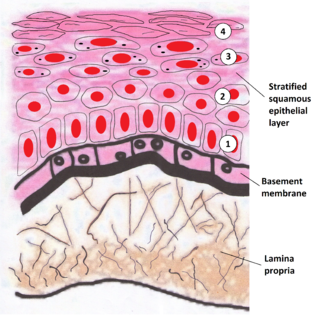
A mucous membrane or mucosa is a membrane that lines various cavities in the body and covers the surface of internal organs. It consists of one or more layers of epithelial cells overlying a layer of loose connective tissue. It is mostly of endodermal origin and is continuous with the skin at various body openings such as the eyes, ears, inside the nose, inside the mouth, lip, vagina, the urethral opening and the anus. Some mucous membranes secrete mucus, a thick protective fluid. The function of the membrane is to stop pathogens and dirt from entering the body and to prevent bodily tissues from becoming dehydrated.

Related Research Articles

Peritoneum serous membrane that forms the lining of the abdominal cavity or the coelom—it covers most of the intra-abdominal (or coelomic) organs—in amniotes and some invertebrates

The peritoneum is the serous membrane forming the lining of the abdominal cavity or coelom in amniotes and some invertebrates, such as annelids. It covers most of the intra-abdominal organs, and is composed of a layer of mesothelium supported by a thin layer of connective tissue. This peritoneal lining of the cavity supports many of the abdominal organs and serves as a conduit for their blood vessels, lymphatic vessels, and nerves.

Epithelium type of animal tissue and human

Epithelium is one of the four basic types of animal tissue, along with connective tissue, muscle tissue and nervous tissue. Epithelial tissues line the outer surfaces of organs and blood vessels throughout the body, as well as the inner surfaces of cavities in many internal organs. An example is the epidermis, the outermost layer of the skin.

Mesothelium membrane composed of simple squamous epithelium that forms the lining of several body cavities: the pleura (thoracic cavity), peritoneum (abdominal cavity including the mesentery), mediastinum and pericardium (heart sac)

The mesothelium is a membrane composed of simple squamous epithelium that forms the lining of several body cavities: the pleura, peritoneum, mediastinum and pericardium. Mesothelial tissue also surrounds the male internal reproductive organs and covers the internal reproductive organs of women. Mesothelium that covers the internal organs is called visceral mesothelium, while the layer that covers the body walls is called the parietal mesothelium. Mesothelium is the epithelial component of serosa.

Lamina propria

The lamina propria is a thin layer of connective tissue that forms part of the moist linings known as mucous membranes or mucosa, which line various tubes in the body, such as the respiratory tract, the gastrointestinal tract, and the urogenital tract.

Loose connective tissue

Loose connective tissue is a category of connective tissue which includes areolar tissue, reticular tissue, and adipose tissue. Loose connective tissue is the most common type of connective tissue in vertebrates. It holds organs in place and attaches epithelial tissue to other underlying tissues. For example, it forms telae, such as the tela submucosa and tela subserosa, which connect mucous and serous membranes to the muscular layer. It also surrounds the blood vessels and nerves. Cells called fibroblasts are widely dispersed in this tissue; they are irregular branching cells that secrete strong fibrous proteins and proteoglycans as an extracellular matrix. The cells of this type of tissue are generally separated by quite some distance by a gelatinous substance primarily made up of collagenous and elastic fibers.

Serous membrane

In anatomy, serous membrane is a smooth tissue membrane consisting of two layers of mesothelium, which secrete serous fluid. The inner layer that covers organs (viscera) in body cavities is called the visceral membrane. A second layer of epithelial cells of the serous membrane, called the parietal layer, lines the body wall. Between the two layers is a potential space, mostly empty except for a few milliliters of lubricating serous fluid that is secreted by the two serous membranes.

Transitional epithelium

Transitional epithelium is a type of stratified epithelium. This tissue consists of multiple layers of epithelial cells which can contract and expand in order to adapt to the degree of distension needed. Transitional epithelium lines the organs of the urinary system and is known here as urothelium. The bladder for example has a need for great distension.

Myometrium

The myometrium is the middle layer of the uterine wall, consisting mainly of uterine smooth muscle cells, but also of supporting stromal and vascular tissue. Its main function is to induce uterine contractions.

Adventitia

Adventitia is the outermost connective tissue covering of an organ, vessel, or other structure. It is also called the tunica adventitia or the tunica externa.

The oral mucosa is the mucous membrane lining the inside of the mouth and consists of stratified squamous epithelium termed oral epithelium and an underlying connective tissue termed lamina propria. The oral cavity has sometimes been described as a mirror that reflects the health of the individual. Changes indicative of disease are seen as alterations in the oral mucosa lining the mouth, which can reveal systemic conditions, such as diabetes or vitamin deficiency, or the local effects of chronic tobacco or alcohol use. The oral mucosa tends to heal faster and with less scar formation compared to the skin. The underlying mechanism remains unknown but research suggest that extracellular vesicles might be involved.

In physiology, the term serous fluid or serosal fluid is any of various body fluids resembling serum, that are typically pale yellow and transparent and of a benign nature. The fluid fills the inside of body cavities. Serous fluid originates from serous glands, with secretions enriched with proteins and water. Serous fluid may also originate from mixed glands, which contain both mucous and serous cells. A common trait of serous fluids is their role in assisting digestion, excretion, and respiration.

Mesenchyme

Mesenchyme, in vertebrate embryology, is a type of connective tissue found mostly during the development of the embryo. It is composed mainly of ground substance with few cells or fibers. It can also refer to a group of mucoproteins found in certain types of cysts (etc.), resembling mucus. It is most easily found as a component of Wharton's jelly.

Perimetrium

The perimetrium is the outer serosa layer of the uterus, equivalent to peritoneum. It is embrionically derived from visceral peritoneum. Perimetrium consists of superficial mesothelium, and a thin layer of loose connective tissue beneath it. The posterior surface of the uterus is completely covered by the perimetrium, but the anterior surface only partially.

Uterine serous carcinoma

Uterine serous carcinoma (USC), also known as uterine papillary serous carcinoma (UPSC) and uterine serous adenocarcinoma, is an uncommon form of endometrial cancer that typically arises in postmenopausal women.

Uterine clear-cell carcinoma (CC) is a rare form of endometrial cancer with distinct morphological features on pathology; it is aggressive and has high recurrence rate. Like uterine papillary serous carcinoma CC does not develop from endometrial hyperplasia and is not hormone sensitive, rather it arises from an atrophic endometrium. Such lesions belong to the type II endometrial cancers.

Fallopian tube

The fallopian tubes, also known as oviducts, uterine tubes, or salpinges are uterine appendages, lined from inside with ciliated simple columnar epithelium, leading from the ovaries of female mammals into the uterus, via the uterotubal junction. They enable the passage of egg cells from the ovaries to the uterus. In non-mammalian vertebrates, the equivalent structures are just called oviducts.

In biology, a tunica is a layer, coat, sheath, or similar covering. The word came to English from the New Latin of science and medicine. Its literal sense is about the same as that of the word tunic, with which it is cognate. In biology one of its senses used to be the taxonomic name of a genus of plants, but the nomenclature has been revised and those plants are now included in the genus Petrorhagia.

Gastrointestinal wall

The gastrointestinal wall surrounding the lumen of the gastrointestinal tract is made up of four layers of specialised tissue – from the lumen outwards:

Anatomical terms of microanatomy

Anatomical terminology is used to describe microanatomical structures. This helps describe precisely the structure, layout and position of an object, and minimises ambiguity. An internationally accepted lexicon is Terminologia Histologica.

References

  1. ACS :: How Is Stomach Cancer Staged?