
The transverse abdominal muscle (TVA), also known as the transverse abdominis, transversalis muscle and transversus abdominis muscle, is a muscle layer of the anterior and lateral abdominal wall which is deep to the internal oblique muscle. It is thought by most fitness instructors to be a significant component of the core.

The lesser omentum is the double layer of peritoneum that extends from the liver to the lesser curvature of the stomach, and to the first part of the duodenum. The lesser omentum is usually divided into these two connecting parts: the hepatogastric ligament, and the hepatoduodenal ligament.

The splenius capitis is a broad, straplike muscle in the back of the neck. It pulls on the base of the skull from the vertebrae in the neck and upper thorax. It is involved in movements such as shaking the head.
The semimembranosus is the most medial of the three hamstring muscles. It is so named because it has a flat tendon of origin. It lies posteromedially in the thigh, deep to the semitendinosus.

The lesser sac, also known as the omental bursa, is the cavity in the abdomen that is formed by the lesser and greater omentum. Usually found in mammals, it is connected with the greater sac via the omental foramen or Foramen of Winslow. In mammals, it is common for the lesser sac to contain considerable amounts of fat..

The sacrotuberous ligament is situated at the lower and back part of the pelvis. It is flat, and triangular in form; narrower in the middle than at the ends.

The falciform ligament is a ligament that attaches the liver to the front body wall, and separates the liver into the left medial lobe and right lateral lobe. The falciform ligament, from Latin 'sickle-shaped', is a broad and thin fold of peritoneum, its base being directed downward and backward and its apex upward and forward. The falciform ligament droops down from the hilum of the liver.

The transversalis fascia is a thin aponeurotic membrane which lies between the inner surface of the transverse abdominal muscle and the parietal peritoneum.

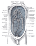
The greater omentum is a large apron-like fold of visceral peritoneum that hangs down from the stomach. It extends from the greater curvature of the stomach, passing in front of the small intestines and doubles back to ascend to the transverse colon before reaching to the posterior abdominal wall. The greater omentum is larger than the lesser omentum, which hangs down from the liver to the lesser curvature. The common anatomical term "epiploic" derives from "epiploon", from the Greek epipleein, meaning to float or sail on, since the greater omentum appears to float on the surface of the intestines. It is the first structure observed when the abdominal cavity is opened anteriorly.

The broad ligament of the uterus is the wide fold of peritoneum that connects the sides of the uterus to the walls and floor of the pelvis.

The porta hepatis or transverse fissure of the liver is a short but deep fissure, about 5 cm long, extending transversely beneath the left portion of the right lobe of the liver, nearer its posterior surface than its anterior border.

The aortic hiatus is a hole in the diaphragm. It is the lowest and most posterior of the large apertures.

The intercostal arteries are a group of arteries that supply the area between the ribs ("costae"), called the intercostal space. The highest intercostal artery is an artery in the human body that usually gives rise to the first and second posterior intercostal arteries, which supply blood to their corresponding intercostal space. It usually arises from the costocervical trunk, which is a branch of the subclavian artery. Some anatomists may contend that there is no supreme intercostal artery, only a supreme intercostal vein.

The spine of the scapula or scapular spine is a prominent plate of bone, which crosses obliquely the medial four-fifths of the scapula at its upper part, and separates the supra- from the infraspinatous fossa.
The median arcuate ligament is a ligament under the diaphragm that connects the right and left crura of diaphragm.

The coronary ligament of the liver refers to parts of the peritoneal reflections that hold the liver to the inferior surface of the diaphragm.
The Hepatophrenic ligament is a ligament connecting the liver to the diaphragm.
The bare area of the liver is a large triangular area on the diaphragmatic surface of the liver, devoid of peritoneal covering. It is attached directly to the diaphragm by loose connective tissue.

In human anatomy, the omental foramen, is the passage of communication, or foramen, between the greater sac, and the lesser sac.
The human liver is divided grossly into four parts or lobes. The four lobes are the right lobe, the left lobe, the caudate lobe, and the quadrate lobe. Seen from the front – the diaphragmatic surface the liver is divided into two lobes the right lobe, and the left lobe. Viewed from the underside – the visceral surface, the other two smaller lobes the caudate lobe, and the quadrate lobe are also visible. The two smaller lobes, the caudate lobe and the quadrate lobe are known as superficial or accessory lobes and both are located on the underside of the right lobe.


