
The peritoneum is the serous membrane forming the lining of the abdominal cavity or coelom in amniotes and some invertebrates, such as annelids. It covers most of the intra-abdominal organs, and is composed of a layer of mesothelium supported by a thin layer of connective tissue. This peritoneal lining of the cavity supports many of the abdominal organs and serves as a conduit for their blood vessels, lymphatic vessels, and nerves.

The abdominal cavity is a large body cavity in humans and many other animals that contain organs. It is a part of the abdominopelvic cavity. It is located below the thoracic cavity, and above the pelvic cavity. Its dome-shaped roof is the thoracic diaphragm, a thin sheet of muscle under the lungs, and its floor is the pelvic inlet, opening into the pelvis.
Articles related to anatomy include:

In human anatomy, the mesentery, an organ that attaches the intestines to the posterior abdominal wall, comprises the double fold of the peritoneum. It helps in storing fat and allowing blood vessels, lymphatics, and nerves to supply the intestines.
The peritoneal cavity is a potential space located between the two layers of the peritoneum—the parietal peritoneum, the serous membrane that lines the abdominal wall, and visceral peritoneum, which surrounds the internal organs. While situated within the abdominal cavity, the term peritoneal cavity specifically refers to the potential space enclosed by these peritoneal membranes. The cavity contains a thin layer of lubricating serous fluid that enables the organs to move smoothly against each other, facilitating the movement and expansion of internal organs during digestion.

In human anatomy, the abdominal aorta is the largest artery in the abdominal cavity. As part of the aorta, it is a direct continuation of the descending aorta.

The rectouterine pouch is the extension of the peritoneum into the space between the posterior wall of the uterus and the rectum in the human female.
The Kocher manoeuvre is a surgical procedure to expose structures in the retroperitoneum behind the duodenum and pancreas. In vascular surgery, it is described as a method to expose the abdominal aorta. It usually has been in contrast to midline laparotomy and right retroperitoneal space dissection. These two procedures have been used for diverse cases, but have approximately equivalent outcomes.

The lesser omentum is the double layer of peritoneum that extends from the liver to the lesser curvature of the stomach, and to the first part of the duodenum. The lesser omentum is usually divided into these two connecting parts: the hepatogastric ligament, and the hepatoduodenal ligament.

In human anatomy, the left gastric artery arises from the celiac artery and runs along the superior portion of the lesser curvature of the stomach before anastomosing with the right gastric artery. It also issues esophageal branches that supply lower esophagus and ascend through the esophageal hiatus to form anastomoses with the esophageal branches of thoracic part of aorta.

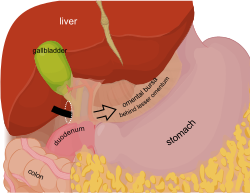
The lesser sac, also known as the omental bursa, is a part of the peritoneal cavity that is formed by the lesser and greater omentum. Usually found in mammals, it is connected with the greater sac via the omental foramen or Foramen of Winslow. In mammals, it is common for the lesser sac to contain considerable amounts of fat.
In human anatomy, the greater sac, also known as the general cavity (of the abdomen) or peritoneum of the peritoneal cavity proper, is the cavity in the abdomen that is inside the peritoneum but outside the lesser sac.

The inferior phrenic artery is a bilaterally paired artery of the abdominal cavity which represents the main source of arterial supply to the diaphragm. Each artery usually arises either from the coeliac trunk or the abdominal aorta, however, their origin is highly variable and the different sites of origin are different for the left artery and right artery. The superior suprarenal artery is a branch of the inferior phrenic artery.

The greater omentum is a large apron-like fold of visceral peritoneum that hangs down from the stomach. It extends from the greater curvature of the stomach, passing in front of the small intestines and doubles back to ascend to the transverse colon before reaching to the posterior abdominal wall. The greater omentum is larger than the lesser omentum, which hangs down from the liver to the lesser curvature. The common anatomical term "epiploic" derives from "epiploon", from the Greek epipleein, meaning to float or sail on, since the greater omentum appears to float on the surface of the intestines. It is the first structure observed when the abdominal cavity is opened anteriorly.

The suspensory ligament of the ovary, also infundibulopelvic ligament, is a fold of peritoneum that extends out from the ovary to the wall of the pelvis.
The testicular artery is a branch of the abdominal aorta that supplies blood to the testicle. It is a paired artery, with one for each of the testicles.

The hepatoduodenal ligament is the portion of the lesser omentum extending between the porta hepatis of the liver and the superior part of the duodenum.
The paracolic gutters are peritoneal recesses – spaces between the colon and the abdominal wall.

The following outline is provided as an overview of and topical guide to human anatomy:

In human anatomy, the liver is divided grossly into four parts or lobes: the right lobe, the left lobe, the caudate lobe, and the quadrate lobe. Seen from the front – the diaphragmatic surface – the liver is divided into two lobes: the right lobe and the left lobe. Viewed from the underside – the visceral surface – the other two smaller lobes, the caudate lobe and the quadrate lobe, are also visible. The two smaller lobes, the caudate lobe and the quadrate lobe, are known as superficial or accessory lobes, and both are located on the underside of the right lobe.


