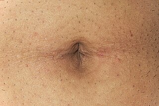
The navel is a protruding, flat, or hollowed area on the abdomen at the attachment site of the umbilical cord. All placental mammals including humans have a navel.

The chorion is the outermost fetal membrane around the embryo in mammals, birds and reptiles. It develops from an outer fold on the surface of the yolk sac, which lies outside the zona pellucida, known as the vitelline membrane in other animals. In insects it is developed by the follicle cells while the egg is in the ovary.
In anatomy, a fissure is a groove, natural division, deep furrow, elongated cleft, or tear in various parts of the body also generally called a sulcus, or in the brain a sulcus.

The anal canal is the terminal part of the large intestine. It is situated between the rectum and anus, below the level of the pelvic diaphragm. In humans it is approximately 2.5 to 4 cm (0.98-1.58 in) long. It lies in the anal triangle of perineum in between the right and left ischioanal fossa.

A sagittal plane, or longitudinal plane, is an anatomical plane which divides the body into right and left parts. The plane may be in the center of the body and split it into two halves (mid-sagittal) or away from the midline and split it into unequal parts (para-sagittal).

The internal iliac artery is the main artery of the pelvis.
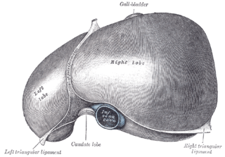
The falciform ligament is a ligament that attaches the liver to the anterior (ventral) body wall, and separates the liver into the left medial lobe and left lateral lobe. The falciform ligament, from Latin, meaning 'sickle-shaped', is a broad and thin fold of peritoneum, its base being directed downward and backward and its apex upward and backward. The falciform ligament droops down from the hilum of the liver.
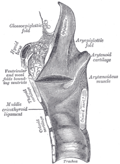
The anterior or lingual surface of the epiglottis is curved forward, and covered on its upper, free part by mucous membrane which is reflected on to the sides and root of the tongue, forming a median and two lateral glossoepiglottic folds; the lateral folds are partly attached to the wall of the pharynx.

The body-stalk, also known as the allantoic stalk, is a band of mesoderm that connects the caudal end of the embryo to the chorion in development. With the formation of the caudal fold, the body-stalk assumes a ventral position; a diverticulum of the yolk-sac extends into the tail fold and is termed the hindgut. With continued development, the body-stalk is later replaced by the umbilical cord.

The round ligament of the liver is a degenerative string of tissue that exists in the free edge of the falciform ligament of the liver. The round ligament divides the left part of the liver into medial and lateral sections.

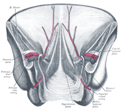
The lateral umbilical fold overlies the inferior epigastric artery and its accompanying veins. Unlike the median and medial umbilical folds, the contents of the lateral umbilical fold remain functional after birth. It originates just medial to the deep inguinal ring to the arcuate line on the posterior surface of the anterior abdominal wall.

The medial umbilical ligament is a paired structure found in human anatomy. It is on the deep surface of the anterior abdominal wall, and is covered by the medial umbilical folds. It should not be confused with the median umbilical ligament, a different structure that represents the remnant of the embryonic urachus.
The lateral inguinal fossa is a structure described in human anatomy. It is a shallow concave stretch of peritoneum on the deep surface of the anterior abdominal wall and is best seen from the greater peritoneal cavity, looking anteriorly.
The medial inguinal fossa is a depression located within the inguinal triangle on the peritoneal surface of the anterior abdominal wall between the ridges formed by the lateral umbilical fold and the medial umbilical ligament, corresponding to the superficial inguinal ring.
The supravesical fossa is a fossa bounded by the medial umbilical fold and median umbilical fold.
The development of the reproductive system is a part of prenatal development, and concerns the sex organs. It is a part of the stages of sexual differentiation. Because its location, to a large extent, overlaps the urinary system, the development of them can also be described together as the development of the urinary and reproductive organs.
Umbilical ligament may refer to:

The human abdomen is divided into regions by anatomists and physicians for purposes of study, diagnosis, and therapy. In the four-region scheme, four quadrants allow localisation of pain and tenderness, scars, lumps, and other items of interest, narrowing in on which organs and tissues may be involved. The quadrants are referred to as the left lower quadrant, left upper quadrant, right upper quadrant and right lower quadrant, as follows below. These terms are not used in comparative anatomy, since most other animals do not stand erect.
The public domain consists of all the creative works to which no exclusive intellectual property rights apply. Those rights may have expired, been forfeited, expressly waived, or may be inapplicable.

Gray's Anatomy is an English language textbook of human anatomy originally written by Henry Gray and illustrated by Henry Vandyke Carter. Earlier editions were called Anatomy: Descriptive and Surgical, Anatomy of the Human Body and Gray's Anatomy: Descriptive and Applied, but the book's name is commonly shortened to, and later editions are titled, Gray's Anatomy. The book is widely regarded as an extremely influential work on the subject, and has continued to be revised and republished from its initial publication in 1858 to the present day. The latest edition of the book, the 41st, was published in September 2015.


