
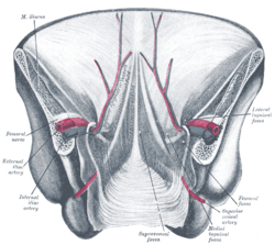
The inguinal canal is a passage in the anterior abdominal wall on each side of the body which in males convey the spermatic cords and in females the round ligament of the uterus. The inguinal canals are larger and more prominent in males.

The rectus abdominis muscle, also known as the "abdominal muscle" or simply the "abs", is a pair of segmented skeletal muscle on the ventral aspect of a person's abdomen. The paired muscles are separated at the midline by a band of dense connective tissue called the linea alba, and the connective tissue defining each lateral margin of the rectus abdominus is the linea semilunaris. The muscle extends from the pubic symphysis, pubic crest and pubic tubercle inferiorly, to the xiphoid process and costal cartilages of the 5th–7th ribs superiorly.

The inguinal ligament, also known as Poupart's ligament or groin ligament, is a band running from the pubic tubercle to the anterior superior iliac spine. It forms the base of the inguinal canal through which an indirect inguinal hernia may develop.

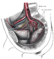
The external iliac arteries are two major arteries which bifurcate off the common iliac arteries anterior to the sacroiliac joint of the pelvis.
The internal iliac artery is the main artery of the pelvis.

In human anatomy, the inguinal triangle is a region of the abdominal wall. It is also known by the eponym Hesselbach's triangle, after Franz Kaspar Hesselbach.

In human anatomy, the inferior epigastric artery is an artery that arises from the external iliac artery. It is accompanied by the inferior epigastric vein; inferiorly, these two inferior epigastric vessels together travel within the lateral umbilical fold The inferior epigastric artery then traverses the arcuate line of rectus sheath to enter the rectus sheath, then anastomoses with the superior epigastric artery within the rectus sheath.

In human anatomy, inferior epigastric vein are 1-2 veins accompanying the inferior epigastric artery. They drain into the external iliac vein just proximal to the inguinal ligament.

The conjoint tendon is a sheath of connective tissue formed from the lower part of the common aponeurosis of the abdominal internal oblique muscle and the transversus abdominis muscle, joining the muscle to the pelvis. It forms the medial part of the posterior wall of the inguinal canal.
In human anatomy, the inguinal region refers to either the groin or the lower lateral regions of the abdomen. It may also refer to:

The transversalis fascia is the fascial lining of the anterolateral abdominal wall situated between the inner surface of the transverse abdominal muscle, and the preperitoneal fascia. It is directly continuous with the iliac fascia, the internal spermatic fascia, and pelvic fascia.
The arcuate line of rectus sheath is a line of demarcation corresponding to the free inferior margin of the posterior layer of the rectus sheath inferior to which only the anterior layer of the rectus sheath is present and the rectus abdominis muscle is therefore in direct contact with the transversalis fascia. The arcuate line is concave inferior-wards.

In human anatomy, the median umbilical ligament is an unpaired midline ligamentous structure upon the lower inner surface of the anterior abdominal wall. It is covered by the median umbilical fold.

The medial umbilical ligament is a paired structure found in human anatomy. It is on the deep surface of the anterior abdominal wall, and is covered by the medial umbilical folds. It is different from the median umbilical ligament, a structure that represents the remnant of the embryonic urachus.

The deep circumflex iliac artery is an artery in the pelvis that travels along the iliac crest of the pelvic bone.
The lateral inguinal fossa is a structure described in human anatomy. It is a shallow concave stretch of peritoneum on the deep surface of the anterior abdominal wall and is best seen from the greater peritoneal cavity, looking anteriorly.
The rectus sheath is a tough fibrous compartment formed by the aponeuroses of the transverse abdominal muscle, and the internal and external oblique muscles. It contains the rectus abdominis and pyramidalis muscles, as well as vessels and nerves.

The medial umbilical fold is an elevation of the peritoneum lining the inner surface of the lower anterior abdominal wall formed by the underlying medial umbilical ligament which the peritoneum covers. Superiorly, the two medial umbilical folds converge towards the midline to meet and terminate at the umbilicus.
Related to the urinary bladder, anteriorly there are the following folds:

The following outline is provided as an overview of and topical guide to human anatomy:
