
The peritoneum is the serous membrane forming the lining of the abdominal cavity or coelom in amniotes and some invertebrates, such as annelids. It covers most of the intra-abdominal organs, and is composed of a layer of mesothelium supported by a thin layer of connective tissue. This peritoneal lining of the cavity supports many of the abdominal organs and serves as a conduit for their blood vessels, lymphatic vessels, and nerves.

A body cavity is any space or compartment, or potential space, in an animal body. Cavities accommodate organs and other structures; cavities as potential spaces contain fluid.
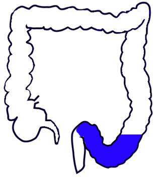
The sigmoid colon is the part of the large intestine that is closest to the rectum and anus. It forms a loop that averages about 35–40 centimetres (14–16 in) in length. The loop is typically shaped like a Greek letter sigma (ς) or Latin letter S. This part of the colon normally lies within the pelvis, but due to its freedom of movement it is liable to be displaced into the abdominal cavity.

The rectouterine pouch is the extension of the peritoneum into the space between the posterior wall of the uterus and the rectum in the human female.

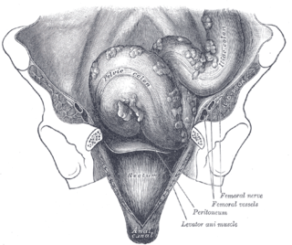
The anal canal is the part that connects the rectum to the anus, located below the level of the pelvic diaphragm. It is located within the anal triangle of the perineum, between the right and left ischioanal fossa. As the final functional segment of the bowel, it functions to regulate release of excrement by two muscular sphincter complexes. The anus is the aperture at the terminal portion of the anal canal.

The internal iliac artery is the main artery of the pelvis.

The broad ligament of the uterus is the wide fold of peritoneum that connects the sides of the uterus to the walls and floor of the pelvis.
The suspensory ligament of the ovary, also infundibulopelvic ligament, is a fold of peritoneum that extends out from the ovary to the wall of the pelvis.

The mesovarium is the portion of the broad ligament of the uterus that suspends the ovaries. The ovary is not covered by the mesovarium; rather, it is covered by germinal epithelium.

The pelvic cavity is a body cavity that is bounded by the bones of the pelvis. Its oblique roof is the pelvic inlet. Its lower boundary is the pelvic floor.

The urethral crest is an anatomical feature present in the urinary system of both males and females.

The supravesical fossa is a depression upon the inner surface of the anterior abdominal wall superior to the bladder formed by a reflection of the peritoneum onto the superior surface of the bladder. It is bounded by the medial umbilical fold and median umbilical fold.

The perimetrium is the outer serosal layer of the uterus, derived from the peritoneum overlying the uterine fundus, and can be considered a visceral peritoneum. It consists of a superficial layer of mesothelium, and a thin layer of loose connective tissue beneath it.
The paracolic gutters are peritoneal recesses – spaces between the colon and the abdominal wall.

The pararectal fossa is an inferior-ward extension of the peritoneum on either side of the rectum. It is formed by a (sacrogenital) fold of peritoneum extending inferior-ward from the posterolateral pelvic wall. It represents a lateral extension of the rectouterine pouch in the female, and of rectovesical pouch in the male. It varies in size with the distension of the rectum.

The rectouterine fold is a bilaterally paired prominent ridge/fold of the peritoneum that represents the lateral boundary of the rectouterine pouch on either side. It is formed by the underlying rectouterine muscle. On either side, the rectouterine fold extends between the sacrum medially, and the base of the broad ligament of the uterus laterally.
In human female anatomy, the vesicouterine pouch, also uterovesicle pouch, is a fold of peritoneum over the uterus and the bladder. Like the rectouterine pouch, it is a female pelvic recess, but shallower and closer to the anterior fornix of the vagina.

The rectovesical pouch is the pocket that lies between the rectum and the bladder in males in humans and other mammals. It is lined by peritoneum.

The ileocecal fold is an anatomical structure of the human abdomen formed by a layer of peritoneum between the ileum and cecum. The upper border of the ileocecal fold is fixed to the ileum opposite its mesenteric attachment, and the lower border passes over the ileocecal junction to join the mesentery of the appendix. Behind the ileocecal fold is the inferior ileocecal fossa.
The iliac colon is the portion of the descending colon which is situated within the left iliac fossa. It is about 12 to 15 cm long.
