Epithelium is one of the four basic types of animal tissue, along with connective tissue, muscle tissue and nervous tissue. Epithelial tissues line the outer surfaces of organs and blood vessels throughout the body, as well as the inner surfaces of cavities in many internal organs. An example is the epidermis, the outermost layer of the skin.

Metaplasia is the transformation of one differentiated cell type to another differentiated cell type. The change from one type of cell to another may be part of a normal maturation process, or caused by some sort of abnormal stimulus. In simplistic terms, it is as if the original cells are not robust enough to withstand their environment, so they transform into another cell type better suited to their environment. If the stimulus causing metaplasia is removed or ceases, tissues return to their normal pattern of differentiation. Metaplasia is not synonymous with dysplasia, and is not considered to be an actual cancer. It is also contrasted with heteroplasia, which is the spontaneous abnormal growth of cytologic and histologic elements. Today, metaplastic changes are usually considered to be an early phase of carcinogenesis, specifically for those with a history of cancers or who are known to be susceptible to carcinogenic changes. Metaplastic change is often viewed as a premalignant condition that requires immediate intervention, either surgical or medical, because metaplasia is associated with cancer.

Squamous metaplasia is a benign non-cancerous change (metaplasia) of surfacing lining cells (epithelium) to a squamous morphology.

Transitional epithelium is a type of stratified epithelium. This tissue consists of multiple layers of epithelial cells which can contract and expand in order to adapt to the degree of distension needed. Transitional epithelium lines the organs of the urinary system and is known here as urothelium. The bladder for example has a need for great distension.
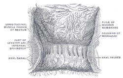
A proctodeum is the back ectodermal part of an alimentary canal. It is created during embryogenesis by a folding of the outer body wall. It will form the lower part of the anal canal, below the pectinate line, which will be lined by stratified squamous non-keratinized and stratified squamous keratinized epithelium. The junction between them is Hilton's white line.
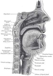
The oral mucosa is the mucous membrane lining the inside of the mouth and consists of stratified squamous epithelium termed oral epithelium and an underlying connective tissue termed lamina propria. The oral cavity has sometimes been described as a mirror that reflects the health of the individual. Changes indicative of disease are seen as alterations in the oral mucosa lining the mouth, which can reveal systemic conditions, such as diabetes or vitamin deficiency, or the local effects of chronic tobacco or alcohol use. The oral mucosa tends to heal faster and with less scar formation compared to the skin. The underlying mechanism remains unknown but research suggest that extracellular vesicles might be involved.
Keratomalacia is an eye disorder that results from vitamin A deficiency. Vitamin A is required to maintain specialized epithelia.
A dentigerous cyst is an odontogenic cyst – thought to be of developmental origin – associated with the crown of an unerupted tooth. The cyst cavity is lined by epithelial cells derived from the reduced enamel epithelium of the tooth forming organ. Regarding its pathogenesis, it has been suggested that the pressure exerted by an erupting tooth on the follicle may obstruct venous flow inducing accumulation of exudate between the reduced enamel epithelium and the tooth crown.
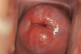
Cervical ectropion is a condition in which the cells from the 'inside' of the cervical canal, known as glandular cells, are present on the 'outside' of the vaginal portion of the cervix. The cells on the 'outside' of the cervix are called squamous epithelial cells. Where the two cells meet is called the transformation zone, also known as the stratified squamous epithelium. Although having this condition is not an abnormality, it is indistinguishable from early cervical cancer. It may be found incidentally when a vaginal examination is done. The area may look red because the glandular cells are red. While many women are born with cervical ectropion, it can be caused by a number of reasons, such as:
An eruption cyst, or eruption hematoma, is a bluish swelling that occurs on the soft tissue over an erupting tooth. It is usually found in children. The fluid in the cyst is sometimes clear creating a pale-coloured cyst although often they are blue. An eruption cyst is a developmental soft-tissue cyst of odontogenic origin that forms over an erupting tooth.
The lateral periodontal cyst is a non-inflammatory developmental cyst that arises from the epithelial post-functional dental lamina, which is a remnant from odontogenesis. It is more common in middle-aged males. Usually asymptomatic, it presents as a regular well-corticated radiolucency on the side of a mandibular canine or premolar root. Histologically, the cyst appears similar to the gingival cyst of the adult, having a non-keratinized squamous epithelial lining. The involved tooth is usually vital and has no indication for root canal treatment unless the signs of non-vital or necrotic pulpal tissue were confirmed. The cysts arise from epithelial rest cells in the periodontal ligament, although it is unknown whether from the cell rests of Malassez, reduced enamel epithelium or dental lamina remnants, and are generally treated by surgical enucleation.

The cavernous portion of the urethra is narrow, and of uniform size in the body of the penis, measuring about 6 mm in diameter; it is dilated behind, within the bulb, and again anteriorly within the glans penis, where it forms the fossa navicularis urethrae.

A squamous cell papilloma is a generally benign papilloma that arises from the stratified squamous epithelium of the skin, lip, oral cavity, tongue, pharynx, larynx, esophagus, cervix, vagina or anal canal. Squamous cell papillomas are typically associated with human papillomavirus (HPV) while for others the cause is unknown.
The nasal vestibule is the most anterior part of the nasal cavity. It is enclosed by the cartilages of nose and lined by the same epithelium of the skin. The other part of the nasal cavity, which is lined by the respiratory epithelium, is called nasal cavity proper. Inside the vestibule are small hairs called vibrissae, which filter dust and other matter that are breathed in. Within the vestibule, the epithelium loses its keratinised nature and undergoes a transition into typical respiratory epithelium before entering the nasal fossa.
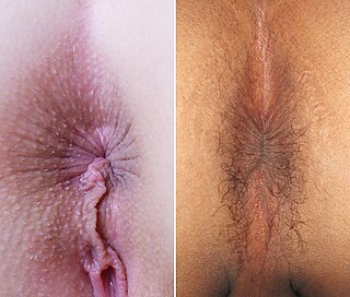
The human anus is the external opening of the rectum. Two sphincters control the exit of feces from the body during an act of defecation, which is the primary function of the anus. These are the internal anal sphincter and the external anal sphincter, which are circular muscles that normally maintain constriction of the orifice and which relaxes as required by normal physiological functioning. The inner sphincter is involuntary and the outer is voluntary. It is located behind the perineum which is located behind the vagina in females and behind the scrotum in males.
The human palatine tonsils (PT) are covered by stratified squamous epithelium that extends into deep and partly branched tonsillar crypts, of which there are about 10 to 30. The crypts greatly increase the contact surface between environmental influences and lymphoid tissue. In an average adult palatine tonsil the estimated epithelial surface area of the crypts is 295 cm2, in addition to the 45 cm2 of epithelium covering the oropharyngeal surface.

The gastrointestinal wall surrounding the lumen of the gastrointestinal tract is made up of four layers of specialised tissue – from the lumen outwards:

Anatomical terminology is used to describe microanatomical structures. This helps describe precisely the structure, layout and position of an object, and minimises ambiguity. An internationally accepted lexicon is Terminologia Histologica.