Knee bursae

Last updated
Knee bursae
Gray350.png
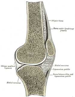
Sagittal section of right knee-joint, thus showing only frontal bursae.
Anatomical terminology

The knee bursae are the fluid-filled sacs and synovial pockets that surround and sometimes communicate with the knee joint cavity. The bursae are thin-walled, and filled with synovial fluid. They represent the weak point of the joint, but also provide enlargements to the joint space. [1] They can be grouped into either communicating and non-communicating bursae or, after their location – frontal, lateral, or medial.

Contents

Frontal

X-ray of the knee of a 12-year-old male, with knee effusion extending into the suprapatellar bursa. Knee effusion, annotated.jpg
X-ray of the knee of a 12-year-old male, with knee effusion extending into the suprapatellar bursa.

In front, there are five bursae:

  1. the suprapatellar bursa or recess between the anterior surface of the lower part of the femur and the deep surface of the quadriceps femoris. [2] It allows for movement of the quadriceps tendon over the distal end of the femur. In about 85% of individuals, this bursa communicates with the knee joint. A distension of this bursa is therefore generally an indication of knee effusion. [3]
  2. the prepatellar bursa between the patella and the skin [2] It allows movement of the skin over the underlying patella.
  3. the deep infrapatellar bursa between the upper part of the tibia and the patellar ligament. [2] It allows for movement of the patellar ligament over the tibia. [4]
  4. the subcutaneous (or superficial) infrapatellar bursa between the patellar ligament and skin. [2]
  5. the pretibial bursa between the tibial tuberosity and the skin. [2] It allows for movement of the skin over the tibial tuberosity. [4]

Lateral

Laterally there are four bursae: [2]

  1. the lateral gastrocnemius (subtendinous) bursa between the lateral head of the gastrocnemius and the joint capsule
  2. the fibular bursa between the lateral (fibular) collateral ligament and the tendon of the biceps femoris
  3. the fibulopopliteal bursa between the fibular collateral ligament and the tendon of the popliteus
  4. and the subpopliteal recess (or bursa) between the tendon of the popliteus and the lateral condyle of the femur

Medial

Medially, there are five bursae:

  1. the medial gastrocnemius (subtendinous) bursa between the medial head of the gastrocnemius and the joint capsule [2]
  2. the anserine bursa between the medial (tibial) collateral ligament and the pes anserinus – the conjoined tendons of the sartorius, gracilis, and semitendinosus muscles. [2]
  3. the bursa semimembranosa between the medial collateral ligament and the tendon of the semimembranosus [2]
  4. there is one between the tendon of the semimembranosus and the head of the tibia [5]
  5. and occasionally there is a bursa between the tendons of the semimembranosus and semitendinosus [5]

See also

Notes

  1. Platzer (2004), p 26
  2. 1 2 3 4 5 6 7 8 9 Burgener (2002), p 390
  3. Daniel J Bell and Brian Gilcrease-Garcia. "Suprapatellar bursa". Radiopaedia .
  4. 1 2 Cipriano (2002), p 358. (A brief description of the "clinically important bursae in the anterior aspect of the knee")
  5. 1 2 Gray's Anatomy (1918)

Related Research Articles

<span class="mw-page-title-main">Human leg</span> Lower extremity or limb of the human body (foot, lower leg, thigh and hip)

The leg is the entire lower limb of the human body, including the foot, thigh or sometimes even the hip or buttock region. The major bones of the leg are the femur, tibia, and adjacent fibula. The thigh is between the hip and knee, while the calf (rear) and shin (front) are between the knee and foot.

<span class="mw-page-title-main">Knee</span> Leg joint in primates

In humans and other primates, the knee joins the thigh with the leg and consists of two joints: one between the femur and tibia, and one between the femur and patella. It is the largest joint in the human body. The knee is a modified hinge joint, which permits flexion and extension as well as slight internal and external rotation. The knee is vulnerable to injury and to the development of osteoarthritis.

<span class="mw-page-title-main">Hamstring</span> Any of the three tendons in the thigh

In human anatomy, a hamstring is any one of the three posterior thigh muscles between the hip and the knee.

<span class="mw-page-title-main">Tibia</span> Leg bone in vertebrates

The tibia, also known as the shinbone or shankbone, is the larger, stronger, and anterior (frontal) of the two bones in the leg below the knee in vertebrates ; it connects the knee with the ankle. The tibia is found on the medial side of the leg next to the fibula and closer to the median plane. The tibia is connected to the fibula by the interosseous membrane of leg, forming a type of fibrous joint called a syndesmosis with very little movement. The tibia is named for the flute tibia. It is the second largest bone in the human body, after the femur. The leg bones are the strongest long bones as they support the rest of the body.

<span class="mw-page-title-main">Popliteal artery</span> Continuation of the femoral artery that supplies the lower leg

The popliteal artery is a deeply placed continuation of the femoral artery opening in the distal portion of the adductor magnus muscle. It courses through the popliteal fossa and ends at the lower border of the popliteus muscle, where it branches into the anterior and posterior tibial arteries.

The biceps femoris is a muscle of the thigh located to the posterior, or back. As its name implies, it consists of two heads; the long head is considered part of the hamstring muscle group, while the short head is sometimes excluded from this characterization, as it only causes knee flexion and is activated by a separate nerve.

The semimembranosus muscle is the most medial of the three hamstring muscles in the thigh. It is so named because it has a flat tendon of origin. It lies posteromedially in the thigh, deep to the semitendinosus muscle. It extends the hip joint and flexes the knee joint.

<span class="mw-page-title-main">Semitendinosus muscle</span> One of the hamstring muscles; posterior part of the thigh

The semitendinosus is a long superficial muscle in the back of the thigh. It is so named because it has a very long tendon of insertion. It lies posteromedially in the thigh, superficial to the semimembranosus.

<span class="mw-page-title-main">Popliteus muscle</span> Muscle responsible for unlocking the knees during walking

The popliteus muscle in the leg is used for unlocking the knees when walking, by laterally rotating the femur on the tibia during the closed chain portion of the gait cycle. In open chain movements, the popliteus muscle medially rotates the tibia on the femur. It is also used when sitting down and standing up. It is the only muscle in the posterior (back) compartment of the lower leg that acts just on the knee and not on the ankle. The gastrocnemius muscle acts on both joints.

The knee examination, in medicine and physiotherapy, is performed as part of a physical examination, or when a patient presents with knee pain or a history that suggests a pathology of the knee joint.

<span class="mw-page-title-main">Stifle joint</span>

The stifle joint is a complex joint in the hind limbs of quadruped mammals such as the sheep, horse or dog. It is the equivalent of the human knee and is often the largest synovial joint in the animal's body. The stifle joint joins three bones: the femur, patella, and tibia. The joint consists of three smaller ones: the femoropatellar joint, medial femorotibial joint, and lateral femorotibial joint.

<span class="mw-page-title-main">Lateral inferior genicular artery</span> Artery in the leg

The lateral inferior genicular is an artery of the leg.

<span class="mw-page-title-main">Lower extremity of femur</span>

The lower extremity of femur is the lower end of the femur in human and other animals, closer to the knee. It is larger than the upper extremity of femur, is somewhat cuboid in form, but its transverse diameter is greater than its antero-posterior; it consists of two oblong eminences known as the lateral condyle and medial condyle.

<span class="mw-page-title-main">Patellar tendon</span> Tendon in the human knee

The patellar tendon is the distal portion of the common tendon of the quadriceps femoris, which is continued from the patella to the tibial tuberosity. It is also sometimes called the patellar ligament as it forms a bone to bone connection when the patella is fully ossified.

<span class="mw-page-title-main">Tuberosity of the tibia</span> Slight protrusion at the top front end

The tuberosity of the tibia, tibial tuberosity or tibial tubercle is an elevation on the proximal, anterior aspect of the tibia, just below where the anterior surfaces of the lateral and medial tibial condyles end.

<span class="mw-page-title-main">Articular capsule of the knee joint</span>

The articular capsule of the knee joint is the wide and lax joint capsule of the knee. It is thin in front and at the side, and contains the patella, ligaments, menisci, and bursae of the knee. The capsule consists of an inner synovial membrane, and an outer fibrous membrane separated by fatty deposits anteriorly and posteriorly.

<span class="mw-page-title-main">Deep fascia of leg</span> Forms a complete investment to the muscles

The deep fascia of leg or crural fascia forms a complete investment to the muscles, and is fused with the periosteum over the subcutaneous surfaces of the bones.

<span class="mw-page-title-main">Pes anserine bursitis</span> Medical condition

Pes anserine bursitis is an inflammatory condition of the medial (inner) knee at the anserine bursa, a sub muscular bursa, just below the pes anserinus.

Posterolateral corner injuries of the knee are injuries to a complex area formed by the interaction of multiple structures. Injuries to the posterolateral corner can be debilitating to the person and require recognition and treatment to avoid long term consequences. Injuries to the PLC often occur in combination with other ligamentous injuries to the knee; most commonly the anterior cruciate ligament (ACL) and posterior cruciate ligament (PCL). As with any injury, an understanding of the anatomy and functional interactions of the posterolateral corner is important to diagnosing and treating the injury.

<span class="mw-page-title-main">Medial knee injuries</span> Medical condition

Medial knee injuries are the most common type of knee injury. The medial ligament complex of the knee consists of:

References