Leptomeningeal collateral circulation

Last updated
Leptomeningeal collateral circulation
Sobo 1909 3 548.png
Arterial supply of the brain
Details
Locationaround the brain
Functionsmall connections ( anastamoses ) between the areas supplied by the major arteries of the brain.
Anatomical terminology

The leptomeningeal collateral circulation (also known as leptomeningeal anastomoses or pial collaterals) is a network of small blood vessels in the brain that connects branches of the middle, anterior and posterior cerebral arteries (MCA, ACA, and PCA), [1] with variation in its precise anatomy between individuals. [2] During a stroke, leptomeningeal collateral vessels allow limited blood flow when other, larger blood vessels provide inadequate blood supply to a part of the brain. [3]

Contents

Structure

Layers of the skull and brain Gray1196.png
Layers of the skull and brain
The middle cerebral artery and its branches MCA angio lateral.jpg
The middle cerebral artery and its branches

Leptomeningeal collaterals lie within the leptomeninges, the two deep layers of the meninges called the pia mater and the arachnoid mater. [4] Their diameter has been measured at approximately 300 micrometers, [5] but there is variability between individuals in the size, quantity and location of these vessels, and between either hemisphere within the same subject. [6]

Inter-territorial end to end anastomoses exist between branches of the anterior cerebral artery and middle cerebral artery, the posterior cerebral artery and middle cerebral artery, the anterior cerebral artery and posterior cerebral artery, and the right and left anterior cerebral arteries. [7] [8] [9] [10] Intra-territorial anastamoses connect adjacent arterial branches within the same arterial territory (between two branches of the same middle cerebral artery, for example). [5]

Inter-territorial leptomeningeal anastamoses relative to branches of the middle cerebral artery [5]
Supplying the Frontal Lobe
Prefrontal arteriesNo anastamoses observed
Orbito-frontal (lateral frontobasal) arteryAnterior and inferior frontal arteries (branches of the anterior cerebral artery)
Precentral (pre-rolandic) arteryPosterior inferior frontal artery (a branch of the anterior cerebral artery)
Central (rolandic) arteryParacentral artery (a branch of the anterior cerebral artery)
Supplying the Parietal Lobe
Anterior parietal arteryPrecuneal artery (a branch of the anterior cerebral artery)
Posterior parietal arteryNo anastamoses observed
Angular arteryParieto-occipital artery (a branch of the posterior cerebral artery)
Temporo-occipitalNo anastamoses observed
Supplying the Temporal Lobe
Posterior temporal arteryNo anastamoses observed
Middle temporal arteryNo anastamoses observed
Anterior temporal arteryAnterior temporal artery (a branch of the posterior cerebral artery)
Temporopolar arteryNo anastamoses observed

Inter-territorial leptomeningeal anastamoses between the posterior cerebral artery and anterior cerebral artery have been observed between the parieto-occipital branch of the posterior cerebral artery, and the precuneal branch or the posterior pericallosal branch of the anterior cerebral artery. [1]

Inter-territoral leptomeningeal anastamoses between the right and left anterior cerebral arteries have been observed between the right and left pericallosal arteries and the right and left callosal marginal arteries. Anastamoses have also been observed between precuneal branches originating from the middle portion of the pericallosal artery, or from the posterior portion of the callosal marginal branch of one side joining the opposite paracentral branch. [1]

There is anatomical variation in collateral circulation from person to person, and as we age, collateral vessels decrease in diameter and number. [2]

Function

Leptomeningeal collateral vessels allow limited cerebral blood flow and brain tissue perfusion when the brain receives insufficient blood supply through an artery, via a series of anastomotic connections between cerebral arteries. [3]

Clinical significance

Stroke

Middle cerebral artery angiography, showing stenosis TOF MRI angiography of right middle cerebral artery stenosis.png
Middle cerebral artery angiography, showing stenosis

During an ischaemic stroke, blood flow through a cerebral artery is compromised. This frequently causes substantial injury to the area of the brain supplied by the artery, but not all of this territory is necessarily affected. A post mortem study of middle cerebral artery strokes demonstrated that the area of brain injury was often smaller than the total area supplied by the middle cerebral artery. Leptomeningeal collateral vessels from the anterior cerebral artery and posterior cerebral artery appeared to allow for perfusion of some brain tissue to persist, partially compensating for the loss of the major vessel. [6] This compensatory effect is however usually inadequate to maintain a normal blood supply. [11]

Therapies that attempt to optimize leptomeningeal collateral circulation appear to improve outcomes following acute ischaemic stroke. [2]

MRI and CT brain imaging is used to determine the severity of a stroke, and help guide treatment. Fluid attenuated inversion recovery (FLAIR) vascular hyperintensity (FVH) is a radiographic marker seen on brain imaging in acute ischaemic stroke. FVH can be used as a proxy for slow leptomeningeal collateral blood flow, and may help reveal which areas of brain tissue are potentially salvageable. [12]

Alzheimer’s disease

The age-related changes that can be seen in leptomeningeal vessels over time appear to be accelerated by Alzheimer's disease, according to mouse models conducted in 2018. [13]

Intracranial haemorrhage

A 2016 study compared patients awaiting carotid artery stenting for unilateral atherosclerotic plaques. Those with leptomeningeal collaterals evident on cranial angiography had a higher incidence of intracranial haemorrhage (ICH) after stenting. The authors argued that the presence of such collaterals on imaging should be considered a risk factor for ICH in patients where carotid stenting is otherwise indicated. [14]

History

Johann Otto Leonhard Heubner Otto Heubner 2.jpg
Johann Otto Leonhard Heubner

The term 'leptomeningeal' derives from the Greek word leptos (λεπτός) meaning thin, in reference to the appearance of the pia mater and arachnoid mater.

Descriptions of leptomenigeal collateral vessels are found in Thomas WillisCerebri Anatome (1664). [15] [16] German physician Otto Heubner first demonstrated their presence in his 1874 work Die luetische Erkrankung Der Hirnaterien. [17] He injected the middle cerebral artery, anterior cerebral artery and posterior cerebral artery in turn, in an attempt to establish the territories these arteries supply. Even when other anastomoses from the circle of Willis were blocked off, the whole cerebral arterial tree could be filled. [1] Later study in the 1950s and 60s by H.M. Vander Eecken and R.D. Adams provided a comprehensive review of the anatomy of the leptomeningeal collateral circulation. [6]

The concept of the ischaemic penumbra, where brain tissue shows capacity to recover if perfusion is quickly restored, was defined in 1981 by Astrup et al. Persistent blood flow through leptomeningeal vessels is a key part of this recovery. [18]

Other animals

Haemodynamic studies of leptomeningeal collaterals have been conducted in primates. [19] Leptomeningeal circulation has been observed in mice and rats during experiments to assess changes associated with disease and ageing in these vessels. [20]

Related Research Articles

A transient ischemic attack (TIA), commonly known as a mini-stroke, is a minor stroke whose noticeable symptoms usually end in less than an hour. TIA causes the same symptoms associated with strokes, such as weakness or numbness on one side of the body, sudden dimming or loss of vision, difficulty speaking or understanding language, slurred speech, or confusion.

Coronary circulation Circulation of blood in the blood vessels of the heart muscle (myocardium)

Coronary circulation is the circulation of blood in the blood vessels that supply the heart muscle (myocardium). Coronary arteries supply oxygenated blood to the heart muscle. Cardiac veins then drain away the blood after it has been deoxygenated. Because the rest of the body, and most especially the brain, needs a steady supply of oxygenated blood that is free of all but the slightest interruptions, the heart is required to function continuously. Therefore its circulation is of major importance not only to its own tissues but to the entire body and even the level of consciousness of the brain from moment to moment. Interruptions of coronary circulation quickly cause heart attacks, in which the heart muscle is damaged by oxygen starvation. Such interruptions are usually caused by coronary ischemia linked to coronary artery disease, and sometimes to embolism from other causes like obstruction in blood flow through vessels.

Circle of Willis Circulatory anastomosis that supplies blood to the brain and surrounding structures

The circle of Willis is a circulatory anastomosis that supplies blood to the brain and surrounding structures in reptiles, birds and mammals, including humans. It is named after Thomas Willis (1621–1675), an English physician.

Cerebrovascular disease Condition that affects the arteries that supply the brain

Cerebrovascular disease includes a variety of medical conditions that affect the blood vessels of the brain and the cerebral circulation. Arteries supplying oxygen and nutrients to the brain are often damaged or deformed in these disorders. The most common presentation of cerebrovascular disease is an ischemic stroke or mini-stroke and sometimes a hemorrhagic stroke. Hypertension is the most important contributing risk factor for stroke and cerebrovascular diseases as it can change the structure of blood vessels and result in atherosclerosis. Atherosclerosis narrows blood vessels in the brain, resulting in decreased cerebral perfusion. Other risk factors that contribute to stroke include smoking and diabetes. Narrowed cerebral arteries can lead to ischemic stroke, but continually elevated blood pressure can also cause tearing of vessels, leading to a hemorrhagic stroke.

Internal carotid artery Artery of the human brain

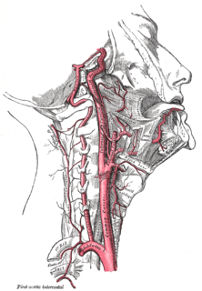
The internal carotid artery is an artery in the neck which supplies the anterior circulation of the brain. In human anatomy, the internal and external carotids arise from the common carotid arteries, where these bifurcate at cervical vertebrae C3 or C4. The internal carotid artery supplies the brain, including the eyes, while the external carotid nourishes other portions of the head, such as the face, scalp, skull, and meninges.

Stroke Death of a region of brain cells due to poor blood flow

A stroke is a medical condition in which poor blood flow to the brain causes cell death. There are two main types of stroke: ischemic, due to lack of blood flow, and hemorrhagic, due to bleeding. Both cause parts of the brain to stop functioning properly. Signs and symptoms of a stroke may include an inability to move or feel on one side of the body, problems understanding or speaking, dizziness, or loss of vision to one side. Signs and symptoms often appear soon after the stroke has occurred. If symptoms last less than one or two hours, the stroke is a transient ischemic attack (TIA), also called a mini-stroke. A hemorrhagic stroke may also be associated with a severe headache. The symptoms of a stroke can be permanent. Long-term complications may include pneumonia and loss of bladder control.

Radial artery Large forearm artery

In human anatomy, the radial artery is the main artery of the lateral aspect of the forearm.

Cerebral circulation Brain blood supply

Cerebral circulation is the movement of blood through a network of cerebral arteries and veins supplying the brain. The rate of cerebral blood flow in an adult human is typically 750 milliliters per minute, or about 15% of cardiac output. Arteries deliver oxygenated blood, glucose and other nutrients to the brain. Veins carry "used or spent" blood back to the heart, to remove carbon dioxide, lactic acid, and other metabolic products. Because the brain would quickly suffer damage from any stoppage in blood supply, the cerebral circulatory system has safeguards including autoregulation of the blood vessels. The failure of these safeguards may result in a stroke. The volume of blood in circulation is called the cerebral blood flow. Sudden intense accelerations change the gravitational forces perceived by bodies and can severely impair cerebral circulation and normal functions to the point of becoming serious life-threatening conditions.

Cerebral angiography Angiography that produces images of blood vessels in and around the brain

Cerebral angiography is a form of angiography which provides images of blood vessels in and around the brain, thereby allowing detection of abnormalities such as arteriovenous malformations and aneurysms. It was pioneered in 1927 by the Portuguese neurologist Egas Moniz at the University of Lisbon, who also helped develop thorotrast for use in the procedure.

Moyamoya disease Disease characterized by constriction of brain arteries

Moyamoya disease is a disease in which certain arteries in the brain are constricted. Blood flow is blocked by constriction and blood clots (thrombosis). A collateral circulation develops around the blocked vessels to compensate for the blockage, but the collateral vessels are small, weak, and prone to bleeding, aneurysm and thrombosis. On conventional angiography, these collateral vessels have the appearance of a "puff of smoke".

Vertebral artery Major arteries of the neck

The vertebral arteries are major arteries of the neck. Typically, the vertebral arteries originate from the subclavian arteries. Each vessel courses superiorly along each side of the neck, merging within the skull to form the single, midline basilar artery. As the supplying component of the vertebrobasilar vascular system, the vertebral arteries supply blood to the upper spinal cord, brainstem, cerebellum, and posterior part of brain.

Anterior cerebral artery

The anterior cerebral artery (ACA) is one of a pair of cerebral arteries that supplies oxygenated blood to most midline portions of the frontal lobes and superior medial parietal lobes of the brain. The two anterior cerebral arteries arise from the internal carotid artery and are part of the circle of Willis. The left and right anterior cerebral arteries are connected by the anterior communicating artery.

Middle cerebral artery

The middle cerebral artery (MCA) is one of the three major paired arteries that supply blood to the cerebrum. The MCA arises from the internal carotid and continues into the lateral sulcus where it then branches and projects to many parts of the lateral cerebral cortex. It also supplies blood to the anterior temporal lobes and the insular cortices.

Carotid endarterectomy

Carotid endarterectomy is a surgical procedure used to reduce the risk of stroke from carotid artery stenosis.

Posterior communicating artery Arteries at the base of the brain that form part of the circle of Willis

In human anatomy, the left and right posterior communicating arteries are arteries at the base of the brain that form part of the circle of Willis. Each posterior communicating artery connects the three cerebral arteries of the same side. Anteriorly, it connects to the internal carotid artery (ICA) prior to the terminal bifurcation of the ICA into the anterior cerebral artery and middle cerebral artery. Posteriorly, it communicates with the posterior cerebral artery.

Anterior communicating artery

In human anatomy, the anterior communicating artery is a blood vessel of the brain that connects the left and right anterior cerebral arteries.

Cerebral infarction Medical condition

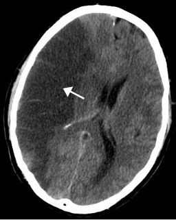
A cerebral infarction is the pathologic process that results in an area of necrotic tissue in the brain. It is caused by disrupted blood supply (ischemia) and restricted oxygen supply (hypoxia), most commonly due to thromboembolism, and manifests clinically as ischemic stroke. In response to ischemia, the brain degenerates by the process of liquefactive necrosis.

Watershed stroke Medical condition

A watershed stroke is defined as a brain ischemia that is localized to the vulnerable border zones between the tissues supplied by the anterior, posterior and middle cerebral arteries. The actual blood stream blockage/restriction site can be located far away from the infarcts. Watershed locations are those border-zone regions in the brain supplied by the major cerebral arteries where blood supply is decreased. Watershed strokes are a concern because they comprise approximately 10% of all ischemic stroke cases. The watershed zones themselves are particularly susceptible to infarction from global ischemia as the distal nature of the vasculature predisposes these areas to be most sensitive to profound hypoperfusion.

Carotid artery dissection Human disease

Carotid artery dissection is a separation of the layers of the artery wall supplying oxygen-bearing blood to the head and brain and is the most common cause of stroke in young adults.

Anterior cerebral artery syndrome Medical condition

Anterior cerebral artery syndrome is a condition whereby the blood supply from the anterior cerebral artery (ACA) is restricted, leading to a reduction of the function of the portions of the brain supplied by that vessel: the medial aspects of the frontal and parietal lobes, basal ganglia, anterior fornix and anterior corpus callosum.

References

  1. 1 2 3 4 Brozici Mariana; van der Zwan Albert; Hillen Berend (2003-11-01). "Anatomy and Functionality of Leptomeningeal Anastomoses". Stroke. 34 (11): 2750–2762. doi: 10.1161/01.STR.0000095791.85737.65 . PMID   14576375.
  2. 1 2 3 Winship, Ian R. (April 2015). "Cerebral collaterals and collateral therapeutics for acute ischemic stroke". Microcirculation. 22 (3): 228–236. doi:10.1111/micc.12177. ISSN   1549-8719. PMID   25351102. S2CID   206189480.
  3. 1 2 Tariq N, Khatri R (October 2008). "Leptomeningeal collaterals in acute ischemic stroke". Journal of Vascular and Interventional Neurology. 1 (4): 91–5. PMC   3317324 . PMID   22518231.
  4. Patel, Neel; Kirmi, Olga (2009). "Anatomy and imaging of the normal meninges". Seminars in Ultrasound, CT and MRI. 30 (6): 559–564. doi:10.1053/j.sult.2009.08.006. ISSN   0887-2171. PMID   20099639.
  5. 1 2 3 Phan, Thanh G.; Hilton, James; Beare, Richard; Srikanth, Velandai; Sinnott, Matthew (2014-09-19). "Computer Modeling of Anterior Circulation Stroke: Proof of Concept in Cerebrovascular Occlusion". Frontiers in Neurology. 5: 176. doi: 10.3389/fneur.2014.00176 . ISSN   1664-2295. PMC   4168699 . PMID   25285093.
  6. 1 2 3 Vander Eecken HM, Adams RD (April 1953). "The anatomy and functional significance of the meningeal arterial anastomoses of the human brain". Journal of Neuropathology and Experimental Neurology. 12 (2): 132–57. doi:10.1097/00005072-195304000-00002. PMID   13053234. S2CID   33711931.
  7. Cipolla, Marilyn J. (2009). Anatomy and Ultrastructure. Morgan & Claypool Life Sciences.
  8. Türe, Uğur; Yaşargil, M. Gazi; Krisht, Ali F. (1996-12-01). "The Arteries of the Corpus Callosum: A Microsurgical Anatomic Study". Neurosurgery. 39 (6): 1075–1084. doi:10.1097/00006123-199612000-00001. ISSN   0148-396X. PMID   8938760.
  9. Kim, D.J.; Krings, T. (2011-09-01). "Whole-Brain Perfusion CT Patterns of Brain Arteriovenous Malformations: A Pilot Study in 18 Patients". American Journal of Neuroradiology. 32 (11): 2061–2066. doi: 10.3174/ajnr.a2659 . ISSN   0195-6108. PMC   7964413 . PMID   21885720.
  10. Coyle, Peter (1994). "Collateral Pial Arteries". In Bevan, Rosemary D.; Bevan, John A. (eds.). The Human Brain Circulation. The Human Brain Circulation: Functional Changes in Disease. Vascular Biomedicine. Humana Press. pp. 237–246. doi:10.1007/978-1-4612-0303-2_18. ISBN   9781461203032.
  11. Derdeyn CP, Powers WJ, Grubb RL (September 1998). "Hemodynamic effects of middle cerebral artery stenosis and occlusion" (PDF). AJNR. American Journal of Neuroradiology. 19 (8): 1463–9. PMC   8338700 . PMID   9763379.
  12. Liu, Dezhi; Scalzo, Fabien; Rao, Neal M; Hinman, Jason D.; Kim, Doojin; Ali, Latisha K; Saver, Jeffrey L.; Sun, Wen; Dai, Qiliang (2016). "FLAIR Vascular Hyperintensity Topography, Novel Imaging Marker for Revascularization in Middle Cerebral Artery Occlusion". Stroke: A Journal of Cerebral Circulation. 47 (11): 2763–2769. doi:10.1161/STROKEAHA.116.013953. ISSN   0039-2499. PMC   5079823 . PMID   27659851.
  13. Zhang, Hua; Jin, Bo; Faber, James E. (2018-12-05). "Mouse models of Alzheimer's disease cause rarefaction of pial collaterals and increased severity of ischemic stroke". Angiogenesis. 22 (2): 263–279. doi:10.1007/s10456-018-9655-0. ISSN   1573-7209. PMC   6475514 . PMID   30519973.
  14. Lee, Kang Ji; Kwak, Hyo Sung; Chung, Gyung Ho; Song, Ji Soo; Hwang, Seung Bae (May 2016). "Leptomeningeal collateral vessels are a major risk factor for intracranial hemorrhage after carotid stenting in patients with carotid atherosclerotic plaque". Journal of NeuroInterventional Surgery. 8 (5): 512–516. doi:10.1136/neurintsurg-2014-011634. ISSN   1759-8486. PMID   25841168. S2CID   24415856.
  15. Lega, Bradley C. (2006). "An Essay Concerning Human Understanding: How the Cerebri Anatome of Thomas Willis Influenced John Locke". Neurosurgery. 58 (3): 567–576. doi:10.1227/01.neu.0000197489.17675.c6. ISSN   0148-396X. PMID   16528199.
  16. Willis, Thomas (1664). Cerebri anatome : cui accessit Nervorum descriptio et usus /. Londini: Typis Tho. Roycroft, impensis Jo. Martyn & Ja. Allestry ...
  17. "Die luetische Erkrankung der Hirnarterien". Wellcome Library. Retrieved 2019-03-10.
  18. Paciaroni, Maurizio; Caso, Valeria; Agnelli, Giancarlo (2009). "The concept of ischemic penumbra in acute stroke and therapeutic opportunities". European Neurology. 61 (6): 321–330. doi: 10.1159/000210544 . ISSN   1421-9913. PMID   19365124.
  19. Symon, L. (1968). "Haemodynamic studies of the leptomeningeal circulation in primates". Proceedings of the Royal Society of Medicine. 61 (6): 610–612. doi:10.1177/003591576806100629. ISSN   0035-9157. PMC   1902586 . PMID   4969977.
  20. Li, Zhaojin; Tremble, Sarah M.; Cipolla, Marilyn J. (2018-12-01). "Implications for understanding ischemic stroke as a sexually dimorphic disease: the role of pial collateral circulations". American Journal of Physiology. Heart and Circulatory Physiology. 315 (6): H1703–H1712. doi:10.1152/ajpheart.00402.2018. ISSN   1522-1539. PMC   6336971 . PMID   30239233.