Veins are blood vessels in the circulatory system of humans and most other animals that carry blood towards the heart. Most veins carry deoxygenated blood from the tissues back to the heart; exceptions are those of the pulmonary and fetal circulations which carry oxygenated blood to the heart. In the systemic circulation, arteries carry oxygenated blood away from the heart, and veins return deoxygenated blood to the heart, in the deep veins.

In human anatomy, the arm refers to the upper limb in common usage, although academically the term specifically means the upper arm between the glenohumeral joint and the elbow joint. The distal part of the upper limb between the elbow and the radiocarpal joint is known as the forearm or "lower" arm, and the extremity beyond the wrist is the hand.

The median nerve is a nerve in humans and other animals in the upper limb. It is one of the five main nerves originating from the brachial plexus.
The forearm is the region of the upper limb between the elbow and the wrist. The term forearm is used in anatomy to distinguish it from the arm, a word which is used to describe the entire appendage of the upper limb, but which in anatomy, technically, means only the region of the upper arm, whereas the lower "arm" is called the forearm. It is homologous to the region of the leg that lies between the knee and the ankle joints, the crus.

The azygos vein is a vein running up the right side of the thoracic vertebral column draining itself towards the superior vena cava. It connects the systems of superior vena cava and inferior vena cava and can provide an alternative path for blood to the right atrium when either of the venae cavae is blocked.
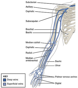
In human anatomy, the cephalic vein is a superficial vein in the arm. It originates from the radial end of the dorsal venous network of hand, and ascends along the radial (lateral) side of the arm before emptying into the axillary vein. At the elbow, it communicates with the basilic vein via the median cubital vein.
The cubital fossa, chelidon or inside of elbow is the area on the anterior side of the upper part between the arm and forearm of a human or other hormid animals. It lies anteriorly to the elbow when in standard anatomical position.
In human anatomy, the median cubital vein is a superficial vein of the arm on the anterior aspect of the elbow. It classically connects the cephalic vein and the basilic vein.
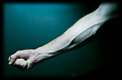
The basilic vein is a large superficial vein of the upper limb that helps drain parts of the hand and forearm. It originates on the medial (ulnar) side of the dorsal venous network of the hand and travels up the base of the forearm, where its course is generally visible through the skin as it travels in the subcutaneous fat and fascia lying superficial to the muscles. The basilic vein terminates by uniting with the brachial veins to form the axillary vein.
The ulnar veins are venae comitantes of the ulnar artery. They drain the superficial venous palmar arch. They arise in the hand and terminate by uniting with the radial veins to form the brachial veins. They mostly drain the medial aspect of the forearm. They receive the venae comitantes of the anterior and posterior interosseous arteries near the elbow, as well as a large branch from the median cubital vein. The ulnar veins are larger than the radial veins.

In human anatomy, the internal thoracic vein is the vein that drains the chest wall and breasts.

The superior sagittal sinus, within the human head, is an unpaired area along the attached margin of the falx cerebri. It allows blood to drain from the lateral aspects of anterior cerebral hemispheres to the confluence of sinuses. Cerebrospinal fluid drains through arachnoid granulations into the superior sagittal sinus and is returned to venous circulation.

The lateral cutaneous nerve of forearm is a sensory nerve representing the continuation of the musculocutaneous nerve beyond the lateral edge of the tendon of the biceps brachii muscle. The lateral cutaneous nerve provides sensory innervation to the skin of the lateral forearm. It pierces the deep fascia of forearm to enter the subcutaneous compartment before splitting into a volar branch and a dorsal branch.

The medial cutaneous nerve of the forearm is a sensory branch of the medial cord of the brachial plexus derived from the ventral rami of spinal nerves C8-T1. It provides sensory innervation to the skin of the medial forearm and skin overlying the olecranon. It descends through the (upper) arm within the brachial fascia alongside the basilic vein, then divides into an anterior branch and a posterior branch upon emerging from the brachial fascia; the two terminal branches travel as far distally as the wrist.

The rectal venous plexus is the venous plexus surrounding the rectum. It consists of an internal and an external rectal plexus. It is drained by the superior, middle, and inferior rectal veins. It forms a portosystemic (portocaval) anastomosis. This allows rectally administered medications to bypass first pass metabolism.

The bicipital aponeurosis is a broad aponeurosis of the biceps brachii, which is located in the cubital fossa of the elbow. It separates superficial from deep structures in much of the fossa.

One or two supratrochlear lymph nodes are placed above the medial epicondyle of the humerus, medial to the basilic vein.

The superficial palmar venous arch consists of a pair of venae comitantes accompanying the superficial palmar arch. It receives the common palmar digital veins. It drains into the superficial ulnar radial and superficial radial veins, and the median antebrachial vein.

The elbow is the region between the upper arm and the forearm that surrounds the elbow joint. The elbow includes prominent landmarks such as the olecranon, the cubital fossa, and the lateral and the medial epicondyles of the humerus. The elbow joint is a hinge joint between the arm and the forearm; more specifically between the humerus in the upper arm and the radius and ulna in the forearm which allows the forearm and hand to be moved towards and away from the body. The term elbow is specifically used for humans and other primates, and in other vertebrates it is not used. In those cases, forelimb plus joint is used.