The carpal bones are the eight small bones that make up the wrist (carpus) that connects the hand to the forearm. The term "carpus" and "carpal" is derived from the Latin carpus and the Greek καρπός (karpós), meaning "wrist". In human anatomy, the main role of the carpal bones is to articulate with the radial and ulnar heads to form a highly mobile condyloid joint, to provide attachments for thenar and hypothenar muscles, and to form part of the rigid carpal tunnel which allows the median nerve and tendons of the anterior forearm muscles to be transmitted to the hand and fingers.
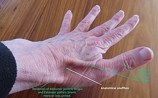
The anatomical snuff box or snuffbox or foveola radialis is a triangular deepening on the radial, dorsal aspect of the hand—at the level of the carpal bones, specifically, the scaphoid and trapezium bones forming the floor. The name originates from the use of this surface for placing and then sniffing powdered tobacco, or "snuff." It is sometimes referred to by its French name tabatière.

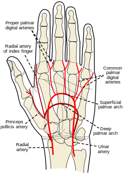
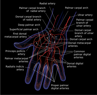
In human anatomy, the radial artery is the main artery of the lateral aspect of the forearm.
The ulnar artery is the main blood vessel, with oxygenated blood, of the medial aspects of the forearm. It arises from the brachial artery and terminates in the superficial palmar arch, which joins with the superficial branch of the radial artery. It is palpable on the anterior and medial aspect of the wrist.

The palmar aponeurosis invests the muscles of the palm, and consists of central, lateral, and medial portions.

The interphalangeal joints of the hand are the hinge joints between the phalanges of the fingers that provide flexion towards the palm of the hand.

The medial cutaneous nerve of the forearm is a sensory branch of the medial cord of the brachial plexus derived from the ventral rami of spinal nerves C8-T1. It provides sensory innervation to the skin of the medial forearm and skin overlying the olecranon. It descends through the (upper) arm within the brachial fascia alongside the basilic vein, then divides into an anterior branch and a posterior branch upon emerging from the brachial fascia; the two terminal branches travel as far distally as the wrist.
The superficial palmar arch is formed predominantly by the ulnar artery, with a contribution from the superficial palmar branch of the radial artery. However, in some individuals the contribution from the radial artery might be absent, and instead anastomoses with either the princeps pollicis artery, the radialis indicis artery, or the median artery, the former two of which are branches from the radial artery.
The deep palmar arch is an arterial network found in the palm. It is usually primarily formed from the terminal part of the radial artery. The ulnar artery also contributes through an anastomosis. This is in contrast to the superficial palmar arch, which is formed predominantly by the ulnar artery.

The palmar metacarpal arteries are three or four arteries that arise from the convexity of the deep palmar arch.

In the palm of the hand the median nerve is covered by the skin and the palmar aponeurosis, and rests on the tendons of the flexor muscles. Immediately after emerging from under the transverse carpal ligament the median nerve becomes enlarged and flattened and splits into a smaller, lateral, and a larger, medial portion.
In the palm of the hand, the median nerve is covered by the skin and the palmar aponeurosis, and rests on the tendons of the Flexor muscles. Immediately after emerging from under the transverse carpal ligament the median nerve becomes enlarged and flattened and splits into a smaller, lateral, and a larger, medial portion.
A circulatory anastomosis is a connection between two blood vessels, such as between arteries, between veins or between an artery and a vein. Anastomoses between arteries and between veins result in a multitude of arteries and veins, respectively, serving the same volume of tissue. Such anastomoses occur normally in the body in the circulatory system, serving as back-up routes in a collateral circulation that allow blood to flow if one link is blocked or otherwise compromised, but may also occur pathologically.
The palmar digital veins on each finger are connected to the dorsal digital veins by oblique intercapitular veins. They drain into a venous plexus which is situated over the thenar and hypothenar eminences and across the front of the wrist.
The palmar metacarpal veins drain the metacarpal region of the palm, eventually draining into the deep veins of the arm.
The superficial palmar venous arch consists of a pair of venae comitantes accompanying the superficial palmar arch. It receives the common palmar digital veins. It drains into the superficial ulnar radial and superficial radial veins, and the median antebrachial vein.
The deep palmar arch, an arterial network is accompanied by a pair of venae comitantes which constitute the deep venous palmar arch. It receives the veins corresponding to the branches of the arterial arch: the palmar metacarpal veins.
The proper palmar digital nerves of the ulnar nerve are nerves of the hand.
The common palmar digital nerves of the ulnar nerve are nerves of the hand. The nerve branches off the superficial branch of the ulnar nerve and runs toward the cleft between the ring and little fingers.

A hand is a prehensile, multi-fingered appendage located at the end of the forearm or forelimb of primates such as humans, chimpanzees, monkeys, and lemurs. A few other vertebrates such as the koala are often described as having "hands" instead of paws on their front limbs. The raccoon is usually described as having "hands" though opposable thumbs are lacking.