Morquio syndrome

Last updated
Morquio syndrome
Other names Mucopolysaccharidosis IV, MPS IV, Morquio-Brailsford syndrome, or Morquio [1]
Mucopolysaccharidosis (Morquio's Disease) 1.jpg
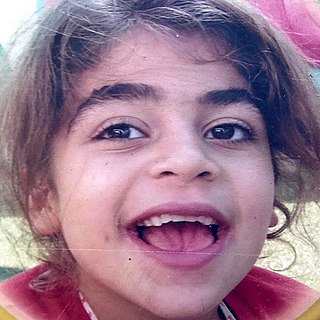
Patient with Morquio syndrome
Specialty Endocrinology   OOjs UI icon edit-ltr-progressive.svg
Complications Skeletal abnormalities, hearing loss, pulmonary failure, heart disease
Usual onsetBirth; condition usually becomes apparent between ages 1 and 3
DurationLifelong
TypesType A and Type B
CausesInherited deficiency of enzymes
TreatmentElosulfase alfa (Vimizim) for Type A; no approved treatment for Type B
Prognosis Reduced lifespan. Usually death occurs in 20s to 30s
Frequency1 in 200,000 to 1 in 300,000

Morquio syndrome, also known as mucopolysaccharidosis type IV (MPS IV), is a rare metabolic disorder in which the body cannot process certain types of sugar molecules called glycosaminoglycans (AKA GAGs, or mucopolysaccharides). In Morquio syndrome, the specific GAG which builds up in the body is called keratan sulfate. This birth defect, which is autosomal recessive, is a type of lysosomal storage disorder. The buildup of GAGs in different parts of the body causes symptoms in many different organ systems. [2] :544 In the US, the incidence rate for Morquio syndrome is estimated at between 1 in 200,000 and 1 in 300,000 live births. [1] [3]

Contents

Signs and symptoms

Corneal clouding in a 30-year-old male with MPS VI. Morquio syndrome and other MPS disorders may also present with corneal clouding. Corneal Clouding in MPS-VI (Maroteaux-Lamy Syndrome).jpg
Corneal clouding in a 30-year-old male with MPS VI. Morquio syndrome and other MPS disorders may also present with corneal clouding.

Patients with Morquio syndrome appear healthy at birth. Types A and B have similar presentations, but Type B generally has milder symptoms. The age of onset is usually between 1 and 3 years of age. Morquio syndrome causes progressive changes to the skeleton of the ribs and chest, which may lead to neurological complications such as nerve compression. Patients may also have hearing loss and clouded corneas. Intelligence is usually normal unless a patient has untreated hydrocephalus.[ citation needed ]

Physical growth slows and often stops around age 8. Skeletal abnormalities include a bell-shaped chest, widely spaced teeth with thin enamel, a flattening or curvature of the spine, shortened long bones, and dysplasia of the hips, knees, ankles, and wrists. The bones that stabilize the connection between the head and neck can be malformed (odontoid hypoplasia); in these cases, a surgical procedure called spinal cervical bone fusion can be lifesaving. These bone abnormalities often lead to abnormalities in gait and a tendency to fall. [4] Restricted breathing, joint stiffness, and heart disease are also common. Children with the more severe form of MPS IV may not live beyond their twenties or thirties.[ citation needed ]

Some additional signs and symptoms of Morquio syndrome include a short stature, scoliosis, kyphosis, hypermobile joints, knock-knees, pectus carinatum, misshapen limbs, unstable vertebrae, cord compression, hepatomegaly, hearing problems, vision problems, and heart problems. [5]

Cause

Morquio syndrome is inherited from an autosomal recessive inherited gene. Every person has two copies of the genes needed to break down keratan sulfate, but only one healthy copy is needed. Both parents pass down one defective copy to their child, resulting in a child with no functional copies of the gene. As such, the body is incapable breaking down keratan sulfate for disposal. The incompletely broken down GAGs remain stored in cells in the body, causing progressive damage. Babies may show little sign of the disease, but as more and more cells become damaged, symptoms start to appear. [6]

Diagnosis

Classification

This syndrome has two forms, A and B, referred to as Morquio A and Morquio B syndrome or MPS IVA and MPS IVB. The two forms are distinguished by the gene product involved; Type A involves a malfunction in the GALNS gene, while Type B involves a malfunction of the GLB1 gene.[ citation needed ]

Genetics of MPS IV
Morquio syndrome typeGeneMissing enzymeChromosomal region
Type A GALNS Galactosamine-6 sulfatase 16q24
Type B GLB1 Beta-galactosidase 3p22

Treatment

The treatment for Morquio syndrome consists of prenatal identification and of enzyme replacement therapy. On 12 February 2014, the US Food and Drug Administration approved the drug elosulfase alfa (Vimizim) for treating Type A. Currently, there is no treatment for Type B. [6]

Prognosis

The lifespan of patients with Morquio syndrome is variable and depends on the subtype. Type A is generally severe, with a life expectancy in the 20s to 30s. [7] In 2016, a man with Morquio syndrome died at the age of 81. [8]

One study found that the mean life expectancy for patients in the United Kingdom was 25.30, with a standard deviation of 17.43 years. On average, female patients lived 4 years longer than male patients. Respiratory failure was the primary cause of death in 63% of patients. Other causes of death were cardiac failure (11%), post-traumatic organ failure (11%), complications of surgery (11%), and heart attack (4%). Life expectancy has been increasing since the 1980s. The average age at death due to respiratory failure improved from 17.42 years old in the 1980s to 30.74 years old in the 2000s. [9]

History

The condition was first described, simultaneously and independently, in 1929, by Luis Morquio (1867–1935), [10] a prominent Uruguayan physician who discovered it in Montevideo, and James Frederick Brailsford (1888–1961), an English radiologist in Birmingham, England. [11] [12] They both recognized the occurrence of corneal clouding, aortic valve disease, and urinary excretion of keratan sulfate. Morquio observed the disorder in four siblings in a family of Swedish descent and reported his observations in French.{{< [13] }}

See also

Related Research Articles

<span class="mw-page-title-main">Weissenbacher–Zweymüller syndrome</span> Medical condition

Weissenbacher–Zweymuller syndrome (WZS), also called Pierre-Robin syndrome with fetal chondrodysplasia, is an autosomal recessive congenital disorder, linked to mutations in the COL11A2 gene, which codes for the α2 strand of collagen type XI. It is a collagenopathy, types II and XI disorder. The condition was first characterized in 1964 by G. Weissenbacher and Ernst Zweymüller.

<span class="mw-page-title-main">Mucopolysaccharidosis</span> Medical condition

Mucopolysaccharidoses are a group of metabolic disorders caused by the absence or malfunctioning of lysosomal enzymes needed to break down molecules called glycosaminoglycans (GAGs). These long chains of sugar carbohydrates occur within the cells that help build bone, cartilage, tendons, corneas, skin and connective tissue. GAGs are also found in the fluids that lubricate joints.

<span class="mw-page-title-main">Lysosomal storage disease</span> Medical condition

Lysosomal storage diseases are a group of over 70 rare inherited metabolic disorders that result from defects in lysosomal function. Lysosomes are sacs of enzymes within cells that digest large molecules and pass the fragments on to other parts of the cell for recycling. This process requires several critical enzymes. If one of these enzymes is defective due to a mutation, the large molecules accumulate within the cell, eventually killing it.
<span class="mw-page-title-main">Sanfilippo syndrome</span> Genetic disorder

Sanfilippo syndrome, also known as mucopolysaccharidosis type III (MPS III), is a rare autosomal recessive lysosomal storage disease that primarily affects the brain and spinal cord. It is caused by a buildup of large sugar molecules called glycosaminoglycans (GAGs, or mucopolysaccharides) in the body's lysosomes.

<span class="mw-page-title-main">Osteogenesis imperfecta</span> Group of genetic disorders

Osteogenesis imperfecta, colloquially known as brittle bone disease, is a group of genetic disorders that all result in bones that break easily. The range of symptoms—on the skeleton as well as on the body's other organs—may be mild to severe. Symptoms found in various types of OI include whites of the eye (sclerae) that are blue instead, short stature, loose joints, hearing loss, breathing problems and problems with the teeth. Potentially life-threatening complications, all of which become more common in more severe OI, include: tearing (dissection) of the major arteries, such as the aorta; pulmonary valve insufficiency secondary to distortion of the ribcage; and basilar invagination.

<span class="mw-page-title-main">Hurler syndrome</span> Genetic disorder

Hurler syndrome, also known as mucopolysaccharidosis Type IH (MPS-IH), Hurler's disease, and formerly gargoylism, is a genetic disorder that results in the buildup of large sugar molecules called glycosaminoglycans (GAGs) in lysosomes. The inability to break down these molecules results in a wide variety of symptoms caused by damage to several different organ systems, including but not limited to the nervous system, skeletal system, eyes, and heart.

<span class="mw-page-title-main">Alagille syndrome</span> Medical condition

Alagille syndrome (ALGS) is a genetic disorder that affects primarily the liver and the heart. Problems associated with the disorder generally become evident in infancy or early childhood. The disorder is inherited in an autosomal dominant pattern, and the estimated prevalence of Alagille syndrome is 1 in every 30,000 to 1 in every 40,000 live births. It is named after the French pediatrician Daniel Alagille, who first described the condition in 1969.

<span class="mw-page-title-main">Hermansky–Pudlak syndrome</span> Medical condition

Heřmanský–Pudlák syndrome is an extremely rare autosomal recessive disorder which results in oculocutaneous albinism, bleeding problems due to a platelet abnormality, and storage of an abnormal fat-protein compound. It is thought to affect around 1 in 500,000 people worldwide, with a significantly higher occurrence in Puerto Ricans, with a prevalence of 1 in 1800. Many of the clinical research studies on the disease have been conducted in Puerto Rico.

<span class="mw-page-title-main">Hunter syndrome</span> X-linked recessive genetic condition

Hunter syndrome, or mucopolysaccharidosis type II, is a rare genetic disorder in which large sugar molecules called glycosaminoglycans build up in body tissues. It is a form of lysosomal storage disease. Hunter syndrome is caused by a deficiency of the lysosomal enzyme iduronate-2-sulfatase (I2S). The lack of this enzyme causes heparan sulfate and dermatan sulfate to accumulate in all body tissues. Hunter syndrome is the only MPS syndrome to exhibit X-linked recessive inheritance.

The GM1 gangliosidoses, usually shortened to GM1, are gangliosidoses caused by mutation in the GLB1 gene resulting in a deficiency of beta-galactosidase. The deficiency causes abnormal storage of acidic lipid materials in cells of the central and peripheral nervous systems, but particularly in the nerve cells, resulting in progressive neurodegeneration. GM1 is a rare lysosomal storage disorder with a prevalence of 1 to every 100,000 to 200,000 live births worldwide, although rates are higher in some regions.

An osteochondrodysplasia, or skeletal dysplasia, is a disorder of the development of bone and cartilage. Osteochondrodysplasias are rare diseases. About 1 in 5,000 babies are born with some type of skeletal dysplasia. Nonetheless, if taken collectively, genetic skeletal dysplasias or osteochondrodysplasias comprise a recognizable group of genetically determined disorders with generalized skeletal affection. These disorders lead to disproportionate short stature and bone abnormalities, particularly in the arms, legs, and spine. Skeletal dysplasia can result in marked functional limitation and even mortality.

<span class="mw-page-title-main">Papillon–Lefèvre syndrome</span> Medical condition

Papillon–Lefèvre syndrome (PLS), also known as palmoplantar keratoderma with periodontitis, is an autosomal recessive genetic disorder caused by a deficiency in cathepsin C.

<span class="mw-page-title-main">Maroteaux–Lamy syndrome</span> Lysosomal storage disease

Maroteaux–Lamy syndrome, or Mucopolysaccharidosis Type VI (MPS-VI), is an inherited disease caused by a deficiency in the enzyme arylsulfatase B (ARSB). ASRB is responsible for the breakdown of large sugar molecules called glycosaminoglycans. In particular, ARSB breaks down dermatan sulfate and chondroitin sulfate. Because people with MPS-VI lack the ability to break down these GAGs, these chemicals build up in the lysosomes of cells. MPS-VI is therefore a type of lysosomal storage disease.

Metaphyseal dysplasia, or Pyle disease, is a disorder of the bones. It is a rare disease in which the outer part of the shafts of long bones is thinner than normal and there is an increased chance of fractures. Its hallmark feature is an abnormality of the long bones in the arms and legs in which the ends (metaphyses) of the bones are abnormally broad; the shape of the bones resembles a boat oar or paddle. The broad metaphyses are due to enlargement of the spongy inner layer of bone. Although trabecular bone is expanded, the dense outermost layer of bone is thinner than normal. As a result, the bones are fragile and fracture easily. The bone abnormalities in the legs commonly cause knock knees in affected individuals.

<span class="mw-page-title-main">Galactosamine-6 sulfatase</span> Protein-coding gene in the species Homo sapiens

N-acetylgalactosamine-6-sulfatase is an enzyme that, in humans, is encoded by the GALNS gene.

<span class="mw-page-title-main">Scheie syndrome</span> Medical condition

Scheie syndrome is a disease caused by a deficiency in the enzyme iduronidase, leading to the buildup of glycosaminoglycans (GAGs) in the body. It is the most mild subtype of mucopolysaccharidosis type I; the most severe subtype of this disease is called Hurler Syndrome.

<span class="mw-page-title-main">Antley–Bixler syndrome</span> Congenital disorder

Antley–Bixler syndrome is a rare, severe autosomal recessive congenital disorder characterized by malformations and deformities affecting the majority of the skeleton and other areas of the body.

<span class="mw-page-title-main">Coarse facial features</span> Medical condition

Coarse facial features is a constellation of facial features that are present in many inborn errors of metabolism.

<span class="mw-page-title-main">Mucopolysaccharidosis type I</span> Medical condition

Mucopolysaccharidosis type I is a spectrum of diseases in the mucopolysaccharidosis family. It results in the buildup of glycosaminoglycans due to a deficiency of alpha-L iduronidase, an enzyme responsible for the degradation of GAGs in lysosomes. Without this enzyme, a buildup of dermatan sulfate and heparan sulfate occurs in the body.

Acromesomelic dysplasia is a rare skeletal disorder that causes abnormal bone and cartilage development, leading to shortening of the forearms, lower legs, hands, feet, fingers, and toes. Five different genetic mutations have been implicated in the disorder. Treatment is individualized but is generally aimed at palliating symptoms, for example, treatment of kyphosis and lumbar hyperlordosis.

References

  1. 1 2 "MPS IV (Morquio syndrome)". MPSSociety.org. National MPS Society. Archived from the original on 21 August 2017. Retrieved 14 January 2015.
  2. James, William D.; Berger, Timothy G. (2006). Andrews' Diseases of the Skin: clinical Dermatology. Saunders Elsevier. ISBN   978-0-7216-2921-6.
  3. Prat C, Lemaire O, Bret J, Zabraniecki L, Fournié B (May 2008). "Morquio syndrome: Diagnosis in an adult". Joint Bone Spine. 75 (4): 495–8. doi:10.1016/j.jbspin.2007.07.021. PMID   18456538.
  4. Thoene, Jess G (Jan 1, 1995). Physicians' Guide to Rare Diseases. Dowden Pub Co. p. 216. ISBN   978-0962871603.
  5. "Morquio Syndrome". Nemours KidsHealth. Retrieved 14 February 2023.
  6. 1 2 "MPS IV (Morquio Syndrome)". Canadian MPS Society . Retrieved 14 June 2019.
  7. "Mucopolysaccharidoses Fact Sheet". National Institute of Neurological Disorders and Stroke. 13 May 2019. Retrieved 14 June 2019.
  8. Blacketer, Rosie (23 September 2016). "Kenneth Dean Martin". Osage County Herald-Chronicle . Retrieved 14 June 2019.
  9. Lavery, Christine; Hendriksz, Chris (10 Apr 2014). "Mortality in Patients with Morquio Syndrome A". Journal of Inherited Metabolic Disease . JIMD Reports. 15: 59–66. doi:10.1007/8904_2014_298. PMC   4270860 . PMID   24718838.
  10. Morquio, L. (1929). "Sur une forme de dystrophie osseuse familiale". Archives de Médecine des Infants. 32. Paris: 129–135. ISSN   0365-4311.
  11. synd/2108 at Who Named It?
  12. Brailsford, J. F. (1929). "Chondro-osteo-dystrophy: Roentgenographic & clinical features of a child with dislocation of vertebrae". American Journal of Surgery. 7 (3). New York: 404–410. doi:10.1016/S0002-9610(29)90496-7.
  13. ref>citation needed|date=November 2020</https://www.whonamedit.com/synd.cfm/2108.html>