Related Research Articles

In neuroscience and psychology, the term language center refers collectively to the areas of the brain which serve a particular function for speech processing and production. Language is a core system which gives humans the capacity to solve difficult problems, and provides them with a unique type of social interaction. Language allows individuals to attribute symbols to specific concepts, and utilize them through sentences and phrases that follow proper grammatical rules. Finally, speech is the mechanism by which language is orally expressed.

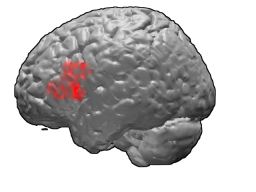
Broca's area, or the Broca area, is a region in the frontal lobe of the dominant hemisphere, usually the left, of the brain with functions linked to speech production.

In human biology, handedness is an individual's preferential use of one hand, known as the dominant hand, due to it being stronger, faster or more dextrous. The other hand, comparatively often the weaker, less dextrous or simply less subjectively preferred, is called the non-dominant hand. In a study from 1975 on 7,688 children in US grades 1-6, left handers comprised 9.6% of the sample, with 10.5% of male children and 8.7% of female children being left-handed. Handedness is often defined by one's writing hand, as it is fairly common for people to prefer to do a particular task with a particular hand. There are people with true ambidexterity, but it is rare—most people prefer using one hand for most purposes.

The corpus callosum, also callosal commissure, is a wide, thick nerve tract, consisting of a flat bundle of commissural fibers, beneath the cerebral cortex in the brain. The corpus callosum is only found in placental mammals. It spans part of the longitudinal fissure, connecting the left and right cerebral hemispheres, enabling communication between them. It is the largest white matter structure in the human brain, about 10 in (250 mm) in length and consisting of 200–300 million axonal projections.

The vertebrate cerebrum (brain) is formed by two cerebral hemispheres that are separated by a groove, the longitudinal fissure. The brain can thus be described as being divided into left and right cerebral hemispheres. Each of these hemispheres has an outer layer of grey matter, the cerebral cortex, that is supported by an inner layer of white matter. In eutherian (placental) mammals, the hemispheres are linked by the corpus callosum, a very large bundle of nerve fibers. Smaller commissures, including the anterior commissure, the posterior commissure and the fornix, also join the hemispheres and these are also present in other vertebrates. These commissures transfer information between the two hemispheres to coordinate localized functions.

The cingulate cortex is a part of the brain situated in the medial aspect of the cerebral cortex. The cingulate cortex includes the entire cingulate gyrus, which lies immediately above the corpus callosum, and the continuation of this in the cingulate sulcus. The cingulate cortex is usually considered part of the limbic lobe.

In neuroanatomy, the central sulcus is a sulcus, or groove, in the cerebral cortex in the brains of vertebrates. It is sometimes confused with the longitudinal fissure.
Split-brain or callosal syndrome is a type of disconnection syndrome when the corpus callosum connecting the two hemispheres of the brain is severed to some degree. It is an association of symptoms produced by disruption of, or interference with, the connection between the hemispheres of the brain. The surgical operation to produce this condition involves transection of the corpus callosum, and is usually a last resort to treat refractory epilepsy. Initially, partial callosotomies are performed; if this operation does not succeed, a complete callosotomy is performed to mitigate the risk of accidental physical injury by reducing the severity and violence of epileptic seizures. Before using callosotomies, epilepsy is instead treated through pharmaceutical means. After surgery, neuropsychological assessments are often performed.

Brodmann area 44, or BA44, is part of the frontal cortex in the human brain. Situated just anterior to premotor cortex (BA6) and on the lateral surface, inferior to BA9.

Brodmann area 45 (BA45), is part of the frontal cortex in the human brain. It is situated on the lateral surface, inferior to BA9 and adjacent to BA46.

Wernicke's area, also called Wernicke's speech area, is one of the two parts of the cerebral cortex that are linked to speech, the other being Broca's area. It is involved in the comprehension of written and spoken language, in contrast to Broca's area, which is primarily involved in the production of language. It is traditionally thought to reside in Brodmann area 22, which is located in the superior temporal gyrus in the dominant cerebral hemisphere, which is the left hemisphere in about 95% of right-handed individuals and 70% of left-handed individuals.
The term laterality refers to the preference most humans show for one side of their body over the other. Examples include left-handedness/right-handedness and left/right-footedness; it may also refer to the primary use of the left or right hemisphere in the brain. It may also apply to animals or plants. The majority of tests have been conducted on humans, specifically to determine the effects on language.

The fusiform gyrus, also known as the lateral occipitotemporal gyrus,is part of the temporal lobe and occipital lobe in Brodmann area 37. The fusiform gyrus is located between the lingual gyrus and parahippocampal gyrus above, and the inferior temporal gyrus below. Though the functionality of the fusiform gyrus is not fully understood, it has been linked with various neural pathways related to recognition. Additionally, it has been linked to various neurological phenomena such as synesthesia, dyslexia, and prosopagnosia.
Expressive language disorder is one of the "specific developmental disorders of speech and language" recognised by the tenth edition of the International Classification of Diseases (ICD-10). As of the eleventh edition, it is considered to be covered by the various categories of developmental language disorder. Transition to the ICD-11 will take place at a different time in different countries.

The inferior frontal gyrus(IFG), (gyrus frontalis inferior), is the lowest positioned gyrus of the frontal gyri, of the frontal lobe, and is part of the prefrontal cortex.

The planum temporale is the cortical area just posterior to the auditory cortex within the Sylvian fissure. It is a triangular region which forms the heart of Wernicke's area, one of the most important functional areas for language. Original studies on this area found that the planum temporale was one of the most asymmetric regions in the brain, with this area being up to ten times larger in the left cerebral hemisphere than the right.

The lateralization of brain function is the tendency for some neural functions or cognitive processes to be specialized to one side of the brain or the other. The median longitudinal fissure separates the human brain into two distinct cerebral hemispheres, connected by the corpus callosum. Although the macrostructure of the two hemispheres appears to be almost identical, different composition of neuronal networks allows for specialized function that is different in each hemisphere.

In human neuroanatomy, brain asymmetry can refer to at least two quite distinct findings:
Emotional lateralization is the asymmetrical representation of emotional control and processing in the brain. There is evidence for the lateralization of other brain functions as well.
Sign language refers to any natural language which uses visual gestures produced by the hands and body language to express meaning. The brain's left side is the dominant side utilized for producing and understanding sign language, just as it is for speech. In 1861, Paul Broca studied patients with the ability to understand spoken languages but the inability to produce them. The damaged area was named Broca's area, and located in the left hemisphere’s inferior frontal gyrus. Soon after, in 1874, Carl Wernicke studied patients with the reverse deficits: patients could produce spoken language, but could not comprehend it. The damaged area was named Wernicke's area, and is located in the left hemisphere’s posterior superior temporal gyrus.
References
- ↑ Coren, S.; Porac, C. (1977-11-11). "Fifty centuries of right-handedness: the historical record". Science. 198 (4317): 631–632. Bibcode:1977Sci...198..631C. doi:10.1126/science.335510. ISSN 0036-8075. PMID 335510.
- 1 2 Carey, David P.; Johnstone, Leah T. (2014). "Quantifying cerebral asymmetries for language in dextrals and adextrals with random-effects meta analysis". Frontiers in Psychology. 5: 1128. doi: 10.3389/fpsyg.2014.01128 . ISSN 1664-1078. PMC 4219560 . PMID 25408673.
- ↑ Carey, David P. (2016), "Broca's and Wernicke's Areas", Encyclopedia of Evolutionary Psychological Science, Springer International Publishing, pp. 1–6, doi:10.1007/978-3-319-16999-6_3339-1, ISBN 9783319169996
- ↑ Beaumont, J. Graham (2008-05-21). Introduction to Neuropsychology, Second Edition. Guilford Press. ISBN 9781606238127.
- ↑ Ocklenburg, Sebastian; Garland, Alexis; Ströckens, Felix; Uber Reinert, Anelisie (2015). "Investigating the neural architecture of handedness". Frontiers in Psychology. 6: 148. doi: 10.3389/fpsyg.2015.00148 . ISSN 1664-1078. PMC 4323997 . PMID 25717316.
- ↑ Aboitiz, Francisco; Scheibel, Arnold B.; Fisher, Robin S.; Zaidel, Eran (December 1992). "Fiber composition of the human corpus callosum". Brain Research. 598 (1–2): 143–153. doi:10.1016/0006-8993(92)90178-c. ISSN 0006-8993. PMID 1486477. S2CID 2378539.
- ↑ Witelson, S. F. (1985-08-16). "The brain connection: the corpus callosum is larger in left-handers". Science. 229 (4714): 665–668. Bibcode:1985Sci...229..665W. doi:10.1126/science.4023705. ISSN 0036-8075. PMID 4023705.
- 1 2 3 4 5 Toga, Arthur W.; Thompson, Paul M. (January 2003). "Mapping brain asymmetry". Nature Reviews Neuroscience. 4 (1): 37–48. doi:10.1038/nrn1009. ISSN 1471-003X. PMID 12511860. S2CID 15867592.
- ↑ Becker, Jill B.; Breedlove, S. Marc; Crews, David; McCarthy, Margaret M. (2002). Behavioral Endocrinology. MIT Press. ISBN 9780262523219.
- ↑ Good, Catriona D.; Johnsrude, Ingrid; Ashburner, John; Henson, Richard N.A.; Friston, Karl J.; Frackowiak, Richard S.J. (2001-09-01). "Cerebral Asymmetry and the Effects of Sex and Handedness on Brain Structure: A Voxel-Based Morphometric Analysis of 465 Normal Adult Human Brains". NeuroImage. 14 (3): 685–700. CiteSeerX 10.1.1.420.7705 . doi:10.1006/nimg.2001.0857. ISSN 1053-8119. PMID 11506541. S2CID 16235256.
- 1 2 Andersen, Kasper Winther; Siebner, Hartwig Roman (2018-04-01). "Mapping dexterity and handedness: recent insights and future challenges". Current Opinion in Behavioral Sciences. 20: 123–129. doi: 10.1016/j.cobeha.2017.12.020 . ISSN 2352-1546.
- ↑ Annett, Marian (2001-12-20). Handedness and Brain Asymmetry. doi:10.4324/9780203759646. ISBN 9780203759646.
- ↑ Herron, Jeannine (2012-12-02). Neuropsychology of Left-Handedness. Elsevier. ISBN 9780323153669.
- ↑ Weiner, Kevin S.; Zilles, Karl (March 2016). "The anatomical and functional specialization of the fusiform gyrus". Neuropsychologia. 83: 48–62. doi:10.1016/j.neuropsychologia.2015.06.033. ISSN 0028-3932. PMC 4714959 . PMID 26119921.
- ↑ Pitcher, David; Walsh, Vincent; Duchaine, Bradley (April 2011). "The role of the occipital face area in the cortical face perception network". Experimental Brain Research. 209 (4): 481–493. doi:10.1007/s00221-011-2579-1. ISSN 1432-1106. PMID 21318346. S2CID 6321920.
- ↑ Frässle, Stefan; Krach, Sören; Paulus, Frieder Michel; Jansen, Andreas (2016-06-02). "Handedness is related to neural mechanisms underlying hemispheric lateralization of face processing". Scientific Reports. 6: 27153. Bibcode:2016NatSR...627153F. doi:10.1038/srep27153. ISSN 2045-2322. PMC 4890016 . PMID 27250879.
- ↑ Edlin, James M.; Leppanen, Marcus L.; Fain, Robin J.; Hackländer, Ryan P.; Hanaver-Torrez, Shelley D.; Lyle, Keith B. (March 2015). "On the use (and misuse?) of the Edinburgh Handedness Inventory". Brain and Cognition. 94: 44–51. doi:10.1016/j.bandc.2015.01.003. ISSN 1090-2147. PMID 25656540. S2CID 24911050.
- ↑ Bruno, Michael A.; Walker, Eric A.; Abujudeh, Hani H. (October 2015). "Understanding and Confronting Our Mistakes: The Epidemiology of Error in Radiology and Strategies for Error Reduction". RadioGraphics. 35 (6): 1668–1676. doi: 10.1148/rg.2015150023 . ISSN 0271-5333. PMID 26466178.
- ↑ Raine, A.; Stoddard, J.; Bihrle, S.; Buchsbaum, M. (1998). "Prefrontal glucose deficits in murderers lacking psychosocial deprivation". Neuropsychiatry, Neuropsychology and Behavioral Neurology. 11 (1): 1–7. PMID 9560822.
- ↑ Raine, A. (2002). "Biosocial studies of antisocial and violent behavior in children and adults: a review". J. Abnorm. Child Psychol. 30 (4): 311–326. doi:10.1023/A:1015754122318. PMID 12108763. S2CID 11608050.
- ↑ Brower, M.; Price, B. (2001). "Neuropsychiatry of frontal lobe dysfunction in violent and criminal behaviour: a critical review". J. Neurol. Neurosurg. Psychiatry. 71 (6): 720–726. doi:10.1136/jnnp.71.6.720. PMC 1737651 . PMID 11723190.
- ↑ Lam, B.Y.H.; Huang, Y.; Gao, Y. (2021). "Gray matter asymmetry in the orbitofrontal cortex in relation to psychopathic traits in adolescents". Journal of Psychiatric Research. 132: 84–96. doi:10.1016/j.jpsychires.2020.10.003. PMC 7736323 . PMID 33068818.
- ↑ Bache, M.A.B.; Naranjo-Orellana, J. (2014). "Laterality and Sports Performance". Arch. Med. Deporte (in Spanish). 31 (3): 200–204.
- ↑ Van der Haegen, L.; Cai, Q. (2012). "Colateralization of Broca's area and the visual word form area in left-handers: fMRI evidence". Brain & Language. 122 (3): 171–178. doi:10.1016/j.bandl.2011.11.004. hdl: 1854/LU-2006353 . PMID 22196742. S2CID 4847527.
- ↑ Sapolsky, R.M. (2004). "Colateralization of Broca's area and the visual word form area in left-handers: fMRI evidence". Phil. Trans. R. Soc. Lond. 359: 1787–1796.