Neutrophilic eccrine hidradenitis

Last updated
Neutrophilic eccrine hidradenitis
Specialty Dermatology   OOjs UI icon edit-ltr-progressive.svg

Neutrophilic eccrine hidradenitis (NEH) usually is a cutaneous complication of chemotherapy, but it can also occur for other reasons. It consists of fever and non specific skin lesions. It is rare, and self-limited. [1] [2]

Contents

Signs and symptoms

Fever and a non specific skin eruption – with reddening (erythema) and swelling (oedema) of the skin – are the most common symptoms of NEH. Patients usually present with the skin eruption 1-2 weeks after use of the cytotoxic drug. Sometimes, the skin eruption can be painful. Skin eruptions can be located on the extremities, trunk, and face. Severe lesions are rare, and can mimic cellulitis. Generalised lesions resembling erythema multiforme have been reported. [3]

Cause

The overwhelming majority of neutrophilic eccrine hidradenitis (NEH) is seen in people with cancer, especially leukaemia, who receive chemotherapy with a cytotoxic drug. These include: Bleomycin, chlorambucil, cyclophosphamide, cytarabine, doxorubicin, lomustine, mitoxantrone, topotecan, and vincristine. [4]

NEH was first described in 1982 in a patient with acute myeloid leukaemia (AML) who had received cytarabine as chemotherapy. [5]

Cancer itself, infections, and other medicinal drugs also can lead to NEH. NEH has been reported in patients with cancer who have not received any form of chemotherapy (i.e., as a paraneoplastic syndrome), in patients with HIV and/or AIDS, and after the use of paracetamol (acetaminophen). Also the use of targeted agents can lead to NEH, e.g. imatinib, a tyrosine kinase inhibitor. [6] [7] [8] [9]

NEH has also been described without any known reason (idiopathic cases), including idiopathic cases in children. [10] [11]

The exact cause of NEH is unknown. In patients receiving chemotherapy, it has been postulated that a high concentration of the cytotoxic drug in sweat has a direct toxic effect on the eccrine glands. [12]

Diagnosis

In all cases of suspected NEH, a skin biopsy should be performed, because the clinical symptoms are non specific, but the histopathological findings on the biopsy are specific. The biopsy shows characteristic changes of the eccrine glands, the major sweat glands of the body. [13]

In NEH, eccrine gland necrosis, and neutrophils surroundings the eccrine glands, are typical findings on biopsy. If the chemotherapy has recently been administered, chemotherapy induced neutropenia may be present, and, as a result, the neutrophils may be absent. But the other characteristic finding, i.e. eccrine gland necrosis, can still be seen. A vacuolar interface dermatitis also is visible in glands and ducts, along with necrosis of the lining cells.

In addition, in patients receiving chemotherapy, keratinocyte atypia can be seen. [14]

Prevention

A single case report suggested that oral dapsone may be useful for prevention. However, the efficacy of oral dapsone as prevention has not been demonstrated very clearly until now. [15]

Treatment

NEH is self-limited and usually resolves without treatment. In the overwhelming majority of the cases, spontaneous resolution occurs within 1–2 weeks.

However, if the patient developed NEH after chemotherapy, the offending cytotoxic drug has to be discontinued, and the patient must avoid this particular cytotoxic drug in the future, because NEH usually re occurs upon re exposure to the same cytotoxic drug. [16]

Despite the fact that NEH is self limited and usually resolves without treatment, some researchers use treatment, mainly systemic corticosteroids, although the efficacy of such a therapy has not been demonstrated in a large randomised controlled clinical trial until now. [17]

See also

Related Research Articles

Hidradenitis suppurativa Human disease

Hidradenitis suppurativa (HS), also known as acne inversa, is a long term skin disease characterized by the occurrence of inflamed and swollen lumps. These are typically painful and break open, releasing fluid or pus. The areas most commonly affected are the underarms, under the breasts, and groin. Scar tissue remains after healing. Self-consciousness or depression may result.

Dapsone chemical compound

Dapsone, also known as diaminodiphenyl sulfone (DDS), is an antibiotic commonly used in combination with rifampicin and clofazimine for the treatment of leprosy. It is a second-line medication for the treatment and prevention of pneumocystis pneumonia and for the prevention of toxoplasmosis in those who have poor immune function. Additionally, it has been used for acne, dermatitis herpetiformis, and various other skin conditions. Dapsone is available both topically and by mouth.

Lichen planus lichen disease that is located in skin, located in tongue or located in oral mucosa, which presents itself in the form of papules, lesions or rashes

Lichen planus (LP) is a chronic inflammatory and immune mediated disease that affects the skin, nails, hair, and mucous membranes. It is characterized by polygonal, flat-topped, violaceous papules and plaques with overlying, reticulated, fine white scale, commonly affecting dorsal hands, flexural wrists and forearms, trunk, anterior lower legs and oral mucosa. Although there is a broad clinical range of LP manifestations, the skin and oral cavity remain as the major sites of involvement. The cause is unknown, but it is thought to be the result of an autoimmune process with an unknown initial trigger. There is no cure, but many different medications and procedures have been used in efforts to control the symptoms.

Dermatofibrosarcoma protuberans A fibrosarcoma that is located in the dermis laryer of the skin and that begins as a hard nodule and grows slowly.

Dermatofibrosarcoma protuberans (DFSP) is a rare tumor of the dermis layer of the skin, and is classified as a sarcoma. There is only about one case per million per year. DFSP is a fibrosarcoma, more precisely a cutaneous soft tissue sarcoma. In many respects, the disease behaves as a benign tumor, but in 2–5% of cases it can metastasize, so it should be considered to have malignant potential. It occurs most often in adults in their thirties; it has been described congenitally, in children, and the elderly. It accounts for approximately 2–6% of soft tissue sarcoma cancers.

Pemphigus rare group of blistering autoimmune diseases that affect the skin and mucous membranes

Pemphigus is a rare group of blistering autoimmune diseases that affect the skin and mucous membranes. The name is derived from the Greek root "pemphix," meaning "pustule."

Livedo reticularis common skin finding consisting of a mottled reticulated vascular pattern that appears as a lace-like purplish discoloration of the skin

Livedo reticularis is a common skin finding consisting of a mottled reticulated vascular pattern that appears as a lace-like purplish discoloration of the skin. The discoloration is caused by swelling of the venules owing to obstruction of capillaries by small blood clots. The blood clots in the small blood vessels can be a secondary effect of a condition that increases a person's risk of forming blood clots, including a wide array of pathological and nonpathological conditions. Examples include hyperlipidemia, microvascular hematological or anemia states, nutritional deficiencies, hyper- and autoimmune diseases, and drugs/toxins.

Febrile neutrophilic dermatosis skin disease

Sweet's syndrome (SS), or acute febrile neutrophilic dermatosis is a skin disease characterized by the sudden onset of fever, an elevated white blood cell count, and tender, red, well-demarcated papules and plaques that show dense infiltrates by neutrophil granulocytes on histologic examination.

Erythema annulare centrifugum

Erythema anulare centrifugum (EAC), is a descriptive term for a class of skin lesion presenting redness (erythema) in a ring form (anulare) that spreads from a center (centrifugum). It was first described by Darier in 1916. Many different terms have been used to classify these types of lesions and it is still controversial on what exactly defines EAC. Some of the types include annular erythema, erythema perstans, erythema gyratum perstans, erythema gyratum repens, darier erythema and erythema figuratum perstans.

Transient acantholytic dermatosis acantholytic

Grover's disease (GD) is a polymorphic, pruritic, papulovesicular dermatosis characterized histologically by acantholysis with or without dyskeratosis. Once confirmed, most cases of Grover's disease last six to twelve months, which is why it was originally called "transient". However it may last much longer. Nevertheless, it is not to be confused with relapsing linear acantholytic dermatosis.

Hidradenitis is any disease in which the histologic abnormality is primarily an inflammatory infiltrate around the eccrine glands. This group includes neutrophilic eccrine hidradenitis and recurrent palmoplantar hidradenitis.

Chemotherapy-induced acral erythema chemotherapy-induced dermal side effects tassociated with the use of cytostatic agents

Chemotherapy-induced acral erythema is reddening, swelling, numbness and desquamation on palms of the hands and soles of the feet that can occur after chemotherapy in patients with cancer. Hand-foot syndrome is also rarely seen in sickle-cell disease. These skin changes usually are well demarcated. Acral erythema typically disappears within a few weeks after discontinuation of the offending drug.

Florid cutaneous papillomatosis (FCP), is an obligate paraneoplastic syndrome.

Drug eruption adverse drug reaction of the skin

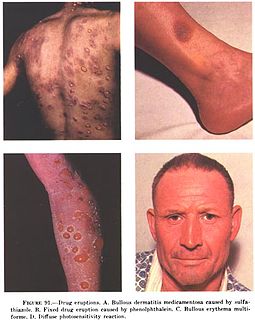
In medicine, a drug eruption is an adverse drug reaction of the skin. Most drug-induced cutaneous reactions are mild and disappear when the offending drug is withdrawn. These are called "simple" drug eruptions. However, more serious drug eruptions may be associated with organ injury such as liver or kidney damage and are categorized as "complex". Drugs can also cause hair and nail changes, affect the mucous membranes, or cause itching without outward skin changes.

Cutaneous small-vessel vasculitis hypersensitivity vasculitis that results in inflammation of small blood vessels, characterized clinically by palpable purpura, which is a slightly elevated purpuric rash over one or more areas of the skin

Cutaneous small-vessel vasculitis (CSVV), also known as hypersensitivity vasculitis, cutaneous leukocytoclastic vasculitis, hypersensitivity angiitis, cutaneous leukocytoclastic angiitis, cutaneous necrotizing vasculitis and cutaneous necrotizing venulitis, is inflammation of small blood vessels, characterized by palpable purpura. It is the most common vasculitis seen in clinical practice.

Papillary eccrine adenoma is a cutaneous condition characterized by an uncommon benign sweat gland neoplasm that presents as a dermal nodule located primarily on the extremities of black patients.

Nevus depigmentosus Human disease

Nevus depigmentosus is a loss of pigment in the skin which can be easily differentiated from vitiligo. Although age factor has not much involvement in the nevus depigmentosus but in about 19% of the cases these are noted at birth. Their size may however grow in proportion to growth of the body. The distribution is also fairly stable and are nonprogressive hypopigmented patches. The exact cause of nevus depigmentosus is still not clearly understood. A sporadic defect in the embryonic development has been suggested to be a causative factor. It has been described as "localised albinism", though this is incorrect.

Porokeratotic eccrine ostial and dermal duct nevus is a skin lesion that resembles a comedonal nevus, but it occurs on the palms and soles where pilosebaceous follicles are normally absent. It is probably transmitted by paradominant transmission.

Neh or NEH may refer to:

Idiopathic pure sudomotor failure (IPSF) is the most common cause of a rare disorder known as acquired idiopathic generalized anhidrosis (AIGA), a clinical syndrome characterized by generalized decrease or absence of sweating without other autonomic and somatic nervous dysfunctions and without persistent organic cutaneous lesions.

References

  1. James, William; Berger, Timothy; Elston, Dirk (2005). Andrews' Diseases of the Skin: Clinical Dermatology. (10th ed.). Saunders. ISBN   0-7216-2921-0. :780
  2. Neutrophilic eccrine hidradenitis. Bachmeyer C, Aractingi S. Clin Dermatol. 2000 May-Jun;18(3):319-30.
  3. Neutrophilic eccrine hidradenitis. Thorisdottir K, Tomecki KJ, Bergfeld WF, Andresen SW. J Am Acad Dermatol. 1993 May;28(5 Pt 1):775-7.
  4. Mucocutaneous reactions to chemotherapy. Susser WS, Whitaker-Worth DL, Grant-Kels JM. J Am Acad Dermatol. 1999;40(3):367-98.
  5. Neutrophilic eccrine hidradenitis. A distinctive type of neutrophilic dermatosis associated with myelogenous leukemia and chemotherapy. Harrist TJ, Fine JD, Berman RS, Murphy GF, Mihm MC Jr. Arch Dermatol. 1982;118(4):263-6.
  6. Unusual manifestations of acute leukemia. Case 2. Leukemia and rash: paraneoplastic or drug-induced? Blank C, Wagner HM, Hohenleutner U, Andreesen R. J Clin Oncol. 2000 Oct 1;18(19):3437-9.
  7. Neutrophilic eccrine hidradenitis: a case report and review of the literature. Bailey DL, Barron D, Lucky AW. Pediatr Dermatol. 1989;6(1):33-8.
  8. Neutrophilic eccrine hidradenitis: a case report and review of the literature. Margolis DJ, Gross PR. Cutis. 1991;48(3):198-200.
  9. Imatinib mesylate and dermatology part 2: a review of the cutaneous side effects of imatinib mesylate. Scheinfeld N. J Drugs Dermatol. 2006 Mar;5(3):228-31.
  10. Generalized idiopathic neutrophilic eccrine hidradenitis in childhood. Lee WJ, Kim CH, Chang SE, Lee MW, Choi JH, Moon KC, Koh JK. Int J Dermatol. 2010 Jan;49(1):75-8.
  11. Idiopathic palmoplantar eccrine hidradenitis in children. Ben-Amitai D, Hodak E, Landau M, Metzker A, Feinmesser M, David M. Eur J Pediatr. 2001 Mar;160(3):189-91.
  12. Neutrophilic eccrine hidradenitis: a distinctive rash associated with cytarabine therapy and acute leukemia. Flynn TC, Harrist TJ, Murphy GF, Loss RW, Moschella SL. J Am Acad Dermatol. 1984;11(4 Pt 1):584-90.
  13. Neutrophilic hidradenitis induced by chemotherapy involves eccrine and apocrine glands. Brehler R, Reimann S, Bonsmann G, Metze D. Am J Dermatopathol. 1997;19(1):73-8.
  14. Smoller BR, Horn TD. Chemotherapy-induced drug eruptions. In: Dermatopathology in systemic disease. Smoller BR, Horn TD (Eds). Oxford University Press. 2001.
  15. Dapsone in prevention of recurrent neutrophilic eccrine hidradenitis. Shear NH, Knowles SR, Shapiro L, Poldre P. J Am Acad Dermatol. 1996;35(5 Pt 2):819-22.
  16. Recurrent neutrophilic eccrine hidradenitis. Bernstein EF, Spielvogel RL, Topolsky DL. Br J Dermatol. 1992;127(5):529-33.
  17. Neutrophilic eccrine hidradenitis associated with Hodgkin’s disease and chemotherapy. A case report. Beutner KR, Packman CH, Markowitch W. Arch Dermatol. 1986;122(7):809-11.
Classification
D
External resources