Related Research Articles
The hock, or gambrel, is the joint between the tarsal bones and tibia of a digitigrade or unguligrade quadrupedal mammal, such as a horse, cat, or dog. This joint may include articulations between tarsal bones and the fibula in some species, while in others the fibula has been greatly reduced and is only found as a vestigial remnant fused to the distal portion of the tibia. It is the anatomical homologue of the ankle of the human foot. While homologous joints occur in other tetrapods, the term is generally restricted to mammals, particularly long-legged domesticated species.

Laminitis is a disease that affects the feet of ungulates and is found mostly in horses and cattle. Clinical signs include foot tenderness progressing to inability to walk, increased digital pulses, and increased temperature in the hooves. Severe cases with outwardly visible clinical signs are known by the colloquial term founder, and progression of the disease will lead to perforation of the coffin bone through the sole of the hoof or being unable to stand up, requiring euthanasia.
Navicular syndrome, often called navicular disease, is a syndrome of lameness problems in horses. It most commonly describes an inflammation or degeneration of the navicular bone and its surrounding tissues, usually on the front feet. It can lead to significant and even disabling lameness.

Bog spavin is a swelling of the tibiotarsal joint of the horse's hock which, in itself, does not cause lameness. The joint becomes distended by excess synovial fluid and/or thickened synovial tissue bringing about a soft, fluctuant swelling on the front of the joint, as well as in the medial and lateral plantar pouches. Bog spavin is generally an indication of underlying pathology within the joint.
Tendinitis/tendonitis is inflammation of a tendon, often involving torn collagen fibers. A bowed tendon is a horseman's term for a tendon after a horse has sustained an injury that causes swelling in one or more tendons creating a "bowed" appearance.

Equine conformation evaluates a horse's bone structure, musculature, and its body proportions in relation to each other. Undesirable conformation can limit the ability to perform a specific task. Although there are several faults with universal disadvantages, a horse's conformation is usually judged by what its intended use may be. Thus "form to function" is one of the first set of traits considered in judging conformation. A horse with poor form for a Grand Prix show jumper could have excellent conformation for a World Champion cutting horse, or to be a champion draft horse. Every horse has good and bad points of its conformation and many horses excel even with conformation faults.
Ringbone is exostosis in the pastern or coffin joint of a horse. In severe cases, the growth can encircle the bones, giving ringbone its name. It has been suggested by some authors that such a colloquial term, whilst commonly used, might be misleading and that it would be better to refer to this condition as osteoarthritis of the inter-phalangeal joints in ungulates.
Sidebone is a common condition of horses, characterized by the ossification of the collateral cartilages of the coffin bone. These are found on either side of the foot protruding above the level of the coronary band. The lateral cartilages support the hoof wall and provide an important role in the support and cushioning provided to the heel. The front feet are most commonly affected.

Fetlock is the common name in horses, large animals, and sometimes dogs for the metacarpophalangeal and metatarsophalangeal joints.

Equine anatomy encompasses the gross and microscopic anatomy of horses, ponies and other equids, including donkeys, mules and zebras. While all anatomical features of equids are described in the same terms as for other animals by the International Committee on Veterinary Gross Anatomical Nomenclature in the book Nomina Anatomica Veterinaria, there are many horse-specific colloquial terms used by equestrians.

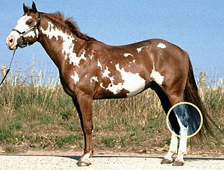
The pastern is a part of the leg of a horse between the fetlock and the top of the hoof. It incorporates the long pastern bone and the short pastern bone, which are held together by two sets of paired ligaments to form the pastern joint. Anatomically homologous to the two largest bones found in the human finger, the pastern was famously mis-defined by Samuel Johnson in his dictionary as "the knee of a horse". When a lady asked Johnson how this had happened, he gave the much-quoted reply: "Ignorance, madam, pure ignorance."

Sesamoiditis is inflammation of the sesamoid bones.
A flexion test is a preliminary veterinary procedure performed on a horse, generally during a prepurchase or a lameness exam. The purpose is to accentuate any pain that may be associated with a joint or soft-tissue structure, allowing the practitioner to localize a lameness to a specific area, or to alert a practitioner to the presence of sub-clinical disease that may be present during a pre-purchase exam.

The skeletal system of the horse has three major functions in the body. It protects vital organs, provides framework, and supports soft parts of the body. Horses typically have 205 bones. The pelvic limb typically contains 19 bones, while the thoracic limb contains 20 bones.

The coffin bone, also known as the pedal bone (U.S.), is the bottommost bone in the front and rear legs of horses, cattle, pigs and other ruminants. In horses it is encased by the hoof capsule. Also known as the distal phalanx, third phalanx, or "P3". The coffin bone meets the short pastern bone or second phalanx at the coffin joint. The coffin bone is connected to the inner wall of the horse hoof by a structure called the laminar layer. The insensitive laminae coming in from the hoof wall connects to the sensitive laminae layer, containing the blood supply and nerves, which is attached to the coffin bone. The lamina is a critical structure for hoof health, therefore any injury to the hoof or its support system can in turn affect the coffin bone.
Lameness is an abnormal gait or stance of an animal that is the result of dysfunction of the locomotor system. In the horse, it is most commonly caused by pain, but can be due to neurologic or mechanical dysfunction. Lameness is a common veterinary problem in racehorses, sport horses, and pleasure horses. It is one of the most costly health problems for the equine industry, both monetarily for the cost of diagnosis and treatment, and for the cost of time off resulting in loss-of-use.
Racehorse injuries and fatalities are a side effect of training and competition. The problem with equine injuries is that they so often result in death. A 2005 study by the United States Department of Agriculture found that injuries are the second leading cause of death in horses, second only to old age. Nureyev's recovery from a broken leg while retired at stud in 1987 typifies the struggle horses have after being injured.
Good conformation in the limbs leads to improved movement and decreased likelihood of injuries. Large differences in bone structure and size can be found in horses used for different activities, but correct conformation remains relatively similar across the spectrum. Structural defects, as well as other problems such as injuries and infections, can cause lameness, or movement at an abnormal gait. Injuries to and problems with horse legs can be relatively minor, such as stocking up, which causes swelling without lameness, or quite serious. Even leg injuries that are not immediately fatal may still be life-threatening to horses, as their bodies are adapted to bear weight on all four legs and serious problems can result if this is not possible.
The treatment of equine lameness is a complex subject. Lameness in horses has a variety of causes, and treatment must be tailored to the type and degree of injury, as well as the financial capabilities of the owner. Treatment may be applied locally, systemically, or intralesionally, and the strategy for treatment may change as healing progresses. The end goal is to reduce the pain and inflammation associated with injury, to encourage the injured tissue to heal with normal structure and function, and to ultimately return the horse to the highest level of performance possible following recovery.

Polysulfated glycosaminoglycan (PSGAG), sold under the brand name Adequan, is an injectable drug for dogs and horses that is used to alleviate the lameness, pain, and lowered range of motion caused by arthritis. It is made of repeat disaccharide units, and is similar to glycosaminoglycans already present in the cartilage; PSGAG thus easily integrates itself there. In vitro studies have shown it to inhibit the enzymes that degrade cartilage and bone, as well as suppress inflammation and stimulate the synthesis of replacement cartilage. While it can cause an increased risk of bleeding, it is relatively safe and has a high LD50. PSGAG is one of the most widely prescribed joint treatments for horses.
References
- ↑ Briggs, Karen (1 September 2000). "Osselets (Traumatic Arthritis of the Fetlock)". TheHorse.com. Retrieved 2017-12-29.
- ↑ Briggs, Karen (1 September 2000). "Osselets (Traumatic Arthritis of the Fetlock)". TheHorse.com. Retrieved 2017-12-29.
- ↑ Christopher E. Kawcak, Myra F. Barrett, in Joint Disease in the Horse (Second Edition), 2016 ISBN 978-1-4557-5969-9
- ↑ Dyce, Sack, and Wensing's Textbook of Veterinary Anatomy 5th Edition ISBN 9780323442640
- ↑ "Osselets". The Merck Veterinary Manual. 2006. Retrieved 2007-07-10.
- ↑ Stashak, Ted S. (2013). "Chapter 2. Conformation and movement. Short upright pastern". Practical Guide to Lameness in Horses (4th ed.). Wiley. ISBN 9781118701133.
- ↑ Briggs, Karen (1 September 2000). "Osselets (Traumatic Arthritis of the Fetlock)". TheHorse.com. Retrieved 2017-12-29.
- ↑ Briggs, Karen (1 September 2000). "Osselets (Traumatic Arthritis of the Fetlock)". TheHorse.com. Retrieved 2017-12-29.
- ↑ "Osselets". The Merck Veterinary Manual. 2006. Retrieved 2007-07-10.