The bones and joints of the equine forelimb distal to the wrist (or carpus): The fetlock (metacarpophalangeal joint) is located between the cannon bone (third metacarpal) and the long pastern bone (proximal phalanx). The pastern joint (proximal interphalangeal joint) is located between the long pastern bone and the short pastern bone (middle phalanx). The coffin joint (distal interphalangeal joint) is located between the short pastern bone and the coffin bone (distal phalanx).
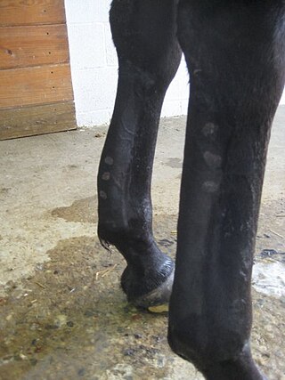
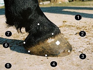
The pastern is a part of the leg of a horse between the fetlock and the top of the hoof. It incorporates the long pastern bone (proximal phalanx) and the short pastern bone (middle phalanx), which are held together by two sets of paired ligaments to form the pastern joint (proximal interphalangeal joint).[1][2] Anatomically homologous to the two largest bones found in the human finger, the pastern was famously mis-defined by Samuel Johnson in his dictionary as "the knee of a horse". When a lady asked Johnson how this had happened, he gave the much-quoted reply: "Ignorance, madam, pure ignorance."[3]
The pastern consists of two bones, the uppermost called the "large pastern bone" or proximal phalanx, which begins just under the fetlock joint, and the lower called the "small pastern bone" or middle phalanx, located between the large pastern bone and the coffin bone, outwardly located at approximately the coronary band.
The joint between these two phalangeal bones is aptly called the "pastern joint". This joint has limited movement, but does help to disperse the concussive forces of the horse's step and also has some influence on the flexion or extension of the entire leg.
The pastern is vital in shock absorption. When the horse's front leg is grounded, the elbow and knee are locked. Therefore, the fetlock and pastern are responsible for all the absorption of concussive forces of a footfall. Together, they effectively distribute it among both the bones of the leg and the tendons and ligaments.[4]
The slope of the shoulder is often the same as the slope of the pastern. The angle of the pastern should also match the angle of the hoof after the latter has been trimmed (the angle will change as the hoof grows and may be off in a few weeks). This keeps the bones of the pastern and coffin joints in proper alignment, with a straight line running through their core. An angle broken forward or back increases the stress on these bones, joints, tendons, and ligaments. If the angle does not match, it could be an indication of poor farrier work, but some horses may have underlying conformational defects that can not be modified through farriery alone.
The pastern joint is evaluated when a horse is studied conformationally, as it will affect the gait of the horse and the soundness of the joints above it. Traditionally, the ideal pastern joint of the front leg was a 45-degree angle. However, this angle has been revised to a slightly steeper angle of 47-55 degrees, as the traditional angle, although it makes for comfortable riding, greatly increases the chance of breakdown. Because there is less need for shock absorption in the hindleg, its pasterns are somewhat more upright than those of the front leg, to increase its strength (about 49-59 degrees). If the hind pasterns are the same angle as the front, or too sloping in general, then they are likely to break down during the horse's career, especially if the horse in employed in strenuous work. The length of the pastern joint is determined by the length of the first phalanx. The short pastern bone is less a determinant because it is smaller, at 2 inches in length, and part of it is encased in the hoof.
Long, sloping pasterns
Long, sloping pasterns are commonly seen in Thoroughbreds and Saddlebreds. A nicely sloped pastern increases the likelihood of a long career. It improves the animal's ability to travel on uneven terrain, helps it withstand the rigors of a competition or race, and makes the gait more comfortable for the rider. They are desired in a riding horse because they increase the shock-absorption ability of the leg, making the horse's gait smoother and more comfortable for the rider. However, this flexibility also increases the risk of certain connective tissue injuries that are not seen in horses with more upright pasterns. This is because many of the tendons and ligaments that go down the back of the leg continue under the back of the fetlock joint, and attach to either the pastern bones or the coffin bone. When the horse puts weight on his leg, the fetlock sinks closer to the ground, which is a needed response as it helps to absorb the shock of the footfall. However, when the pasterns are too long or sloping it does not support the fetlock enough, and the fetlock may hyper-extend, possibly to the point where the ergot touches the ground. This stresses the soft tissues that run under the fetlock because they are stretched longer. If stretched too much, they may tear or rupture.
Medical problems that are more common in horses with long, sloping pasterns include:
A fracture of the sesamoid bones found at the back of the fetlock, should the joint hyperextend to the point where it touches the ground. This is especially likely if the horse is tired, such as at the end of a race.
Injury to the suspensory ligament
Ringbone, due to excessive stress on the pastern joint[5]
Short, upright pasterns
Draft horse with upright (65-degree) pasterns
Short, upright pasterns are beneficial in that they decrease the chance that the horse will suffer from soft-tissue injury. However, upright pasterns increase concussion by transmitting more of the shock of footfalls to the bones rather than the tendons. This not only makes the gaits uncomfortable due to the jarring, but also increases the chance of arthritis and may shorten the animal's career. A short, upright pastern also decreases the stride length of the gait, which again makes the gait more uncomfortable and decreases the efficiency of the horse's movement (since he must take more strides per meter than a longer-strided horse).
Medical problems linked to short, upright pasterns are usually a result of excess concussion. They include:
Short, upright pasterns are often seen in draft horses. This is because draft horses bred for pulling rather than riding (and so they were not selected for smooth gaits of a saddle horse), and because upright pasterns give more leverage to dig into the ground as the horse pulls a heavy load. Short, upright pasterns are also commonly seen in Quarter Horses, Warmbloods, and Paint Horses. However, riding horses are more likely to have problems with upright pasterns than draft horses because they tend to work at faster speeds. Due to the lack of shock absorption, horses that have upright pasterns should be kept off hard surfaces whenever possible.
Notes
↑ Hadden, WA; Rogers, C; Wilcox, GJ, eds. (2005). "Chapter 3: Pastern". Horseman's veterinary encyclopedia (Reviseded.). Guilford, Connecticut: The Lyons Press. pp.87–100. ISBN978-1-59228-527-3– via Google Books.
The foot is an anatomical structure found in many vertebrates. It is the terminal portion of a limb which bears weight and allows locomotion. In many animals with feet, the foot is a separate organ at the terminal part of the leg made up of one or more segments or bones, generally including claws and/or nails.
The human leg is the entire lower limb of the human body, including the foot, thigh or sometimes even the hip or buttock region. The major bones of the leg are the femur, tibia, and adjacent fibula. The thigh is between the hip and knee, while the calf (rear) and shin (front) are between the knee and foot.
Laminitis is a disease that affects the feet of ungulates and is found mostly in horses and cattle. Clinical signs include foot tenderness progressing to inability to walk, increased digital pulses, and increased temperature in the hooves. Severe cases with outwardly visible clinical signs are known by the colloquial term founder, and progression of the disease will lead to perforation of the coffin bone through the sole of the hoof or being unable to stand up, requiring euthanasia.
Navicular syndrome, often called navicular disease, is a syndrome of lameness problems in horses. It most commonly describes an inflammation or degeneration of the navicular bone and its surrounding tissues, usually on the front feet. It can lead to significant and even disabling lameness.
Tendinitis/tendonitis is inflammation of a tendon, often involving torn collagen fibers. A bowed tendon is a horseman's term for a tendon after a horse has sustained an injury that causes swelling in one or more tendons creating a "bowed" appearance.
Equine conformation evaluates a horse's bone structure, musculature, and its body proportions in relation to each other. Undesirable conformation can limit the ability to perform a specific task. Although there are several faults with universal disadvantages, a horse's conformation is usually judged by what its intended use may be. Thus "form to function" is one of the first set of traits considered in judging conformation. A horse with poor form for a Grand Prix show jumper could have excellent conformation for a World Champion cutting horse, or to be a champion draft horse. Every horse has good and bad points of its conformation and many horses excel even with conformation faults.
Ringbone is exostosis in the pastern or coffin joint of a horse. In severe cases, the growth can encircle the bones, giving ringbone its name. It has been suggested by some authors that such a colloquial term, whilst commonly used, might be misleading and that it would be better to refer to this condition as osteoarthritis of the inter-phalangeal joints in ungulates.
Fetlock is the common name in horses, large animals, and sometimes dogs for the metacarpophalangeal and metatarsophalangeal joints.
A horse hoof is the lower extremity of each leg of a horse, the part that makes contact with the ground and carries the weight of the animal. It is both hard and flexible. It is a complex structure surrounding the distal phalanx of the 3rd digit of each of the four limbs, which is covered by soft tissue and keratinised (cornified) matter. The arteries that supply the hoof with blood are, the vena plantaris externa and vena plantaris interna, which branch off the tibialis posterior. The horse hoof encapsules one of the three metatarsus bones that are found in the hoof and heel area.
Equine anatomy encompasses the gross and microscopic anatomy of horses, ponies and other equids, including donkeys, mules and zebras. While all anatomical features of equids are described in the same terms as for other animals by the International Committee on Veterinary Gross Anatomical Nomenclature in the book Nomina Anatomica Veterinaria, there are many horse-specific colloquial terms used by equestrians.
Sesamoiditis is inflammation of the sesamoid bones.
Osselet is arthritis in the fetlock joint of a horse, caused by trauma. Osselets usually occur in the front legs of the horse, because there is more strain and concussion on the fetlock there than in the hind legs. The arthritis will occur at the joint between the cannon bone and large pastern bone, at the front of the fetlock.
A flexion test is a preliminary veterinary procedure performed on a horse, generally during a prepurchase or a lameness exam. The purpose is to accentuate any pain that may be associated with a joint or soft-tissue structure, allowing the practitioner to localize a lameness to a specific area, or to alert a practitioner to the presence of sub-clinical disease that may be present during a pre-purchase exam.
The skeletal system of the horse is a skeletal system of a horse that has three major functions in the body. It protects vital organs, provides framework, and supports soft parts of the body. Horses typically have 205 bones. The pelvic limb typically contains 19 bones, while the thoracic limb contains 20 bones.
Lameness is an abnormal gait or stance of an animal that is the result of dysfunction of the locomotor system. In the horse, it is most commonly caused by pain, but can be due to neurologic or mechanical dysfunction. Lameness is a common veterinary problem in racehorses, sport horses, and pleasure horses. It is one of the most costly health problems for the equine industry, both monetarily for the cost of diagnosis and treatment, and for the cost of time off resulting in loss-of-use.
Comparative foot morphology involves comparing the form of distal limb structures of a variety of terrestrial vertebrates. Understanding the role that the foot plays for each type of organism must take account of the differences in body type, foot shape, arrangement of structures, loading conditions and other variables. However, similarities also exist among the feet of many different terrestrial vertebrates. The paw of the dog, the hoof of the horse, the manus (forefoot) and pes (hindfoot) of the elephant, and the foot of the human all share some common features of structure, organization and function. Their foot structures function as the load-transmission platform which is essential to balance, standing and types of locomotion.
Racehorse injuries and fatalities are a side effect of the training and competition of horse racing. Racehorse injuries are considered especially difficult to treat, as they frequently result in the death of the horse. A 2005 study by the United States Department of Agriculture found that injuries are the second leading cause of death in horses, second only to old age. The recovery of the well-known horse Nureyev from a broken leg while retired at stud in 1987 typifies the struggle horses have after being injured.
The limbs of the horse are structures made of dozens of bones, joints, muscles, tendons, and ligaments that support the weight of the equine body. They include two apparatuses: the suspensory apparatus, which carries much of the weight, prevents overextension of the joint and absorbs shock, and the stay apparatus, which locks major joints in the limbs, allowing horses to remain standing while relaxed or asleep. The limbs play a major part in the movement of the horse, with the legs performing the functions of absorbing impact, bearing weight, and providing thrust. In general, the majority of the weight is borne by the front legs, while the rear legs provide propulsion. The hooves are also important structures, providing support, traction and shock absorption, and containing structures that provide blood flow through the lower leg. As the horse developed as a cursorial animal, with a primary defense mechanism of running over hard ground, its legs evolved to the long, sturdy, light-weight, one-toed form seen today.
The treatment of equine lameness is a complex subject. Lameness in horses has a variety of causes, and treatment must be tailored to the type and degree of injury, as well as the financial capabilities of the owner. Treatment may be applied locally, systemically, or intralesionally, and the strategy for treatment may change as healing progresses. The end goal is to reduce the pain and inflammation associated with injury, to encourage the injured tissue to heal with normal structure and function, and to ultimately return the horse to the highest level of performance possible following recovery.
This page is based on this Wikipedia article Text is available under the CC BY-SA 4.0 license; additional terms may apply. Images, videos and audio are available under their respective licenses.