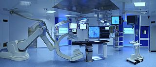
Patient registration

Last updated

Patient registration is used to correlate the reference position of a virtual 3D dataset gathered by computer medical imaging with the reference position of the patient. This procedure is crucial in computer assisted surgery, in order to insure the reproducitibility of the preoperative registration and the clinical situation during surgery. The use of the term "patient registration" out of this context can lead to a confusion with the procedure of registering a patient into the files of a medical institution.

Contents

In computer assisted surgery, the first step is to gather a 3D dataset that reproduces with great accuracy the geometry of the normal and pathological tissues in the region that has to be operated on. This is mainly obtained by using CT or MRI scans of that region. The role of patient registration is to obtain a close-to-ideal reference reproducibility of the dataset – in order to correlate the position (offset) of the gathered dataset with the patient's position during the surgical intervention. Patient registration (1) eliminates the necessity of maintaining the same strict position of the patient during both preoperative scanning and surgery, and (2) provides the surgical robot the necessary reference information to act accurately on the patient, even if he has (been) moved during the intervention.

Application

Patient registration was used mostly in head surgery – oral and maxillofacial surgery, neurosurgery, otolaryngology. With the advent of marker- and markerless-registration, the concept has been extended for abdominal surgery.

Using headframes

The first attempts in 3D mapping of human tissues were made by V. Horsley and R. Clarke in 1906. [1] They have built a rectangular stereotactic headframe that had to be fixed to the head. It was based on cartesian principles and allowed them to accurately and reproductibly guide needle-like electrodes for neurophysiological experiments. They have experimented animals and were able to contribute to the mapping of the cerebellum. Improved versions of the Horsley–Clarke apparatus are still in used today in experimental neurosurgery.[ citation needed ]

The first stereotactic device for humans was also developed in neurosurgery, by E. Spiegel and H. Wycis in 1947. [2] It was used for surgical treatment of Parkinson's disease and, during time, its applicability was extended for the surgical treatment of tumors, vascular malformations, functional neurosurgery etc. The system was based both on headframes and X-ray images taken for all three planes of space.

Further development of stereotactic surgery was made by Brown, Roberts and Wells in 1980. [3] They have developed a halo ring that was applied on the skull, during a CT scan and neurosurgical interventions. This method provided improved surgical guidance and was in fact the first development of computer guided surgery.

Patient registration for the head area has developed for nearly two decades on the same principle of combining CT scans with mechanical reference devices such as headframes or halo rings. But the clinical experience showed that headgear is very uncomfortable to wear and even impossible to apply on little children, because their lack of cooperation; furthermore, the headframes can create artifacts in preoperative data gathering, or during surgery.[ citation needed ]

Reference markers

Skin

In 1986, a different approach was developed by Roberts und Strohbehn. [4] They have used as landmarks several markers on the patient's skin both preoperative CT registration, and intraoperatively. This was a new current of the time in patient registration. Still, the method is time-consuming, and the exact reproducitibility of the marker positions is questionable.

Bone

The bony structures can provide a much better stability and reproducibility of the landmarks for patient registration. Based on this concept, a further technique was used: to implant temporary markers into bone structures that are superficial to the skin, under local anestesia. [5] This was also combined with surface markers and CT registration. [6] The technique has the disadvantage of a further minimal surgical procedure of placing the bone implants, with some risk of infection for the patient.

Surgical planning using bone segment navigation for the osteotomy of the jaw bones, based on models fixed into an articulator (registration based on infrared devices) SurgicalPlanningArtikulator.jpg
Surgical planning using bone segment navigation for the osteotomy of the jaw bones, based on models fixed into an articulator (registration based on infrared devices)

Dental splint markers

Dental splints have been traditionally used for transferring and reproducing 3D reference landmarks for positioning cast models in articulators – in dental prosthetics, orthodontics and orthognathic surgery. By applying several infrared markers on the splints and using an infrared camera, a better registration was obtained. [7]

Markerless patient registration

Anatomical landmarks

The first attempts, based on the identification of anatomical landmarks were made by Caversaccio and Zulliger. [8] The method was based on identifying certain antropometrical points and other anatomical landmarks on the skull, in correlation with the CT registration. But the landmarks cannot be exactly pointed out and reproduced during patient dataset registration and surgery, therefore the method is not precise enough.

Surface registration

Schematic representation of the SSN system SSNSchemaEnglish.jpg
Schematic representation of the SSN system
Actual usage of the SSN system in the operating room SSNimOP.jpg
Actual usage of the SSN system in the operating room

Since 1998, new procedures have been developed by Marmulla and co-workers, using a different approach to the problem. [9] [10] Both during CT dataset gathering and surgical intervention, the patient registration was made by registering complete areas and surfaces, instead of distinctive surface markers. This was achieved by using laser scanners and a small guiding transmitter. The precision of the patient registration was significantly improved with this method.

Based on this concept, several registration and navigation systems were built by the same team. The Surgical Segment Navigator (SSN and SSN++) is such a system, developed for the first time for oral and maxillofacial surgery. This system correlates three different coordinate sets: CT data set, surface laser scan data set and the dataset produced by a small guiding transmitter, placed on the patient's head. The Laboratory Unit for Computer-Assisted Surgery (LUCAS) is used for planning surgery in the laboratory. This technological and surgical advance has permitted the elimination of mechanical guidance systems and improved the accuracy of the determinations, and thus the surgical act.

A research group at Ryerson University (now Toronto Metropolitan University) developed a method to use optical topographical imaging (OTI) to create a 3D model of the surface of open surgical sites and perform surface registration to CT and MRI data sets for neurosurgical navigation. [11] [12] The OTI technology is being licensed by 7D Surgical for their navigation platform. [12]

Related Research Articles

<span class="mw-page-title-main">Neurosurgery</span> Medical specialty of disorders which affect any portion of the nervous system

Neurosurgery or neurological surgery, known in common parlance as brain surgery, is the medical specialty concerned with the surgical treatment of disorders which affect any portion of the nervous system including the brain, spinal cord and peripheral nervous system.

Lars Leksell (1907–1986) was a Swedish physician and Professor of Neurosurgery at the Karolinska Institute in Stockholm, Sweden. He was the inventor of radiosurgery.

<span class="mw-page-title-main">Stereotactic surgery</span> Medical procedure

Stereotactic surgery is a minimally invasive form of surgical intervention that makes use of a three-dimensional coordinate system to locate small targets inside the body and to perform on them some action such as ablation, biopsy, lesion, injection, stimulation, implantation, radiosurgery (SRS), etc.

Image-guided surgery (IGS) is any surgical procedure where the surgeon uses tracked surgical instruments in conjunction with preoperative or intraoperative images in order to directly or indirectly guide the procedure. Image guided surgery systems use cameras, ultrasonic, electromagnetic or a combination of fields to capture and relay the patient's anatomy and the surgeon's precise movements in relation to the patient, to computer monitors in the operating room or to augmented reality headsets. This is generally performed in real-time though there may be delays of seconds or minutes depending on the modality and application.

<span class="mw-page-title-main">Jean Talairach</span>

Jean Talairach was a psychiatrist and neurosurgeon who practiced at the Sainte-Anne Hospital Center in Paris, and who is noted for the Talairach coordinates, which are relevant in stereotactic neurosurgery.

Hypophysectomy is the surgical removal of the hypophysis. It is most commonly performed to treat tumors, especially craniopharyngioma tumors. Sometimes it is used to treat Cushing's syndrome due to pituitary adenoma or Simmond's disease It is also applied in neurosciences to understand the functioning of hypophysis. There are various ways a hypophysectomy can be carried out. These methods include transsphenoidal hypophysectomy, open craniotomy, and stereotactic radiosurgery.

Neuronavigation is the set of computer-assisted technologies used by neurosurgeons to guide or "navigate” within the confines of the skull or vertebral column during surgery, and used by psychiatrists to accurately target rTMS. The set of hardware for these purposes is referred to as a neuronavigator.

<span class="mw-page-title-main">Surgical segment navigator</span>

The surgical segment navigator (SSN) is a computer-based system for use in surgical navigation. It is integrated into a common platform, together with the surgical tool navigator (STN), the surgical microscope navigator (SMN) and the 6DOF manipulator (MKM), developed by Carl Zeiss.

Bone segment navigation is a surgical method used to find the anatomical position of displaced bone fragments in fractures, or to position surgically created fragments in craniofacial surgery. Such fragments are later fixed in position by osteosynthesis. It has been developed for use in craniofacial and oral and maxillofacial surgery.

Computer-assisted surgery (CAS) represents a surgical concept and set of methods, that use computer technology for surgical planning, and for guiding or performing surgical interventions. CAS is also known as computer-aided surgery, computer-assisted intervention, image-guided surgery, digital surgery and surgical navigation, but these are terms that are more or less synonymous with CAS. CAS has been a leading factor in the development of robotic surgery.

<span class="mw-page-title-main">Surgical planning</span>

Surgical planning is the preoperative method of pre-visualising a surgical intervention, in order to predefine the surgical steps and furthermore the bone segment navigation in the context of computer-assisted surgery. The surgical planning is most important in neurosurgery and oral and maxillofacial surgery. The transfer of the surgical planning to the patient is generally made using a medical navigation system.

<span class="mw-page-title-main">Laboratory Unit for Computer Assisted Surgery</span>

Laboratory Unit for Computer Assisted Surgery is a system used for virtual surgical planning. Starting with 1998, LUCAS was developed at the University of Regensburg, Germany, with the support of the Carl Zeiss Company. The resulting surgical planning is then reproduced onto the patient by using a navigation system. In fact, LUCAS is integrated into the same platform together with the Surgical Segment Navigator (SSN), the Surgical Tool Navigator (STN), the Surgical Microscope Navigator (SMN) and the 6DOF Manipulator, also from the Carl Zeiss Company.

<span class="mw-page-title-main">Cone beam computed tomography</span> Medical imaging technique

Cone beam computed tomography is a medical imaging technique consisting of X-ray computed tomography where the X-rays are divergent, forming a cone.

<span class="mw-page-title-main">Rotational angiography</span> Medical imaging technique based on x-ray,

Rotational angiography is a medical imaging technique based on x-ray, that allows to acquire CT-like 3D volumes during hybrid surgery or during a catheter intervention using a fixed C-Arm. The fixed C-Arm thereby rotates around the patient and acquires a series of x-ray images that are then reconstructed through software algorithms into a 3D image. Synonyms for rotational angiography include flat-panel volume CT and cone-beam CT.

<span class="mw-page-title-main">Hybrid operating room</span> Type of surgical theatre

A hybrid operating room is a surgical theatre that is equipped with advanced medical imaging devices such as fixed C-Arms, X-ray computed tomography (CT) scanners or magnetic resonance imaging (MRI) scanners. These imaging devices enable minimally-invasive surgery. Minimally-invasive surgery is intended to be less traumatic for the patient and minimize incisions on the patient and perform surgery procedure through one or several small cuts.

Cybermed Inc., located in Seoul, South Korea, has been active in the field of 3D image processing and dental software since its conception in 1998. Its U.S. counterpart, OnDemand3D Technology Inc., is currently headquartered in Irvine, California.

<span class="mw-page-title-main">Brainlab</span>

Brainlab is a privately held German medical technology company headquartered in Munich, Bavaria. Brainlab develops software and hardware for radiotherapy and radiosurgery, and the surgical fields of neurosurgery, ENT and craniomaxillofacial, spine surgery, and traumatic interventions. Their products focus on image-guided surgery and radiosurgery, digital operating room integration technologies, and cloud-based data sharing.

<span class="mw-page-title-main">Surgical sealant film</span>

A surgical sealant film is an implantable medical device used during surgery. It is a preformed flexible patch that is applied to supplement sutures and surgical staples to seal tissues and prevent leaks of fluid and air.

The Dextroscope is a medical equipment system that creates a virtual reality (VR) environment in which surgeons can plan neurosurgical and other surgical procedures.

Rüdiger Marmulla is a German cranio-maxillofacial surgeon.

References

  1. Clarke RH, Horsley V (1906). "On a method of investigating the deep ganglia and tracts of the central nervous system (cerebellum)". British Medical Journal. 2: 1799–1800.
  2. Spiegel EA, Wycis HT, Marks M, Lee AJ (October 1947). "Stereotaxic Apparatus for Operations on the Human Brain". Science. 106 (2754): 349–50. Bibcode:1947Sci...106..349S. doi:10.1126/science.106.2754.349. PMID   17777432.
  3. Heilbrun MP, Roberts TS, Apuzzo ML, Wells TH, Sabshin JK (August 1983). "Preliminary experience with Brown-Roberts-Wells (BRW) computerized tomography stereotaxic guidance system". Journal of Neurosurgery. 59 (2): 217–22. doi:10.3171/jns.1983.59.2.0217. PMID   6345727.
  4. Roberts DW, Strohbehn JW, Hatch JF, Murray W, Kettenberger H (October 1986). "A frameless stereotaxic integration of computerized tomographic imaging and the operating microscope". Journal of Neurosurgery. 65 (4): 545–9. doi:10.3171/jns.1986.65.4.0545. PMID   3531430.
  5. Alp MS, Dujovny M, Misra M, Charbel FT, Ausman JI (January 1998). "Head registration techniques for image-guided surgery". Neurological Research. 20 (1): 31–7. doi:10.1080/01616412.1998.11740481. PMID   9471100.
  6. Maurer Jr CR, Aboutanos GB, Dawant BM, Margolin RA, Maciunas RJ, Fitzpatrick JM (May 1995). "Registration of CT and MR brain images using a combination of points and surfaces.". Medical Imaging. Vol. 2434. International Society for Optics and Photonics. pp. 109–123.
  7. Hassfeld S, Mühling J, Zöller J (February 1995). "Intraoperative navigation in oral and maxillofacial surgery". International Journal of Oral and Maxillofacial Surgery. 24 (1 Pt 2): 111–9. doi:10.1016/s0901-5027(05)80871-9. PMID   7782645.
  8. Caversaccio M, Zulliger D, Bächler R, Nolte LP, Häusler R (November 2000). "Practical aspects for optimal registration (matching) on the lateral skull base with an optical frameless computer-aided pointer system". The American Journal of Otology. 21 (6): 863–70. PMID   11078077.
  9. Marmulla R, Niederdellmann H (December 1998). "Computer-assisted bone segment navigation". Journal of Cranio-Maxillo-Facial Surgery. 26 (6): 347–59. doi:10.1016/s1010-5182(98)80067-x. PMID   10036650.
  10. Marmulla R, Lüth T, Mühling J, Hassfeld S (July 2004). "Markerless laser registration in image-guided oral and maxillofacial surgery". Journal of Oral and Maxillofacial Surgery. 62 (7): 845–51. doi:10.1016/j.joms.2004.01.014. PMID   15218564.
  11. Jakubovic R, Guha D, Gupta S, Lu M, Jivraj J, Standish BA, et al. (October 2018). "High Speed, High Density Intraoperative 3D Optical Topographical Imaging with Efficient Registration to MRI and CT for Craniospinal Surgical Navigation". Scientific Reports. 8 (1): 14894. Bibcode:2018NatSR...814894J. doi:10.1038/s41598-018-32424-z. PMC   6173775 . PMID   30291261.
  12. 1 2 Guha D, Jakubovic R, Alotaibi NM, Deorajh R, Gupta S, Fehlings MG, et al. (August 2019). "Optical Topographic Imaging for Spinal Intraoperative 3-Dimensional Navigation in the Cervical Spine: Initial Preclinical and Clinical Feasibility". Clinical Spine Surgery. 32 (7): 303–308. doi: 10.1097/BSD.0000000000000795 . PMID   30839418.