
The acetabulum, also called the cotyloid cavity, is a concave surface of the pelvis. The head of the femur meets with the pelvis at the acetabulum, forming the hip joint.
In human anatomy, and in mammals in general, the mons pubis or pubic mound is a rounded mass of fatty tissue found over the pubic symphysis of the pubic bones.

The pubic symphysis is a secondary cartilaginous joint between the left and right superior rami of the pubis of the hip bones. It is in front of and below the urinary bladder. In males, the suspensory ligament of the penis attaches to the pubic symphysis. In females, the pubic symphysis is attached to the suspensory ligament of the clitoris. In most adults, it can be moved roughly 2 mm and with 1 degree rotation. This increases for women at the time of childbirth.

The pectineus muscle is a flat, quadrangular muscle, situated at the anterior (front) part of the upper and medial (inner) aspect of the thigh. The pectineus muscle is the most anterior adductor of the hip. The muscle's primary action is hip flexion; it also produces adduction and internal rotation of the hip.
The linea aspera is a ridge of roughened surface on the posterior surface of the shaft of the femur. It is the site of attachments of muscles and the intermuscular septum.
The adductor muscles of the hip are a group of muscles in the medial compartment of the thigh mostly used for bringing the thighs together.
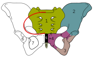
The ischium forms the lower and back region of the hip bone.
In vertebrates, the pubis or pubic bone forms the lower and anterior part of each side of the hip bone. The pubis is the most forward-facing of the three bones that make up the hip bone. The left and right pubic bones are each made up of three sections; a superior ramus, an inferior ramus, and a body.

The transversalis fascia is the fascial lining of the anterolateral abdominal wall situated between the inner surface of the transverse abdominal muscle, and the preperitoneal fascia. It is directly continuous with the iliac fascia, the internal spermatic fascia, and pelvic fascia.
The linea terminalis or innominate line consists of the pubic crest, pectineal line, the arcuate line, the sacral ala, and the sacral promontory.

Medial to the anterior inferior iliac spine is a broad, shallow groove, over which the iliacus and psoas major muscles pass. This groove is bounded medially by an eminence, the iliopubic eminence, which marks the point of union of the ilium and pubis.

The iliac fascia is the fascia overlying the iliacus muscle.

The lacunar ligament, also named Gimbernat's ligament, is a ligament in the inguinal region. It connects the inguinal ligament to the pectineal ligament, near the point where they both insert on the pubic tubercle.
The pectineal ligament, sometimes known as the inguinal ligament of Cooper, is an extension of the lacunar ligament. It runs on the pectineal line of the pubic bone. The pectineal ligament is the posterior border of the femoral ring.
Pectineal line may refer to:

The hypogastrium is a region of the abdomen located below the umbilical region.
The iliopectineal line is the border of the iliopubic eminence. It can be defined as a compound structure of the arcuate line and pectineal line. With the sacral promontory, it makes up the linea terminalis.

The hip bone is a large flat bone, constricted in the center and expanded above and below. In some vertebrates it is composed of three parts: the ilium, ischium, and the pubis.

In human anatomy, the adductor minimus is a small and flat skeletal muscle in the thigh which constitutes the upper, lateral part of the adductor magnus muscle. It adducts and laterally rotates the femur.

