Pelvic outlet

Last updated
Pelvic outlet
Gray239.png
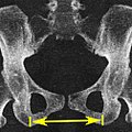
Diameters of inferior aperture of lesser pelvis (female)
1116 Muscle of the Female Perineum.png
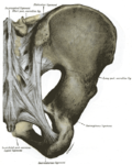
Muscles of the female perineum
Details
Identifiers
Latin apertura pelvis inferior
TA98 A02.5.02.009
TA2 1290
FMA 17273
Anatomical terms of bone

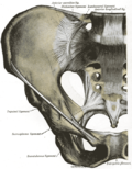
The lower circumference of the lesser pelvis is very irregular; the space enclosed by it is named the inferior aperture or pelvic outlet. It is an important component of pelvimetry.

Contents

Boundaries

It has the following boundaries:

Notches

These eminences are separated by three notches:

In situ

When the ligaments are in situ, the inferior aperture of the pelvis is lozenge-shaped, bounded as follows:

See also

Additional images

Related Research Articles

<span class="mw-page-title-main">Perineum</span> Region of the body including the perineal body and surrounding structures

The perineum in humans is the space between the anus and scrotum in the male, or between the anus and the vulva in the female. The perineum is the region of the body between the pubic symphysis and the coccyx, including the perineal body and surrounding structures. There is some variability in how the boundaries are defined. The perineal raphe is visible and pronounced to varying degrees. The perineum is an erogenous zone.

<span class="mw-page-title-main">Levator ani</span> Broad, thin muscle group, situated on either side of the pelvis

The levator ani is a broad, thin muscle group, situated on either side of the pelvis. It is formed from three muscle components: the pubococcygeus, the iliococcygeus, and the puborectalis.

<span class="mw-page-title-main">Sacrum</span> Triangular-shaped bone at the bottom of the spine

The sacrum, in human anatomy, is a large, triangular bone at the base of the spine that forms by the fusing of the sacral vertebrae (S1–S5) between ages 18 and 30.

<span class="mw-page-title-main">Pubic symphysis</span> Cartilaginous joint that sits between and joins the left and right superior rami of the pubic bones

The pubic symphysis is a secondary cartilaginous joint between the left and right superior rami of the pubis of the hip bones. It is in front of and below the urinary bladder. In males, the suspensory ligament of the penis attaches to the pubic symphysis. In females, the pubic symphysis is close to the clitoris. In most adults it can be moved roughly 2 mm and with 1 degree rotation. This increases for women at the time of childbirth.

<span class="mw-page-title-main">Ischium</span> Lower and back region of the hip bone

The ischium forms the lower and back region of the hip bone.

<span class="mw-page-title-main">Pubis (bone)</span> Most forward-facing of the three main regions making up the os coxa

In vertebrates, the pubic region is the most forward-facing of the three main regions making up the coxal bone. The left and right pubic regions are each made up of three sections, a superior ramus, inferior ramus, and a body.

<span class="mw-page-title-main">Sacrotuberous ligament</span>

The sacrotuberous ligament is situated at the lower and back part of the pelvis. It is flat, and triangular in form; narrower in the middle than at the ends.

<span class="mw-page-title-main">Sacrospinous ligament</span>

The sacrospinous ligament is a thin, triangular ligament in the human pelvis. The base of the ligament is attached to the outer edge of the sacrum and coccyx, and the tip of the ligament attaches to the spine of the ischium, a bony protuberance on the human pelvis. Its fibres are intermingled with the sacrotuberous ligament.

<span class="mw-page-title-main">Obturator artery</span>

The obturator artery is a branch of the internal iliac artery that passes antero-inferiorly on the lateral wall of the pelvis, to the upper part of the obturator foramen, and, escaping from the pelvic cavity through the obturator canal, it divides into both an anterior and a posterior branch.

<span class="mw-page-title-main">Lesser sciatic foramen</span> Opening between pelvis and thigh in human anatomy

The lesser sciatic foramen is an opening between the pelvis and the back of the thigh. The foramen is formed by the sacrotuberous ligament which runs between the sacrum and the ischial tuberosity and the sacrospinous ligament which runs between the sacrum and the ischial spine.

<span class="mw-page-title-main">Ischial tuberosity</span> Bones used to sit

The ischial tuberosity, also known colloquially as the sit bones or sitz bones, or as a pair the sitting bones, is a large swelling posteriorly on the superior ramus of the ischium. It marks the lateral boundary of the pelvic outlet.

<span class="mw-page-title-main">Pelvic cavity</span> Body cavity bounded by the bones of the pelvis

The pelvic cavity is a body cavity that is bounded by the bones of the pelvis. Its oblique roof is the pelvic inlet. Its lower boundary is the pelvic floor.

<span class="mw-page-title-main">Wing of ilium</span>

The wing(ala)of ilium is the large expanded portion of the ilium, the bone which bounds the greater pelvis laterally. It presents for examination two surfaces—an external and an internal—a crest, and two borders—an anterior and a posterior.

<span class="mw-page-title-main">Sacrococcygeal symphysis</span>

The sacrococcygeal symphysis is an amphiarthrodial joint, formed between the oval surface at the apex of the sacrum, and the base of the coccyx.

<span class="mw-page-title-main">Perineal membrane</span> Anatomical term

The perineal membrane is an anatomical term for a fibrous membrane in the perineum. The term "inferior fascia of urogenital diaphragm", used in older texts, is considered equivalent to the perineal membrane.

<span class="mw-page-title-main">Anal triangle</span>

The anal triangle is the posterior part of the perineum. It contains the anal canal.

<span class="mw-page-title-main">Outline of human anatomy</span> Overview of and topical guide to human anatomy

The following outline is provided as an overview of and topical guide to human anatomy:

<span class="mw-page-title-main">Hip bone</span>

The hip bone is a large flat bone, constricted in the center and expanded above and below. In some vertebrates it is composed of three parts: the ilium, ischium, and the pubis.

<span class="mw-page-title-main">Pelvis</span> Lower part of the trunk of the human body between the abdomen and the thighs

The pelvis is the lower part of the trunk, between the abdomen and the thighs, together with its embedded skeleton.

<span class="mw-page-title-main">Vaginal support structures</span> Structures that maintain the position of the vagina within the pelvic cavity

The vaginal support structures are those muscles, bones, ligaments, tendons, membranes and fascia, of the pelvic floor that maintain the position of the vagina within the pelvic cavity and allow the normal functioning of the vagina and other reproductive structures in the female. Defects or injuries to these support structures in the pelvic floor leads to pelvic organ prolapse. Anatomical and congenital variations of vaginal support structures can predispose a woman to further dysfunction and prolapse later in life. The urethra is part of the anterior wall of the vagina and damage to the support structures there can lead to incontinence and urinary retention.

References

  1. Sara Paterson-Brown, Phillip Bennett, Catherine Williamson (2010). Basic Science in Obstetrics and Gynaecology (Fourth Edition) - Obstetric pelvic definitions and dimensions. Elsevier Ltd. ISBN   978-0-443-10281-3.{{cite book}}: CS1 maint: multiple names: authors list (link)