Sacrospinous ligament

Last updated
Sacrospinous ligament
Greater sciatic foramen.png
Articulations of pelvis, anterior view, with greater sciatic foramen (labeled in red) and its boundaries.
Details
From Ischial spine
To Sacrum
Identifiers
Latin ligamentum sacrospinale
TA98 A03.6.03.007
TA2 1852
FMA 21485
Anatomical terminology

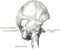
The sacrospinous ligament (small or anterior sacrosciatic ligament) is a thin, triangular ligament in the human pelvis. The base of the ligament is attached to the outer edge of the sacrum and coccyx, and the tip of the ligament attaches to the spine of the ischium, a bony protuberance on the human pelvis. Its fibres are intermingled with the sacrotuberous ligament.

Contents

Structure

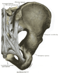
The sacrotuberous ligament passes behind the sacrospinous ligament. In its entire length, the sacrospinous ligament covers the equally triangular coccygeus muscle, to which its closely connected. [1]

Function

The presence of the ligament in the greater sciatic notch creates an opening (foramen), the greater sciatic foramen, and also converts the lesser sciatic notch into the lesser sciatic foramen. [2] The greater sciatic foramen lies above the ligament, and the lesser sciatic foramen lies below it.

The pudendal vessels and nerve pass behind the sacrospinous ligament directly medially and inferiorly to the ischial spine. The inferior gluteal artery, from a branch of the internal iliac artery, pass behind the sciatic nerve and the sacrospinous ligament and is left uncovered in a small opening above the top of the sacrospinous ligament. The coccygeal branch of the inferior gluteal artery passes behind the mid-portion of the sacrospinous ligament and pierces the sacrotuberous ligament at multiple locations. The main body of the inferior gluteal artery leaves the pelvis posteriorly to the upper border of the sacrospinous ligament, to follow the inferior portion of the sciatic nerve out of the greater sciatic foramen. [3]

The main function of the ligament is to prevent rotation of the ilium past the sacrum. Laxity of this ligament and the sacrotuberous ligament allows this rotation to occur. Stresses to these ligaments occur most often when leaning forward or getting out of a chair.[ citation needed ]

Clinical significance

Vaginal prolapse or uterine prolapse may occur in women when other pelvic ligaments and supportive structures are weakened. One treatment is sacrospinous fixation. In this surgery, the apex of the vagina is sutured to the sacrospinous ligament, which may offer a sturdier support than weakened pelvic ligaments, ideally preventing further prolapse. [4]

Additional images

Notes

  1. Gray's Anatomy 1918
  2. Platzer (2004), p 188
  3. Thompson et al. (1999)
  4. Vasavada et al. (2004), p 661

Related Research Articles

Human leg Lower extremity or limb of the human body (foot, lower leg, thigh and hip)

The human leg, in the general word sense, is the entire lower limb of the human body, including the foot, thigh and even the hip or gluteal region. However, the definition in human anatomy refers only to the section of the lower limb extending from the knee to the ankle, also known as the crus or, especially in non-technical use, the shank. Legs are used for standing, and all forms of locomotion including recreational such as dancing, and constitute a significant portion of a person's mass. Female legs generally have greater hip anteversion and tibiofemoral angles, but shorter femur and tibial lengths than those in males.

Sacrum Triangular-shaped bone at the bottom of the spine

The sacrum, in human anatomy, is a large, triangular bone at the base of the spine that forms by the fusing of the sacral vertebrae (S1–S5) between ages 18 and 30.

Internal pudendal artery

The internal pudendal artery is one of the three pudendal arteries. It branches off the internal iliac artery, and provides blood to the external genitalia.

Piriformis muscle One of six small hip muscles in the lateral rotator group

The piriformis muscle is a muscle in the gluteal region of the lower limbs. It is one of the six muscles in the lateral rotator group.

Internal obturator muscle One of six small hip muscles in the lateral rotator group

The internal obturator muscle or obturator internus muscle originates on the medial surface of the obturator membrane, the ischium near the membrane, and the rim of the pubis.

Internal iliac artery

The internal iliac artery is the main artery of the pelvis.

Ischium Lower and back region of the hip bone

The ischium forms the lower and back region of the hip bone.

Sacrotuberous ligament

The sacrotuberous ligament is situated at the lower and back part of the pelvis. It is flat, and triangular in form; narrower in the middle than at the ends.

Superior gluteal artery

The superior gluteal artery is the largest and final branch of the internal iliac artery. It is the continuation of the posterior division of that vessel. It is a short artery which runs backward between the lumbosacral trunk and the first sacral nerve. It divides into a superficial and a deep branch after passing out of the pelvis above the upper border of the piriformis muscle.

Inferior gluteal artery

The inferior gluteal artery, the smaller of the two terminal branches of the anterior trunk of the internal iliac artery, is distributed chiefly to the buttock and back of the thigh.

Lesser sciatic foramen

The lesser sciatic foramen is an opening between the pelvis and the back of the thigh. The foramen is formed by the sacrotuberous ligament which runs between the sacrum and the ischial tuberosity and the sacrospinous ligament which runs between the sacrum and the ischial spine.

Greater sciatic foramen Part of the pelvis

The greater sciatic foramen is an opening in the posterior human pelvis. It is formed by the sacrotuberous and sacrospinous ligaments. The piriformis muscle passes through the foramen and occupies most of its volume. The greater sciatic foramen is wider in women than in men.

Wing of ilium

The wing of ilium is the large expanded portion which bounds the greater pelvis laterally. It presents for examination two surfaces—an external and an internal—a crest, and two borders—an anterior and a posterior.

Internal iliac vein Large blood vessel of the pelvis

The internal iliac vein begins near the upper part of the greater sciatic foramen, passes upward behind and slightly medial to the internal iliac artery and, at the brim of the pelvis, joins with the external iliac vein to form the common iliac vein.

Greater sciatic notch

The greater sciatic notch is a notch in the ilium, one of the bones that make up the human pelvis. It lies between the posterior inferior iliac spine (above), and the ischial spine (below). The sacrospinous ligament changes this notch into an opening, the greater sciatic foramen.

Lesser sciatic notch

Below the ischial spine is a small notch, the lesser sciatic notch; it is smooth, coated in the recent state with cartilage, the surface of which presents two or three ridges corresponding to the subdivisions of the tendon of the Obturator internus, which winds over it.

Outline of human anatomy Overview of and topical guide to human anatomy

The following outline is provided as an overview of and topical guide to human anatomy:

Hip bone

The hip bone is a large irregular bone, constricted in the center and expanded above and below. In some vertebrates it is composed of three major regions: the ilium, ischium, and the pubis.

Pelvis Lower part of the trunk of the human body between the abdomen and the thighs

The pelvis is the lower part of the trunk, between the abdomen and the thighs, together with its embedded skeleton.

Gluteal lines

The gluteal lines are three curved lines outlined from three bony ridges on the exterior surface of the ilium in the gluteal region. They are the anterior gluteal line; the inferior gluteal line, and the posterior gluteal line.

References

PD-icon.svgThis article incorporates text in the public domain from page 309 of the 20th edition of Gray's Anatomy (1918)