
In anatomy, the atlas (C1) is the most superior (first) cervical vertebra of the spine.

The coccyx, commonly referred to as the tailbone, is the final segment of the vertebral column in all apes, and analogous structures in certain other mammals such as horses. In tailless primates since Nacholapithecus, the coccyx is the remnant of a vestigial tail. In animals with bony tails, it is known as tailhead or dock, in bird anatomy as tailfan. It comprises three to five separate or fused coccygeal vertebrae below the sacrum, attached to the sacrum by a fibrocartilaginous joint, the sacrococcygeal symphysis, which permits limited movement between the sacrum and the coccyx.

The sacroiliac joint or SI joint (SIJ) is the joint between the sacrum and the ilium bones of the pelvis, which are connected by strong ligaments. In humans, the sacrum supports the spine and is supported in turn by an ilium on each side. The joint is strong, supporting the entire weight of the upper body. It is a synovial plane joint with irregular elevations and depressions that produce interlocking of the two bones. The human body has two sacroiliac joints, one on the left and one on the right, that often match each other but are highly variable from person to person.

The dimples of Venus are sagittally symmetrical indentations sometimes visible on the human lower back, just superior to the gluteal cleft. They are directly superficial to the two sacroiliac joints, the sites where the sacrum attaches to the ilium of the pelvis. An imaginary line joining both dimples of Venus passes over the spinous process of the second sacral vertebra.

The ischium forms the lower and back part of the hip bone.

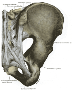
The sacrotuberous ligament is situated at the lower and back part of the pelvis. It is flat, and triangular in form; narrower in the middle than at the ends.

The erector spinae or spinal erectors is a set of muscles that straighten and rotate the back.

The lateral parts of the occipital bone are situated at the sides of the foramen magnum; on their under surfaces are the condyles for articulation with the superior facets of the atlas.

The wing of ilium is the large expanded portion which bounds the greater pelvis laterally. It presents for examination two surfaces—an external and an internal—a crest, and two borders—an anterior and a posterior.
The crest of the ilium is the superior border of the wing of ilium and the superiolateral margin of the greater pelvis.

The iliolumbar ligament is a strong ligament passing from the tip of the transverse process of the fifth lumbar vertebra to the posterior part of the inner lip of the iliac crest.

The anterior sacroiliac ligament consists of numerous thin bands, which connect the anterior surface of the lateral part of the sacrum to the margin of the auricular surface of the ilium and to the preauricular sulcus.
The interosseous sacroiliac ligament is a ligament of the sacroiliac joint that lies deep to the posterior ligament, and consists of a series of short, strong fibers connecting the tuberosities of the sacrum and ilium. It is the strongest ligament in the body. The major function of the interosseous sacroiliac ligament is to keep the sacrum and ilium together and therefore prevent abduction or distraction of the sacroiliac joint. It also helps to bear the weight of the thorax, upper limbs, head, and neck. This is performed by the nearly horizontal direction of the fibers running perpendicular from the sacrum to the ilium.

The posterior sacrococcygeal ligament or dorsal sacrococcygeal ligament is a ligament which stretches from the sacrum to the coccyx and thus dorsally across the sacrococcygeal symphysis shared by these two bones.
The superior pubic ramus is a part of the pubic bone which forms a portion of the obturator foramen. The obturator foramen, along with the ilium and other fused bones, forms part of either side of the pelvis.

The hip bone is a large irregular bone, constricted in the center and expanded above and below. In some vertebrates it is composed of three parts: the ilium, ischium, and the pubis.
Sacroiliac joint dysfunction generally refers to pain in the sacroiliac joint region that is caused by abnormal motion in the sacroiliac joint, either too much motion or too little motion. It typically results in inflammation of the sacroiliac joint, and can be debilitating.

The pelvis is either the lower part of the trunk of the human body between the abdomen and the thighs or the skeleton embedded in it.

In the vertebrate spinal column, each vertebra is an irregular bone with a complex structure composed of bone and some hyaline cartilage, the proportions of which vary according to the segment of the backbone and the species of vertebrate.
The public domain consists of all the creative works to which no exclusive intellectual property rights apply. Those rights may have expired, been forfeited, expressly waived, or may be inapplicable.

Gray's Anatomy is an English language textbook of human anatomy originally written by Henry Gray and illustrated by Henry Vandyke Carter. Earlier editions were called Anatomy: Descriptive and Surgical, Anatomy of the Human Body and Gray's Anatomy: Descriptive and Applied, but the book's name is commonly shortened to, and later editions are titled, Gray's Anatomy. The book is widely regarded as an extremely influential work on the subject, and has continued to be revised and republished from its initial publication in 1858 to the present day. The latest edition of the book, the 41st, was published in September 2015.

