Nuchal ligament

Last updated
Nuchal ligament
Nuchal ligament.PNG
Muscles connecting the arm to the spine seen from behind (nuchal ligament labeled in red at center)
Gray89.png
Details
System Skeletal
From External occipital protuberance
To Spinous process of cervical vertebra 7
Identifiers
Latin ligamentum nuchae
TA98 A03.2.01.006
TA2 1678
FMA 13427
Anatomical terminology

The nuchal ligament is a ligament at the back of the neck that is continuous with the supraspinous ligament.

Contents

Structure

The nuchal ligament extends from the external occipital protuberance on the skull and median nuchal line to the spinous process of the seventh cervical vertebra in the lower part of the neck. [1]

From the anterior border of the nuchal ligament, a fibrous lamina is given off. This is attached to the posterior tubercle of the atlas, and to the spinous processes of the cervical vertebrae, and forms a septum between the muscles on either side of the neck.

The trapezius and splenius capitis muscle attach to the nuchal ligament.

Function

It is a tendon-like structure that has developed independently in humans and other animals well adapted for running. [2] In some four-legged animals, particularly ungulates and canids, the nuchal ligament serves to sustain the weight of the head.

Clinical significance

In Chiari malformation treatment, decompression and duraplasty with a harvested nuchal ligament showed similar outcomes to pericranial and artificial grafts. [3]

Other animals

In sheep and cattle, it is known as the paxwax. [4] It relieves the animal of the weight of its head.

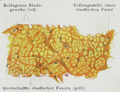
The nuchal ligament is unusual in being a ligament with an elastic component, allowing for stretch. [5] Most ligaments are mostly made of highly aligned collagen fibres which do not permit stretching.[ citation needed ]

Structurally, the nuchal ligament is formed with the association of both elastin proteins as well as type III collagen (45%). The collagen fibrils share a consistent size as well as helical pattern which gives the ligament its tensile strength. The elastin on the other hand is a protein that allows for flexibility. These two elements of the nuchal ligament maintain a complex balance which allows the constant weight bearing of the head along with multidirectional movement without damaging the durability of the ligament through over-use/stretching. [6]

In most other mammals, including the great apes, the nuchal ligament is absent or present only as a thin fascia. [2] As it is required for running, not all animals have one. [7]

All dogs (and all living Canidae - wolves, foxes, and wild dogs) possess a similar ligament connecting the spinous process of their first thoracic (or chest) vertebrae to the back of the axis bone (second cervical or neck bone), which supports the weight of the head without active muscle exertion, thus saving energy. [8] This ligament is analogous in function (but different in exact structural detail) to the nuchal ligament found in ungulates. [8] This ligament allows dogs to carry their heads while running long distances, such as while following scent trails with their nose to the ground, without expending much energy. [8]

In horses, the parasite Culicoides can spread onchocerca cervicalis by living in the nuchal ligament, causing dermatitis. [9]

Meat industry

In the meat industry, the nuchal ligament is referred to as paddywhack (also spelled pandywack; also called back strap or paxwax). [10]

The word is mentioned in a dictionary of South-west Lincolnshire dialect as a synonym of paxwax (originally faxwax; Old English compound of "hair" + "to grow"). [11] Hence, paddywack has been in use with this meaning since at least 1886. [12]

Dried paddywhack is commonly packaged and sold as a dog treat, [10] hence the phrase, "Knick-knack, paddywhack, give the dog a bone" in the nursery rhyme, This Old Man [ citation needed ]. Paddywack is unpalatable as a human food because it cannot be softened or tenderised, but it makes a good natural dog chew. [13] It is classed as offal by the meat industry. [10]

Additional images

See also

Related Research Articles

<span class="mw-page-title-main">Atlas (anatomy)</span> First cervical vertebra of the spine which supports the skull

In anatomy, the atlas (C1) is the most superior (first) cervical vertebra of the spine and is located in the neck.

<span class="mw-page-title-main">Trapezius</span> Muscle between the lower spine and the shoulder blade

The trapezius is a large paired trapezoid-shaped surface muscle that extends longitudinally from the occipital bone to the lower thoracic vertebrae of the spine and laterally to the spine of the scapula. It moves the scapula and supports the arm.

<span class="mw-page-title-main">Omohyoid muscle</span> Human neck muscle

The omohyoid muscle is a muscle in the neck. It is one of the infrahyoid muscles. It consists of two bellies separated by an intermediate tendon. Its inferior belly is attached to the scapula; its superior belly is attached to the hyoid bone. Its intermediate tendon is anchored to the clavicle and first rib by a fascial sling. The omohyoid is innervated by the ansa cervicalis of the cervical plexus. It acts to depress the hyoid bone.

<span class="mw-page-title-main">Cervical vertebrae</span> Vertebrae of the neck

In tetrapods, cervical vertebrae are the vertebrae of the neck, immediately below the skull. Truncal vertebrae lie caudal of cervical vertebrae. In sauropsid species, the cervical vertebrae bear cervical ribs. In lizards and saurischian dinosaurs, the cervical ribs are large; in birds, they are small and completely fused to the vertebrae. The vertebral transverse processes of mammals are homologous to the cervical ribs of other amniotes. Most mammals have seven cervical vertebrae, with the only three known exceptions being the manatee with six, the two-toed sloth with five or six, and the three-toed sloth with nine.

<span class="mw-page-title-main">Obliquus capitis inferior muscle</span>

The obliquus capitis inferior muscle is a muscle in the upper back of the neck. It is one of the suboccipital muscles. Its inferior attachment is at the spinous process of the axis; its superior attachment is at the transverse process of the atlas. It is innervated by the suboccipital nerve. The muscle rotates the head to its side.

<span class="mw-page-title-main">Obliquus capitis superior muscle</span>

The obliquus capitis superior muscle is a small muscle in the upper back part of the neck. It is one of the suboccipital muscles. It attaches inferiorly at the transverse process of the atlas ; it attaches superiorly at the external surface of the occipital bone. The muscle is innervated by the suboccipital nerve.

<span class="mw-page-title-main">Semispinalis muscles</span> Group of three muscles belonging to the transversospinales

The semispinalis muscles are a group of three muscles belonging to the transversospinales. These are the semispinalis capitis, the semispinalis cervicis and the semispinalis thoracis.

<span class="mw-page-title-main">Splenius capitis muscle</span>

The splenius capitis is a broad, straplike muscle in the back of the neck. It pulls on the base of the skull from the vertebrae in the neck and upper thorax. It is involved in movements such as shaking the head.

<span class="mw-page-title-main">Rectus capitis posterior major muscle</span> Tendon

The rectus capitis posterior major is a muscle in the upper back part of the neck. It is one of the suboccipital muscles. Its inferior attachment is at the spinous process of the axis ; its superior attachment is onto the outer surface of the occipital bone on and around the side part of the inferior nuchal line. The muscle is innervated by the suboccipital nerve. The muscle acts to extend the head and rorate the head to its side.

<span class="mw-page-title-main">Rectus capitis posterior minor muscle</span> Tendon

The rectus capitis posterior minor is a muscle in the upper back part of the neck. It is one of the suboccipital muscles. Its inferior attachment is at the posterior arch of atlas; its superior attachment is onto the occipital bone at and below the inferior nuchal line. The muscle is innervated by the suboccipital nerve. The muscle acts as a weak extensor of the head.

Wobbler disease is a catchall term referring to several possible malformations of the cervical vertebrae that cause an unsteady (wobbly) gait and weakness in dogs and horses. A number of different conditions of the cervical (neck) spinal column cause similar clinical signs. These conditions may include malformation of the vertebrae, intervertebral disc protrusion, and disease of the interspinal ligaments, ligamenta flava, and articular facets of the vertebrae. Wobbler disease is also known as cervical vertebral instability (CVI), cervical spondylomyelopathy (CSM), and cervical vertebral malformation (CVM). In dogs, the disease is most common in large breeds, especially Great Danes and Doberman Pinschers. In horses, it is not linked to a particular breed, though it is most often seen in tall, race-bred horses of Thoroughbred or Standardbred ancestry. It is most likely inherited to at least some extent in dogs and horses.

<span class="mw-page-title-main">Erector spinae muscles</span> Human muscle group

The erector spinae or spinal erectors is a set of muscles that straighten and rotate the back. The spinal erectors work together with the glutes to maintain stable posture standing or sitting.

<span class="mw-page-title-main">Nuchal lines</span> Part of the skulls occipital bone

The nuchal lines are four curved lines on the external surface of the occipital bone:

<span class="mw-page-title-main">Deep cervical fascia</span>

The deep cervical fascia lies under cover of the platysma, and invests the muscles of the neck; it also forms sheaths for the carotid vessels, and for the structures situated in front of the vertebral column. Its attachment to the hyoid bone prevents the formation of a dewlap.

<span class="mw-page-title-main">Ligamenta flava</span> Ligaments connecting the laminae of adjacent vertebrae

The ligamenta flava are a series of ligaments that connect the ventral parts of the laminae of adjacent vertebrae. They help to preserve upright posture, preventing hyperflexion, and ensuring that the vertebral column straightens after flexion. Hypertrophy can cause spinal stenosis.

<span class="mw-page-title-main">Investing layer of deep cervical fascia</span>

The investing layer of deep cervical fascia is the most superficial part of the deep cervical fascia, and encloses the whole neck.

The term forehand refers to the front half of a horse's body.

<span class="mw-page-title-main">Vertebral column</span> Bony structure found in vertebrates

The vertebral column, also known as the spinal column, spine or backbone, is the core part of the axial skeleton in vertebrate animals. The vertebral column is the defining and eponymous characteristic of the vertebrate endoskeleton, where the notochord found in all chordates has been replaced by a segmented series of mineralized irregular bones called vertebrae, separated by fibrocartilaginous intervertebral discs. The dorsal portion of the vertebral column houses the spinal canal, an elongated cavity formed by alignment of the vertebral neural arches that encloses and protects the spinal cord, with spinal nerves exiting via the intervertebral foramina to innervate each body segments.

<span class="mw-page-title-main">Vertebra</span> Bone in the vertebral column

Each vertebra is an irregular bone with a complex structure composed of bone and some hyaline cartilage, that make up the vertebral column or spine, of vertebrates. The proportions of the vertebrae differ according to their spinal segment and the particular species.

References

PD-icon.svgThis article incorporates text in the public domain from page 290 of the 20th edition of Gray's Anatomy (1918)

  1. Drake, Richard L.; Vogl, Wayne; Tibbitts, Adam W.M. Mitchell; illustrations by Richard; Richardson, Paul (2005). Gray's anatomy for students (Pbk. ed.). Philadelphia: Elsevier/Churchill Livingstone. p. 45. ISBN   978-0-443-06612-2.
  2. 1 2 Swindler, D. R., and C. D. Wood. 1973 An Atlas of Primate Gross Anatomy. Seattle: University of Washington Press[ page needed ]
  3. Cools MJ, Quinsey CS, Elton SW (April 2018). "Chiari decompression outcomes using ligamentum nuchae harvest and duraplasty in pediatric patients with Chiari malformation type I". J Neurosurg Pediatr. 22 (1): 47–51. doi:10.3171/2018.1.PEDS17670. PMID   29652242. S2CID   4793248.
  4. Merriam Webster Dictionary . Retrieved 4 October 2021.
  5. Virginia Studdert, Clive Gay, Douglas C. Blood, Saunders Comprehensive Veterinary Dictionary, p. 1252, Elsevier Health Sciences, 2011 ISBN   0702047449.
  6. Morocutti, M; Raspanti, M; Ottani, V; Govoni, P; Ruggeri (1991). "Ultrastructure of the bovine nuchal ligament". Journal of Anatomy. 178: 145–54. PMC   1260543 . PMID   1810923.
  7. Bramble, Dennis M.; Lieberman, Daniel E. (2004). "Endurance running and the evolution of Homo" (PDF). Nature. 432 (7015): 345–52. Bibcode:2004Natur.432..345B. doi:10.1038/nature03052. PMID   15549097. S2CID   2470602.
  8. 1 2 3 Wang, Xiaoming and Tedford, Richard H. Dogs: Their Fossil Relatives and Evolutionary History. New York: Columbia University Press, 2008. pp.97-8
  9. Haussler, Kevin K.; Jeffcott, Leo B. (2014). "21 - Back and pelvis". Equine sports medicine and surgery : basic and clinical sciences of the equine athlete (2nd ed.). Edinburgh: Saunders. pp. 419–456. doi:10.1016/B978-0-7020-4771-8.00021-1. ISBN   978-0-7020-4771-8. OCLC   865010322.
  10. 1 2 3 Herzmann, Peter (2017). "It's all edible: Four views of offal". In McWilliams, Mark (ed.). Offal: Rejected and Reclaimed Food: Proceedings of the 2016 Oxford Symposium on Food and Cookery. Oxford Symposium. pp. 21–39 [31]. ISBN   978-1-909248-55-7.
  11. "paxwax", Oxford English Dictionary Online, retrieved 10 October 2021 (subscription required).
  12. Robert Eden George Cole, A Glossary of Words Used in South-west Lincolnshire, English Dialect Society, 1886 OCLC   249538445.
  13. Ballard, Miranda (2021). Cluck, Oink, Baa, Moo. Ryland Peters & Small. ISBN   978-1-78879-395-7.[ page needed ]