Cruciate ligament of atlas

Last updated
Cruciate ligament of atlas
Gray307.png
Membrana tectoria, transverse, and alar ligaments. ("Transverse ligament" and "vertical portion" visible intersecting at center.)
Details
System skeletal
Frommedial tubercles of atlas bone (C1), anterior side of foramen magnum of occipital bone of skull, body of axis bone (C2)
Identifiers
Latin ligamentum cruciforme atlantis
TA98 A03.2.04.004
TA2 1699
FMA 25018
Anatomical terminology

The cruciate ligament of the atlas (cruciform ligament) is a cross-shaped (thus the name) ligament in the neck forming part of the atlanto-axial joint. It consists of the transverse ligament of atlas, a superior longitudinal band, and an inferior longitudinal band.

Contents

The cruciate ligament of the atlas prevents abnormal movement of the atlanto-axial joint.

It may be torn, such as by fractures of the atlas bone.

C: Cruciate ligament of atlas Anatomy of the Neck Sagittal Color MRI.png
C: Cruciate ligament of atlas

Structure

The cruciate ligament of the atlas consists of the transverse ligament of the atlas, a superior longitudinal band, and an inferior longitudinal band. [1] [2] The superior longitudinal band connects the transverse ligament to the anterior side of the foramen magnum (near the basilar part) in the occipital bone of the skull. The inferior longitudinal band connects the transverse ligament to the body of the axis bone (C2). [1]

Variation

The inferior longitudinal band may be absent in some people; the rest of the ligament is invariably present. [3]

Gerber's ligament

In about half of individuals, an additional band - Gerber's ligament - arises at the junction of the transverse ligament and superior band, and is situated deep to the superior band. [4] [ better source needed ]

Function

The cruciate ligament of the atlas prevents abnormal movements of the atlanto-axial joint. [1] The longitudinal bands prevent hyperflexion and hyperextension of the occipital bone, and hold the transverse ligament of the atlas in a normal position. [1]

Clinical significance

Any part of the cruciate ligament of the atlas may tear, which is a significant injury.[ citation needed ] This may be caused by fractures of the atlas bone. [3] Ligament tears may be imaged with radiography, a CT scan, or magnetic resonance imaging. [3]

Ossification

Very rarely, the cruciate ligament of the atlas may ossify. [5] This may lead to cervical myelopathy, a deficit in the spinal cord. [5]

Etymology

The terms "cruciform" and "cruciate" refer to the cross shape of the ligament. [1] Both terms are frequently used, although the term "cruciate" may be confusing due to confusion with the anterior cruciate ligament and the posterior cruciate ligament of the knee. [3]

Related Research Articles

<span class="mw-page-title-main">Atlas (anatomy)</span> First cervical vertebra of the spine which supports the skull

In anatomy, the atlas (C1) is the most superior (first) cervical vertebra of the spine and is located in the neck.

Articles related to anatomy include:

<span class="mw-page-title-main">Axis (anatomy)</span> Second cervical vertebra of the spine

In anatomy, the axis is the second cervical vertebra (C2) of the spine, immediately inferior to the atlas, upon which the head rests.

<span class="mw-page-title-main">Cervical vertebrae</span> Vertebrae of the neck

In tetrapods, cervical vertebrae are the vertebrae of the neck, immediately below the skull. Truncal vertebrae lie caudal of cervical vertebrae. In sauropsid species, the cervical vertebrae bear cervical ribs. In lizards and saurischian dinosaurs, the cervical ribs are large; in birds, they are small and completely fused to the vertebrae. The vertebral transverse processes of mammals are homologous to the cervical ribs of other amniotes. Most mammals have seven cervical vertebrae, with the only three known exceptions being the manatee with six, the two-toed sloth with five or six, and the three-toed sloth with nine.

<span class="mw-page-title-main">Obliquus capitis inferior muscle</span>

The obliquus capitis inferior muscle is a muscle in the upper back of the neck. It is one of the suboccipital muscles. Its inferior attachment is at the spinous process of the axis; its superior attachment is at the transverse process of the atlas. It is innervated by the suboccipital nerve. The muscle rotates the head to its side.

<span class="mw-page-title-main">Obliquus capitis superior muscle</span>

The obliquus capitis superior muscle is a small muscle in the upper back part of the neck. It is one of the suboccipital muscles. It attaches inferiorly at the transverse process of the atlas ; it attaches superiorly at the external surface of the occipital bone. The muscle is innervated by the suboccipital nerve.

<span class="mw-page-title-main">Alar ligament</span>

In anatomy, the alar ligaments are ligaments which connect the dens to tubercles on the medial side of the occipital condyle.

<span class="mw-page-title-main">Rectus capitis posterior major muscle</span> Tendon

The rectus capitis posterior major is a muscle in the upper back part of the neck. It is one of the suboccipital muscles. Its inferior attachment is at the spinous process of the axis ; its superior attachment is onto the outer surface of the occipital bone on and around the side part of the inferior nuchal line. The muscle is innervated by the suboccipital nerve. The muscle acts to extend the head and rorate the head to its side.

<span class="mw-page-title-main">Rectus capitis posterior minor muscle</span> Tendon

The rectus capitis posterior minor is a muscle in the upper back part of the neck. It is one of the suboccipital muscles. Its inferior attachment is at the posterior arch of atlas; its superior attachment is onto the occipital bone at and below the inferior nuchal line. The muscle is innervated by the suboccipital nerve. The muscle acts as a weak extensor of the head.

<span class="mw-page-title-main">Atlanto-axial joint</span>

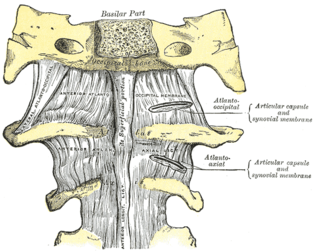
The atlanto-axial joint is a joint in the upper part of the neck between the atlas bone and the axis bone, which are the first and second cervical vertebrae. It is a pivot joint.

<span class="mw-page-title-main">Transverse ligament of atlas</span>

In anatomy, the transverse ligament of the atlas is a broad, though ligament which arches across the ring of the atlas posterior to the dens to keep the dens in contact with the atlas. It forms the transverse component of the cruciform ligament of atlas

<span class="mw-page-title-main">Posterior longitudinal ligament</span> Ligament connecting vertebral bodies of all of the vertebrae

The posterior longitudinal ligament is a ligament connecting the posterior surfaces of the vertebral bodies of all of the vertebrae of humans. It weakly prevents hyperflexion of the vertebral column. It also prevents posterior spinal disc herniation, although problems with the ligament can cause it.

<span class="mw-page-title-main">Tectorial membrane of atlanto-axial joint</span> Ligament in the spinal canal in the head joint area

The tectorial membrane of atlanto-axial joint is a tough membrane/broad, strong band representing the superior-ward prolongation of the posterior longitudinal ligament.

<span class="mw-page-title-main">Occipital condyles</span> Undersurface protuberances of the occipital bone in vertebrates

The occipital condyles are undersurface protuberances of the occipital bone in vertebrates, which function in articulation with the superior facets of the atlas vertebra.

<span class="mw-page-title-main">Clivus (anatomy)</span> Bony part of the skull base

The clivus, or Blumenbach clivus, is a bony part of the cranium at the base of the skull. It is a shallow depression behind the dorsum sellae of the sphenoid bone. It slopes gradually to the anterior part of the basilar occipital bone at its junction with the sphenoid bone. It extends to the foramen magnum. It is related to the pons and the abducens nerve.

Atlanto-occipital dislocation, orthopedic decapitation, or internal decapitation describes ligamentous separation of the spinal column from the skull base. It is possible for a human to survive such an injury; however, 70% of cases result in immediate death. It should not be confused with atlanto-axial dislocation, which describes ligamentous separation between the first and second cervical vertebra.

<span class="mw-page-title-main">Anterior atlantooccipital membrane</span>

The anterior atlantooccipital membrane is a broad, dense membrane extending between the anterior margin of the foramen magnum (superiorly), and the anterior arch of atlas (inferiorly).

<span class="mw-page-title-main">Posterior atlantooccipital membrane</span> Membrane at the base of the skull

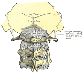
The posterior atlantooccipital membrane is a broad but thin membrane extending between the to the posterior margin of the foramen magnum above, and posterior arch of atlas below. It forms the floor of the suboccipital triangle.

<span class="mw-page-title-main">Outline of human anatomy</span> Overview of and topical guide to human anatomy

The following outline is provided as an overview of and topical guide to human anatomy:

<span class="mw-page-title-main">Vertebra</span> Bone in the vertebral column

Each vertebra is an irregular bone with a complex structure composed of bone and some hyaline cartilage, that make up the vertebral column or spine, of vertebrates. The proportions of the vertebrae differ according to their spinal segment and the particular species.

References

PD-icon.svgThis article incorporates text in the public domain from page 293 of the 20th edition of Gray's Anatomy (1918)

  1. 1 2 3 4 5 Cramer, Gregory D. (2014). "5 - The Cervical Region". Clinical anatomy of the spine, spinal cord, and ANS (3rd ed.). St. Louis: Elsevier Health Sciences, Mosby. pp. 135–209. doi:10.1016/B978-0-323-07954-9.00005-0. ISBN   978-0-323-07954-9. OCLC   830314791.
  2. Federative Committee on Anatomical Terminology (1998). Terminologia anatomica: international anatomical terminology. Thieme. pp. 27–. ISBN   978-3-13-114361-7 . Retrieved 17 June 2010.
  3. 1 2 3 4 Tubbs, R. Shane; Iwanaga, Joe; Loukas, Marios; Kassem, Mohammad D. (2019-01-04). "The Cruciform Ligament". Clinical Anatomy of the Ligaments of the Craniocervical Junction. Cambridge Scholars Publishing. pp. 155–160. ISBN   978-1-5275-2418-7.
  4. Ishak, Basem; Gnanadev, Raja; Dupont, Graham; Kikuta, Shogo; Altafulla, Juan; Iwanaga, Joe; Tubbs, R. Shane (2019-04-01). "Gerber's Ligament—A Forgotten Structure of the Craniocervical Junction". World Neurosurgery . Elsevier. 124: e707–e709. doi:10.1016/j.wneu.2018.12.198. ISSN   1878-8750. PMID   30660889. S2CID   58649895 via ScienceDirect.
  5. 1 2 Baqai, Muhammad Waqas Saeed; Javed, Gohar; Baig, Mirza Zain (2019). "Ossification of the Cruciform Ligament of Atlas; a Rare Cause of Cervical Myelopathy: Case Report and Review of Literature". Asian Journal of Neurosurgery. 14 (3): 999–1003. doi: 10.4103/ajns.AJNS_76_19 . ISSN   1793-5482. PMC   6703042 . PMID   31497151.