Transverse ligament of atlas

Last updated
Transverse ligament of the atlas
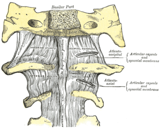
Gray306.png
Articulation between odontoid process and atlas. (Transverse ligament visible at center.)
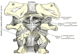
Gray307.png
Membrana tectoria, transverse, and alar ligaments. (Transverse ligament visible at center.)
Details
Identifiers
Latin ligamentum transversum atlantis
TA98 A03.2.04.006
TA2 1703
FMA 24961
Anatomical terminology

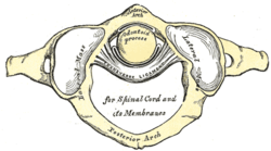
In anatomy, the transverse ligament of the atlas is a broad, tough ligament which arches across the ring of the atlas (first cervical vertebra) posterior to the dens [1] to keep the dens (odontoid process) in contact with the atlas.[ citation needed ] It forms the transverse component of the cruciform ligament of atlas.

Contents

Structure

The length of the ligament is variable; its mean length is 2 cm. [1]

The ligament broadens [1] and thickens [2] medially. The anterior medial aspect of the ligament is lined by a thin layer of articular cartilage. [1] The neck of the odontoid process is constricted where it is embraced posteriorly by the transverse ligament [2] so it retains the dens in position even after all other ligaments have been sectioned. [1]

Attachments

The ligament attaches on either side onto a small yet prominent tubercle upon the medial aspect of either lateral mass of atlas. [1]

Cruciate ligament

A strong median band (crus superius [2] ) extends superiorly from the superior margin of the ligament to attach onto the basilar part of occipital bone (between the attachments of the apical ligament of dens, and tectorial membrane). A weaker and somewhat inconsistent median band (crus inferius [2] ) extends inferiorly from the ligament to attach onto the posterior aspect of the [1] body of [2] axis. The ligament and the two median bands together constitute the cruciate ligament of atlas. [1]

Relations

The transverse ligament divides the vertebral foramen of the axis into an anterior portion (constituting one third of its lumen) which contains the dens, and a posterior portion (constituting two thirds of the foramen's lumen) which contains the spinal cord and its coverings [1] as well as the two accessory nerves (CN XI). [2]

Clinical significance

Excessive laxity of the posterior transverse ligament can lead to atlantoaxial instability, a common complication in patients with Down's Syndrome. Laxity has also been hypothesized as the cause of degenerative hypertrophy and mechanical atlantoaxial stress. [3] Degenerative processes can give rise to transverse ligament cysts, resulting in progressive cervical myelopathy. The treatment of choice for transverse ligament cysts with progressive neurological decline is surgical resection and cervical fusion. [4] Conservative treatment with external neck immobilization is less commonly reported , but may be very useful in select cases where immediate surgical intervention is not indicated. [5] [6]

See also

Related Research Articles

<span class="mw-page-title-main">Atlas (anatomy)</span> First cervical vertebra of the spine which supports the skull

In anatomy, the atlas (C1) is the most superior (first) cervical vertebra of the spine and is located in the neck.

Articles related to anatomy include:

<span class="mw-page-title-main">Lumbar vertebrae</span> Five vertebrae between the pelvis and the rib cage

The lumbar vertebrae are, in human anatomy, the five vertebrae between the rib cage and the pelvis. They are the largest segments of the vertebral column and are characterized by the absence of the foramen transversarium within the transverse process and by the absence of facets on the sides of the body. They are designated L1 to L5, starting at the top. The lumbar vertebrae help support the weight of the body, and permit movement.

<span class="mw-page-title-main">Axis (anatomy)</span> Second cervical vertebra of the spine

In anatomy, the axis is the second cervical vertebra (C2) of the spine, immediately inferior to the atlas, upon which the head rests.

<span class="mw-page-title-main">Spondylosis</span> Degeneration of the vertebral column

Spondylosis is the degeneration of the vertebral column from any cause. In the more narrow sense it refers to spinal osteoarthritis, the age-related degeneration of the spinal column, which is the most common cause of spondylosis. The degenerative process in osteoarthritis chiefly affects the vertebral bodies, the neural foramina and the facet joints. If severe, it may cause pressure on the spinal cord or nerve roots with subsequent sensory or motor disturbances, such as pain, paresthesia, imbalance, and muscle weakness in the limbs.

<span class="mw-page-title-main">Cervical vertebrae</span> Vertebrae of the neck

In tetrapods, cervical vertebrae are the vertebrae of the neck, immediately below the skull. Truncal vertebrae lie caudal of cervical vertebrae. In sauropsid species, the cervical vertebrae bear cervical ribs. In lizards and saurischian dinosaurs, the cervical ribs are large; in birds, they are small and completely fused to the vertebrae. The vertebral transverse processes of mammals are homologous to the cervical ribs of other amniotes. Most mammals have seven cervical vertebrae, with the only three known exceptions being the manatee with six, the two-toed sloth with five or six, and the three-toed sloth with nine.

<span class="mw-page-title-main">Vertebral artery</span> Major arteries of the neck

The vertebral arteries are major arteries of the neck. Typically, the vertebral arteries originate from the subclavian arteries. Each vessel courses superiorly along each side of the neck, merging within the skull to form the single, midline basilar artery. As the supplying component of the vertebrobasilar vascular system, the vertebral arteries supply blood to the upper spinal cord, brainstem, cerebellum, and posterior part of brain.

<span class="mw-page-title-main">Alar ligament</span>

In anatomy, the alar ligaments are ligaments which connect the dens to tubercles on the medial side of the occipital condyle.

<span class="mw-page-title-main">Pubis (bone)</span> Most forward-facing of the three main regions making up the os coxa

In vertebrates, the pubis or pubic bone forms the lower and anterior part of each side of the hip bone. The pubis is the most forward-facing of the three bones that make up the hip bone. The left and right pubic bones are each made up of three sections, a superior ramus, inferior ramus, and a body.
<span class="mw-page-title-main">Sacrospinous ligament</span>

The sacrospinous ligament is a thin, triangular ligament in the human pelvis. The base of the ligament is attached to the outer edge of the sacrum and coccyx, and the tip of the ligament attaches to the spine of the ischium, a bony protuberance on the human pelvis. Its fibres are intermingled with the sacrotuberous ligament.
<span class="mw-page-title-main">Atlanto-axial joint</span>

The atlanto-axial joint is a joint in the upper part of the neck between the atlas bone and the axis bone, which are the first and second cervical vertebrae. It is a pivot joint.

<span class="mw-page-title-main">Occipital condyles</span> Undersurface protuberances of the occipital bone in vertebrates

The occipital condyles are undersurface protuberances of the occipital bone in vertebrates, which function in articulation with the superior facets of the atlas vertebra.

<span class="mw-page-title-main">Apical ligament of dens</span>

The ligament of apex dentis is a ligament that spans between the second cervical vertebra in the neck and the skull. It lies as a fibrous cord in the triangular interval between the alar ligaments, which extends from the tip of the odontoid process on the axis to the anterior margin of the foramen magnum, being intimately blended with the deep portion of the anterior atlantooccipital membrane and superior crus of the transverse ligament of the atlas.

<span class="mw-page-title-main">Cruciate ligament of atlas</span> Ligament forming part of theatlanto-axial joint

The cruciate ligament of the atlas is a cross-shaped ligament in the neck forming part of the atlanto-axial joint. It consists of the transverse ligament of atlas, a superior longitudinal band, and an inferior longitudinal band.

<span class="mw-page-title-main">Outline of human anatomy</span> Overview of and topical guide to human anatomy

The following outline is provided as an overview of and topical guide to human anatomy:

<span class="mw-page-title-main">Facet syndrome</span> Medical condition

Facet syndrome is a syndrome in which the facet joints cause painful symptoms. In conjunction with degenerative disc disease, a distinct but functionally related condition, facet arthropathy is believed to be one of the most common causes of lower back pain.

<span class="mw-page-title-main">Denticulate ligaments</span>

Denticulate ligaments are lateral projections of the spinal pia mater forming triangular-shaped ligaments that anchor the spinal cord along its length to the dura mater on each side. There are usually 21 denticulate ligaments on each side, with the uppermost pair occurring just below the foramen magnum, and the lowest pair occurring between spinal nerve roots of T12 and L1. The denticulate ligaments are traditionally believed to provide stability for the spinal cord against motion within the vertebral column.

<span class="mw-page-title-main">Vertebral column</span> Bony structure found in vertebrates

The vertebral column, also known as the backbone or spine, is the core part of the axial skeleton in vertebrate animals. The vertebral column is the defining characteristic of vertebrate endoskeleton in which the notochord found in all chordates has been replaced by a segmented series of mineralized irregular bones called vertebrae, separated by fibrocartilaginous intervertebral discs. The dorsal portion of the vertebral column houses the spinal canal, a cavity formed by alignment of the neural arches that encloses and protects the spinal cord.

<span class="mw-page-title-main">Vertebra</span> Bone in the vertebral column

Each vertebra is an irregular bone with a complex structure composed of bone and some hyaline cartilage, that make up the vertebral column or spine, of vertebrates. The proportions of the vertebrae differ according to their spinal segment and the particular species.

<span class="mw-page-title-main">Rheumatoid disease of the spine</span> Medical condition

Rheumatoid disease of the spine is a morbid consequence of untreated longstanding severe cervical spinal rheumatoid arthritis (RA)–an inflammatory autoimmune disease that attacks the ligaments, joints, and bones of the neck. Although the anterior subluxation of the atlantoaxial joint is the most common manifestation of the disorder, subluxation can also occur with posterior or vertical movement, and subaxial joints can also be involved.
References

  1. 1 2 3 4 5 6 7 8 9 Standring, Susan (2020). Gray's Anatomy: The Anatomical Basis of Clinical Practice (42th ed.). New York. p. 841. ISBN   978-0-7020-7707-4. OCLC   1201341621.{{cite book}}: CS1 maint: location missing publisher (link)
  2. 1 2 3 4 5 6 Gray's anatomy, 1918
  3. Takeuchi, Mikinobu; Yasuda, Muneyoshi; Takahashi, Emiko; Funai, Mikiko; Joko, Masahiro; Takayasu, Msakazu (December 2011). "A large retro-odontoid cystic mass caused by transverse ligament degeneration with atlantoaxial subluxation leading to granuloma formation and chronic recurrent microbleeding case report". The Spine Journal. 11 (12): 1152–1156. doi:10.1016/j.spinee.2011.11.007. ISSN   1878-1632. PMID   22177924.
  4. Van Gompel, Jamie J.; Morris, Jonathan M.; Kasperbauer, Jan L.; Graner, Darlene E.; Krauss, William E. (April 2011). "Cystic deterioration of the C1-2 articulation: clinical implications and treatment outcomes". Journal of Neurosurgery. Spine. 14 (4): 437–443. doi:10.3171/2010.12.SPINE10302. ISSN   1547-5646. PMID   21314283.
  5. Sagiuchi, Takao; Shimizu, Satoru; Tanaka, Ryusui; Tachibana, Shigekuni; Fujii, Kiyotaka (August 2006). "Regression of an atlantoaxial degenerative articular cyst associated with subluxation during conservative treatment. Case report and review of the literature". Journal of Neurosurgery. Spine. 5 (2): 161–164. doi:10.3171/spi.2006.5.2.161. ISSN   1547-5654. PMID   16925084.
  6. Oushy, Soliman; Carlstrom, Lucas P.; Krauss, William E. (2018-03-05). "Spontaneous Regression of a Retroodontoid Transverse Ligament Cyst: A Case Report". Neurosurgery. 84 (2): E112–E115. doi:10.1093/neuros/nyy036. ISSN   1524-4040. PMID   29518219.