Related Research Articles

The peritoneum is the serous membrane forming the lining of the abdominal cavity or coelom in amniotes and some invertebrates, such as annelids. It covers most of the intra-abdominal organs, and is composed of a layer of mesothelium supported by a thin layer of connective tissue. This peritoneal lining of the cavity supports many of the abdominal organs and serves as a conduit for their blood vessels, lymphatic vessels, and nerves.

The abdominal cavity is a large body cavity in humans and many other animals that contain organs. It is a part of the abdominopelvic cavity. It is located below the thoracic cavity, and above the pelvic cavity. Its dome-shaped roof is the thoracic diaphragm, a thin sheet of muscle under the lungs, and its floor is the pelvic inlet, opening into the pelvis.

In a multicellular organism, an organ is a collection of tissues joined in a structural unit to serve a common function. In the hierarchy of life, an organ lies between tissue and an organ system. Tissues are formed from same type cells to act together in a function. Tissues of different types combine to form an organ which has a specific function. The intestinal wall for example is formed by epithelial tissue and smooth muscle tissue. Two or more organs working together in the execution of a specific body function form an organ system, also called a biological system or body system.

Abdominal pain, also known as a stomach ache, is a symptom associated with both non-serious and serious medical issues. Since the abdomen contains most of the body's vital organs, it can be an indicator of a wide variety of diseases. Given that, approaching the examination of a person and planning of a differential diagnosis is extremely important.

Chilaiditi syndrome is a rare condition when pain occurs due to transposition of a loop of large intestine in between the diaphragm and the liver, visible on plain abdominal X-ray or chest X-ray.

The lesser omentum is the double layer of peritoneum that extends from the liver to the lesser curvature of the stomach, and to the first part of the duodenum. The lesser omentum is usually divided into these two connecting parts: the hepatogastric ligament, and the hepatoduodenal ligament.

In the anatomy of humans and homologous primates, the ascending colon is the part of the colon located between the cecum and the transverse colon.

The abdomen is the front part of the torso between the thorax (chest) and pelvis in humans and in other vertebrates. The area occupied by the abdomen is called the abdominal cavity. In arthropods, it is the posterior tagma of the body; it follows the thorax or cephalothorax.

In human anatomy, the transverse colon is the longest and most movable part of the colon.

The greater omentum is a large apron-like fold of visceral peritoneum that hangs down from the stomach. It extends from the greater curvature of the stomach, passing in front of the small intestines and doubles back to ascend to the transverse colon before reaching to the posterior abdominal wall. The greater omentum is larger than the lesser omentum, which hangs down from the liver to the lesser curvature. The common anatomical term "epiploic" derives from "epiploon", from the Greek epipleein, meaning to float or sail on, since the greater omentum appears to float on the surface of the intestines. It is the first structure observed when the abdominal cavity is opened anteriorly.

The round ligament of the liver, ligamentum teres or ligamentum teres hepatis is a ligament that forms part of the free edge of the falciform ligament of the liver. It connects the liver to the umbilicus. It is the remnant of the left umbilical vein. The round ligament divides the left part of the liver into medial and lateral sections.

The hepatogastric ligament or gastrohepatic ligament connects the liver to the lesser curvature of the stomach. It contains the right and the left gastric arteries. In the abdominal cavity, it separates the greater and lesser sacs on the right. It is sometimes cut during surgery in order to access the lesser sac. The hepatogastric ligament consists of a dense cranial portion and the caudal portion termed the pars flaccida.
The paracolic gutters are peritoneal recesses – spaces between the colon and the abdominal wall.

The bare area of the liver is a large triangular area on the diaphragmatic surface of the liver. It is the only part of the liver with no peritoneal covering, although it is still covered by Glisson's capsule. It is attached directly to the diaphragm by loose connective tissue. The bare area of the liver is relevant to the portacaval anastomosis, encloses the right extraperitoneal subphrenic space, and can be a site of spread of infection from the abdominal cavity to the thoracic cavity
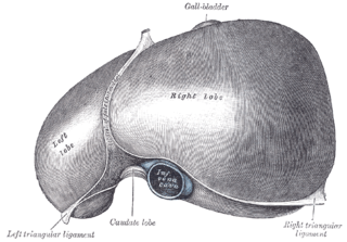
The left triangular ligament is a large peritoneal fold. It connects the posterior part of the upper surface of the left lobe of the liver to the thoracic diaphragm.
The right triangular ligament is situated at the right extremity of the bare area, and is a small fold which passes to the diaphragm, being formed by the apposition of the upper and lower layers of the coronary ligament.

In human anatomy, the omental foramen is the passage of communication, or foramen, between the greater sac, and the lesser sac of the peritoneal cavity.
The development of the digestive system in the human embryo concerns the epithelium of the digestive system and the parenchyma of its derivatives, which originate from the endoderm. Connective tissue, muscular components, and peritoneal components originate in the mesoderm. Different regions of the gut tube such as the esophagus, stomach, duodenum, etc. are specified by a retinoic acid gradient that causes transcription factors unique to each region to be expressed. Differentiation of the gut and its derivatives depends upon reciprocal interactions between the gut endoderm and its surrounding mesoderm. Hox genes in the mesoderm are induced by a Hedgehog signaling pathway secreted by gut endoderm and regulate the craniocaudal organization of the gut and its derivatives. The gut system extends from the oropharyngeal membrane to the cloacal membrane and is divided into the foregut, midgut, and hindgut.

The human abdomen is divided into quadrants and regions by anatomists and physicians for the purposes of study, diagnosis, and treatment. The division into four quadrants allows the localisation of pain and tenderness, scars, lumps, and other items of interest, narrowing in on which organs and tissues may be involved. The quadrants are referred to as the left lower quadrant, left upper quadrant, right upper quadrant and right lower quadrant. These terms are not used in comparative anatomy, since most other animals do not stand erect.

In human anatomy, the liver is divided grossly into four parts or lobes: the right lobe, the left lobe, the caudate lobe, and the quadrate lobe. Seen from the front – the diaphragmatic surface – the liver is divided into two lobes: the right lobe and the left lobe. Viewed from the underside – the visceral surface – the other two smaller lobes, the caudate lobe and the quadrate lobe, are also visible. The two smaller lobes, the caudate lobe and the quadrate lobe, are known as superficial or accessory lobes, and both are located on the underside of the right lobe.
References
- ↑ "Abdominal cavity" . Encyclopædia Britannica. Vol. I: A-Ak - Bayes (15th ed.). Chicago, IL: Encyclopædia Britannica, Inc. 2010. pp. 19–20. ISBN 978-1-59339-837-8.