The sensory nervous system is a part of the nervous system responsible for processing sensory information. A sensory system consists of sensory neurons, neural pathways, and parts of the brain involved in sensory perception and interoception. Commonly recognized sensory systems are those for vision, hearing, touch, taste, smell, balance and visceral sensation. Sense organs are transducers that convert data from the outer physical world to the realm of the mind where people interpret the information, creating their perception of the world around them.

In physiology, a stimulus is a detectable change in the physical or chemical structure of an organism's internal or external environment. The ability of an organism or organ to detect external stimuli, so that an appropriate reaction can be made, is called sensitivity (excitability). Sensory receptors can receive information from outside the body, as in touch receptors found in the skin or light receptors in the eye, as well as from inside the body, as in chemoreceptors and mechanoreceptors. When a stimulus is detected by a sensory receptor, it can elicit a reflex via stimulus transduction. An internal stimulus is often the first component of a homeostatic control system. External stimuli are capable of producing systemic responses throughout the body, as in the fight-or-flight response. In order for a stimulus to be detected with high probability, its level of strength must exceed the absolute threshold; if a signal does reach threshold, the information is transmitted to the central nervous system (CNS), where it is integrated and a decision on how to react is made. Although stimuli commonly cause the body to respond, it is the CNS that finally determines whether a signal causes a reaction or not.
Stimulus modality, also called sensory modality, is one aspect of a stimulus or what is perceived after a stimulus. For example, the temperature modality is registered after heat or cold stimulate a receptor. Some sensory modalities include: light, sound, temperature, taste, pressure, and smell. The type and location of the sensory receptor activated by the stimulus plays the primary role in coding the sensation. All sensory modalities work together to heighten stimuli sensation when necessary.
A mechanoreceptor, also called mechanoceptor, is a sensory receptor that responds to mechanical pressure or distortion. Mechanoreceptors are innervated by sensory neurons that convert mechanical pressure into electrical signals that, in animals, are sent to the central nervous system.

Sensory neurons, also known as afferent neurons, are neurons in the nervous system, that convert a specific type of stimulus, via their receptors, into action potentials or graded receptor potentials. This process is called sensory transduction. The cell bodies of the sensory neurons are located in the dorsal ganglia of the spinal cord.
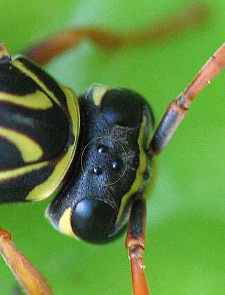
A simple eye refers to a form of eye or an optical arrangement composed of a single lens and without an elaborate retina such as occurs in most vertebrates. In this sense "simple eye" is distinct from a multi-lensed "compound eye", and is not necessarily at all simple in the usual sense of the word.
Neural adaptation or sensory adaptation is a gradual decrease over time in the responsiveness of the sensory system to a constant stimulus. It is usually experienced as a change in the stimulus. For example, if a hand is rested on a table, the table's surface is immediately felt against the skin. Subsequently, however, the sensation of the table surface against the skin gradually diminishes until it is virtually unnoticeable. The sensory neurons that initially respond are no longer stimulated to respond; this is an example of neural adaptation.
Johnston's organ is a collection of sensory cells found in the pedicel of the antennae in the class Insecta. Johnston's organ detects motion in the flagellum. It consists of scolopidia arrayed in a bowl shape, each of which contains a mechanosensory chordotonal neuron. The number of scolopidia varies between species. In homopterans, the Johnston's organs contain 25 - 79 scolopidia. The presence of Johnston's organ is a defining characteristic which separates the class Insecta from the other hexapods belonging to the group Entognatha. Johnston's organ was named after the physician Christopher Johnston, father of the physician and Assyriologist Christopher Johnston.

Campaniform sensilla are a class of mechanoreceptors found in insects, which respond to local stress and strain within the animal's cuticle. Campaniform sensilla function as proprioceptors that detect mechanical load as resistance to muscle contraction, similar to mammalian Golgi tendon organs. Sensory feedback from campaniform sensilla is integrated in the control of posture and locomotion.
Chordotonal organs are stretch receptor organs found only in insects and crustaceans. They are located at most joints and are made up of clusters of scolopidia that either directly or indirectly connect two joints and sense their movements relative to one another. They can have both extero- and proprioceptive functions, for example sensing auditory stimuli or leg movement. The word was coined by Vitus Graber in 1882, though he interpreted them as being stretched between two points like a string, sensing vibrations through resonance.

The ability to sense infrared thermal radiation evolved independently in two different groups of snakes, one consisting of the families Boidae (boas) and Pythonidae (pythons), the other of the family Crotalinae. What is commonly called a pit organ allows these animals to essentially "see" radiant heat at wavelengths between 5 and 30 μm. The more advanced infrared sense of pit vipers allows these animals to strike prey accurately even in the absence of light, and detect warm objects from several meters away. It was previously thought that the organs evolved primarily as prey detectors, but recent evidence suggests that it may also be used in thermoregulation and predator detection, making it a more general-purpose sensory organ than was supposed.

Proprioception is the sense of self-movement, force, and body position.
A sense is a biological system used by an organism for sensation, the process of gathering information about the world through the detection of stimuli. Although in some cultures five human senses were traditionally identified as such, many more are now recognized. Senses used by non-human organisms are even greater in variety and number. During sensation, sense organs collect various stimuli for transduction, meaning transformation into a form that can be understood by the brain. Sensation and perception are fundamental to nearly every aspect of an organism's cognition, behavior and thought.

The jamming avoidance response is a behavior of some species of weakly electric fish. It occurs when two electric fish with wave discharges meet – if their discharge frequencies are very similar, each fish shifts its discharge frequency to increase the difference between the two. By doing this, both fish prevent jamming of their sense of electroreception.
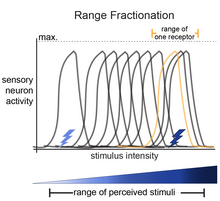
Feature detection is a process by which the nervous system sorts or filters complex natural stimuli in order to extract behaviorally relevant cues that have a high probability of being associated with important objects or organisms in their environment, as opposed to irrelevant background or noise.
During every moment of an organism's life, sensory information is being taken in by sensory receptors and processed by the nervous system. Sensory information is stored in sensory memory just long enough to be transferred to short-term memory. Humans have five traditional senses: sight, hearing, taste, smell, touch. Sensory memory (SM) allows individuals to retain impressions of sensory information after the original stimulus has ceased. A common demonstration of SM is a child's ability to write letters and make circles by twirling a sparkler at night. When the sparkler is spun fast enough, it appears to leave a trail which forms a continuous image. This "light trail" is the image that is represented in the visual sensory store known as iconic memory. The other two types of SM that have been most extensively studied are echoic memory, and haptic memory; however, it is reasonable to assume that each physiological sense has a corresponding memory store. For example, children have been shown to remember specific "sweet" tastes during incidental learning trials but the nature of this gustatory store is still unclear. However, sensory memories might be related to a region of the thalamus, which serves as a source of signals encoding past experiences in the neocortex.

The subgenual organ is an organ in insects that is involved in the perception of sound. The name refers to the location of the organ just below the knee in the tibia of all legs in most insects.
Hair-plates are a type of proprioceptor found in the folds of insect joints. They consist of a cluster of hairs, in which each hair is innervated by a single mechanosensory neuron. Functionally, hair-plates operate as "limit-detectors" by signaling the extreme ranges of motion of a joint.

The femoral chordotonal organ is a group of mechanosensory neurons found in an insect leg that detects the movements and the position of the femur/tibia joint. It is thought to function as a proprioceptor that is critical for precise control of leg position by sending the information regarding the femur/tibia joint to the motor circuits in the ventral nerve cord and the brain