
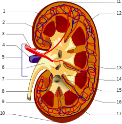
In humans, the kidneys are two reddish-brown bean-shaped blood-filtering organs that are a multilobar, multipapillary form of mammalian kidneys, usually without signs of external lobulation. They are located on the left and right in the retroperitoneal space, and in adult humans are about 12 centimetres in length. They receive blood from the paired renal arteries; blood exits into the paired renal veins. Each kidney is attached to a ureter, a tube that carries excreted urine to the bladder.

The nephron is the minute or microscopic structural and functional unit of the kidney. It is composed of a renal corpuscle and a renal tubule. The renal corpuscle consists of a tuft of capillaries called a glomerulus and a cup-shaped structure called Bowman's capsule. The renal tubule extends from the capsule. The capsule and tubule are connected and are composed of epithelial cells with a lumen. A healthy adult has 1 to 1.5 million nephrons in each kidney. Blood is filtered as it passes through three layers: the endothelial cells of the capillary wall, its basement membrane, and between the foot processes of the podocytes of the lining of the capsule. The tubule has adjacent peritubular capillaries that run between the descending and ascending portions of the tubule. As the fluid from the capsule flows down into the tubule, it is processed by the epithelial cells lining the tubule: water is reabsorbed and substances are exchanged ; first with the interstitial fluid outside the tubules, and then into the plasma in the adjacent peritubular capillaries through the endothelial cells lining that capillary. This process regulates the volume of body fluid as well as levels of many body substances. At the end of the tubule, the remaining fluid—urine—exits: it is composed of water, metabolic waste, and toxins.
Articles related to anatomy include:

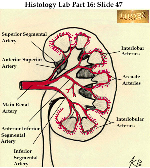
The renal medulla is the innermost part of the kidney. The renal medulla is split up into a number of sections, known as the renal pyramids. Blood enters into the kidney via the renal artery, which then splits up to form the segmental arteries which then branch to form interlobar arteries. The interlobar arteries each in turn branch into arcuate arteries, which in turn branch to form interlobular arteries, and these finally reach the glomeruli. At the glomerulus the blood reaches a highly disfavourable pressure gradient and a large exchange surface area, which forces the serum portion of the blood out of the vessel and into the renal tubules. Flow continues through the renal tubules, including the proximal tubule, the loop of Henle, through the distal tubule and finally leaves the kidney by means of the collecting duct, leading to the renal pelvis, the dilated portion of the ureter.

The glomerulus is a network of small blood vessels (capillaries) known as a tuft, located at the beginning of a nephron in the kidney. Each of the two kidneys contains about one million nephrons. The tuft is structurally supported by the mesangium, composed of intraglomerular mesangial cells. The blood is filtered across the capillary walls of this tuft through the glomerular filtration barrier, which yields its filtrate of water and soluble substances to a cup-like sac known as Bowman's capsule. The filtrate then enters the renal tubule of the nephron.

In the kidney, the macula densa is an area of closely packed specialized cells lining the wall of the distal tubule where it touches the glomerulus. Specifically, the macula densa is found in the terminal portion of the distal straight tubule, after which the distal convoluted tubule begins.

The renal arteries are paired arteries that supply the kidneys with blood. Each is directed across the crus of the diaphragm, so as to form nearly a right angle.

The renal veins in the renal circulation, are large-calibre veins that drain blood filtered by the kidneys into the inferior vena cava. There is one renal vein draining each kidney. Each renal vein is formed by the convergence of the interlobar veins of one kidney.
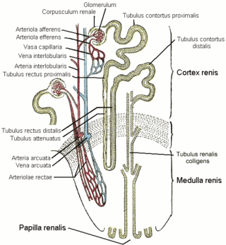
The vasa recta of the kidney, are the straight arterioles, and the straight venules of the kidney, – a series of blood vessels in the blood supply of the kidney that enter the medulla as the straight arterioles, and leave the medulla to ascend to the cortex as the straight venules.. They lie parallel to the loop of Henle.

The afferent arterioles are a group of blood vessels that supply the nephrons in many excretory systems. They play an important role in the regulation of blood pressure as a part of the tubuloglomerular feedback mechanism.
The efferent arterioles are blood vessels that are part of the urinary tract of organisms. Efferent means "outgoing", in this case meaning carrying blood out away from the glomerulus. The efferent arterioles form a convergence of the capillaries of the glomerulus, and carry blood away from the glomerulus that has already been filtered. They play an important role in maintaining the glomerular filtration rate despite fluctuations in blood pressure.
The stellate veins join to form the interlobular veins, which pass inward between the rays, receive branches from the plexuses around the convoluted tubules, and, having arrived at the bases of the renal pyramids, join with the venae rectae.
Cortical radial arteries, formerly known as interlobular arteries, are renal blood vessels given off at right angles from the side of the arcuate arteries looking toward the cortical substance. The interlobular arteries pass directly outward between the medullary rays to reach the fibrous tunic, where they end in the capillary network of this part.
The stellate veins are veins that lie beneath the fibrous tunic of the kidney. They are stellate in arrangement and are derived from the capillary network, into which the terminal branches of the interlobular arteries break up. These join to form the interlobular veins, which pass inward between the rays.

The arcuate arteries of the kidney, also known as arciform arteries, are vessels of the renal circulation. They are located at the border of the renal cortex and renal medulla.
The arcuate vein is a vessel of the renal circulation. It is located at the border of the renal cortex and renal medulla. Arcuate veins pass around the renal pyramids at the border between the renal cortex and renal medulla in an arch shape. Arcuate veins receive blood from cortical radiate veins, and in turn deliver blood into the arcuate veins.

The interlobar arteries are vessels of the renal circulation which supply the renal lobes. The interlobar arteries branch from the lobar arteries which branch from the segmental arteries, from the renal artery. They give rise to arcuate arteries.
The interlobar veins are veins of the renal circulation which drain the renal lobes. They collect blood from the arcuate veins. The interlobar veins unite to form a renal vein. Each interlobar vein passes along the edge of the renal pyramids.

In the circulatory system of vertebrates, a portal venous system occurs when a capillary bed pools into another capillary bed through veins, without first going through the heart. Both capillary beds and the blood vessels that connect them are considered part of the portal venous system.

The mammalian kidneys are a pair of excretory organs of the urinary system of mammals, a type of metanephric kidney. The kidneys in mammals are usually bean-shaped, located behind the peritoneum (retroperitoneally) on the back (dorsal) wall of the body. Each kidney consists of a renal capsule, peripheral cortex, internal medulla, calices, and renal pelvis, although the calices or renal pelvis may be absent in some species. Urine is excreted from the kidney through the ureter. The structure of the kidney may differ between species depending on the environment, in particular on its aridity. The cortex is responsible for filtering the blood, this part of the kidney is similar to the typical kidneys of less developed vertebrates. Nitrogen-containing waste products are excreted by the kidneys in mammals mainly in the form of urea.