Ureter

Last updated

Ureter
Urinary system.svg
The ureters (labeled 4) are tubes that carry urine and connect the kidneys to the bladder.
Details
Precursor Ureteric bud
System Urinary system
Artery Superior vesical artery, vaginal artery, ureteral branches of renal artery
Nerve Ureteric plexus
Identifiers
Latin ureter
MeSH D014513
TA98 A08.2.01.001
TA2 3394
FMA 9704
Anatomical terminology

The ureters are tubes composed of smooth muscle that transport urine from the kidneys to the urinary bladder. In an adult human, the ureters typically measure 20 to 30 centimeters in length and about 3 to 4 millimeters in diameter. They are lined with urothelial cells, a form of transitional epithelium, and feature an extra layer of smooth muscle in the lower third to aid in peristalsis. The ureters can be affected by a number of diseases, including urinary tract infections and kidney stone. Stenosis is when a ureter is narrowed, due to for example chronic inflammation. Congenital abnormalities that affect the ureters can include the development of two ureters on the same side or abnormally placed ureters. Additionally, reflux of urine from the bladder back up the ureters is a condition commonly seen in children.

Contents

The ureters have been identified for at least two thousand years, with the word "ureter" stemming from the stem uro- relating to urinating and seen in written records since at least the time of Hippocrates. It is, however, only since the 1500s that the term "ureter" has been consistently used to refer to the modern structure, and only since the development of medical imaging in the 1900s that techniques such as X-ray, CT, and ultrasound have been able to view the ureters. The ureters are also seen from the inside using a flexible camera, called ureteroscopy, which was first described in 1964.

Structure

Structures that are near the ureters. 1. Human urinary system: 2. Kidney, 3. Renal pelvis, 4. Ureter, 5. Urinary bladder, 6. Urethra. (Left side with frontal section), 7. Adrenal gland
Vessels:
8. Renal artery and vein, 9. Inferior vena cava, 10. Abdominal aorta, 11. Common iliac artery and vein
With transparency:
12. Liver, 13. Large intestine, 14. Pelvis Urinary system.svg
Structures that are near the ureters. 1. Human urinary system: 2. Kidney, 3. Renal pelvis, 4. Ureter, 5. Urinary bladder, 6. Urethra. (Left side with frontal section), 7. Adrenal gland
Vessels:
8. Renal artery and vein, 9. Inferior vena cava, 10. Abdominal aorta, 11. Common iliac artery and vein
With transparency:
12. Liver, 13. Large intestine, 14. Pelvis

The ureters are tubular structures, approximately 20–30 cm (8–12 in) in adults, [1] that pass from the pelvis of each kidney into the bladder. From the renal pelvis, they descend on top of the psoas major muscle to reach the brim of the pelvis. Here, they cross in front of the common iliac arteries. They then pass down along the sides of the pelvis and finally curve forward and enter the bladder from its left and right sides at the back of the bladder. [2] The ureters are 1.5–6 mm (0.06–0.24 in) in diameter [1] and surrounded by a layer of smooth muscle for 1–2 cm (0.4–0.8 in) near their ends just before they enter the bladder. [2]

The ureters enter the bladder from its back surface, traveling 1.5–2 cm (0.6–0.8 in) before opening into the bladder at an angle on its outer back surface at the slit-like ureteric orifices. [2] [3] This location is also called the vesicoureteric junction. [4] In the contracted bladder, they are about 25 mm (1 in) apart and about the same distance from the internal urethral orifice; in the distended bladder, these measurements may be increased to about 50 mm (2 in). [2]

A number of structures pass by, above, and around the ureters on their path down from the kidneys to the bladder. [2] In its upper part, the ureter travels on the psoas major muscle and sits just behind the peritoneum. As it passes down the muscle, it travels over the genitofemoral nerve. The inferior vena cava and the abdominal aorta sit to the midline of the right and left ureters, respectively. [2] In the lower part of the abdomen, the right ureter sits behind the lower mesentery and the terminal ileum, and the left ureter sits behind the jejunum and the sigmoid colon. [2] As the ureters enter the pelvis, they are surrounded by connective tissue, and travel backward and outward, passing in front of the internal iliac arteries and internal iliac veins. They then travel inward and forward, crossing the umbilical, inferior vesical, and middle rectal arteries. [2] From here, in males, they cross under the vas deferens and in front of the seminal vesicles to enter the bladder near the trigone. [2] In females, the ureters pass behind the ovaries and then travel in the lower midline section of the broad ligament of the uterus. For a short part, the uterine arteries travel on top for a short (2.5 cm (1 in)) period. They then pass by the cervix, traveling inward towards the bladder. [2]

Blood and lymphatic supply

The arteries which supply the ureter vary along its course. The upper third of the ureter, closest to the kidney, is supplied by the renal arteries. [2] The middle part of the ureter is supplied by the common iliac arteries, direct branches from the abdominal aorta, and gonadal arteries; [1] the gonadal arteries being the testicular artery in men and the ovarian artery in women. [2] The lower third of the ureter, closest to the bladder, is supplied by branches from the internal iliac arteries, mainly the superior and inferior vesical arteries. [1] The arterial supply can be variable, with arteries that contribute include the middle rectal artery, branches directly from the aorta, [1] and, in women, the uterine and vaginal arteries. [2]

The arteries that supply the ureters end in a network of vessels within the adventitia of the ureters. [1] There are many connections ( anastamoses ) between the arteries of the ureter, [2] particularly in the adventitia, [5] which means damage to a single vessel does not compromise the blood supply of the ureter. [2] [5] Venous drainage mostly parallels that of the arterial supply; [5] [2] that is, it begins as a network of smaller veins in the adventitia; with the renal veins draining the upper ureters, and the vesicular and gonadal veins draining the lower ureters. [1]

Lymphatic drainage depends on the position of lymphatic vessels in the ureter. [1] Lymph collects in submucosal, intramuscular and adventitial lymphatic vessels. [2] Those vessels closer to the kidney drain into renal collecting vessels, and from here into the lateral aortic nodes near the gonadal vessels. [2] The middle part of the ureter drains into the right paracaval and interaortocaval nodes on the right, and the left paraaortic nodes on the left. [1] In the lower ureter, lymph may drain into the common iliac lymph nodes, or lower down in the pelvis to the common, external, or internal iliac lymph nodes. [2]

Nerve supply

The ureters are richly supplied by nerves that form a network ( plexus ) of nerves, the ureteric plexus that lies in the adventitia of the ureters. [2] This plexus is formed from a number of nerve roots directly (T9-12, L1, and S2-4), as well as branches from other nerve plexuses and nerves; specifically, the upper third of the ureter receives nerve branches from the renal plexus and aortic plexus, the middle part receives branches from the upper hypogastric plexus and nerve, and the lower ureter receives branches from the lower hypogastric plexus and nerve. [2] The plexus is in the adventitia. These nerves travel in individual bundles and along small blood vessels to form the ureteric plexus. [2] Sensation supplied is sparse close to the kidneys and increases closer to the bladder. [2]

Sensation to the ureters is provided by nerves that come from T11 - L2 segments of the spinal cord. [2] When pain is caused, for example by spasm of the ureters or by a stone, the pain may be referred to the dermatomes of T11 - L2, namely the back and sides of the abdomen, the scrotum (males) or labia majora (females) and upper part of the front of the thigh. [2]

Microanatomy

Microscopic cross-section of the ureter, showing the epithelium (purple cells) sitting next to the lumen. A large amount of muscle fibres can be seen surrounding the epithelium, and the adventitia sits beyond this 2607 Ureter.jpg
Microscopic cross-section of the ureter, showing the epithelium (purple cells) sitting next to the lumen. A large amount of muscle fibres can be seen surrounding the epithelium, and the adventitia sits beyond this

The ureter is lined by urothelium, a type of transitional epithelium that is capable of responding to stretches in the ureters. The transitional epithelium may appear as a layer of column-shaped cells when relaxed, and of flatter cells when distended. Below the epithelium sits the lamina propria. The lamina propria is made up of loose connective tissue with many elastic fibers interspersed with blood vessels, veins and lymphatics. The ureter is surrounded by two muscular layers, an inner longitudinal layer of muscle, and an outer circular or spiral layer of muscle. [6] [7] The lower third of the ureter has a third muscular layer. [7] Beyond these layers sits an adventitia containing blood vessels, lymphatic vessels, and veins. [7]

Development

Image showing the bottom part of an embryo 4 - 5 weeks old. Here, the ureter (in orange) can be seen emerging from the bottom of the mesonephric duct (labelled "Wolffian duct"), connected to the primitive bladder. Image from Gray's Anatomy 1918 edition. Gray1116.png
Image showing the bottom part of an embryo 4 - 5 weeks old. Here, the ureter (in orange) can be seen emerging from the bottom of the mesonephric duct (labelled "Wolffian duct"), connected to the primitive bladder. Image from Gray's Anatomy 1918 edition.

The ureters develop from the ureteric buds, which are outpouchings from the mesonephric duct. This is a duct, derived from mesoderm, found in the early embryo. [8] Over time, the buds elongate, moving into surrounding mesodermal tissue, dilate, and divide into left and right ureters. Eventually, successive divisions from these buds form not only the ureters, but also the pelvis, major and minor calyces, and collecting ducts of the kidneys. [8]

The mesonephric duct is connected with the cloaca, which over the course of development splits into a urogenital sinus and the anorectal canal. [8] The urinary bladder forms from the urogenital sinus. Over time, as the bladder enlarges, it absorbs the surrounding parts of the primitive ureters. [8] Finally, the entry points of the ureters into the bladder move upwards, owing to the upward migration of the kidneys in the developing embryo. [8]

Function

The ureters are a component of the urinary system. Urine, produced by the kidneys, travels along the ureters to the bladder. It does this through regular contractions called peristalsis. [2]

Clinical significance

Ureteral stones

A giant ureteral stone with dimensions of approximately 6 x 5 x 4 cm and weighing 61 grams extracted from the left ureter of a 19-year old male A-giant-ureter-stone.jpg
A giant ureteral stone with dimensions of approximately 6 × 5 × 4 cm and weighing 61 grams extracted from the left ureter of a 19-year old male

A kidney stone can move from the kidney and become lodged inside the ureter, which can block the flow of urine, as well as cause a sharp cramp in the back, side, or lower abdomen. [9] Pain often comes in waves lasting up to two hours, then subsides, called renal colic. [10] The affected kidney could then develop hydronephrosis, should a part of the kidney become swollen due to blocked flow of urine. [9] It is classically described that there are three sites in the ureter where a kidney stone will commonly become stuck: where the ureter meets the renal pelvis; where the iliac blood vessels cross the ureters; and where the ureters enter the urinary bladder, [9] however a retrospective case study, which is a primary source, of where stones lodged based on medical imaging did not show many stones at the place where the iliac blood vessels cross. [11]

Most stones are compounds containing calcium such as calcium oxalate and calcium phosphate. The first recommended investigation is a CT scan of the abdomen because it can detect almost all stones. Management includes analgesia, often with nonsteroidal antiinflammatories. [10] Small stones (< 4mm) may pass themselves; larger stones may require lithotripsy, and those with complications such as hydronephrosis or infection may require surgery to remove. [10]

Reflux

Vesicoureteral reflux refers to the reflux of fluid from the bladder into the ureters. [12] This condition can be associated with urinary tract infections, particularly in children, and is present in up to 28–36% of children to some degree. [12] A number of forms of medical imaging are available for diagnosis of the condition, with modalities including doppler urinary tract ultrasound.Factors that affect which of these are selected depends if a child is able to receive a urinary catheter, and whether a child is toilet trained. [12] Whether these investigations are performed at the first time a child has an illness, or later and depending on other factors (such as if the causal bacteria is E. coli ) differ between US, EU and UK guidelines. [12]

Management is also variable, with differences between international guidelines on issues such as whether prophylactic antibiotics should be used, and whether surgery is recommended. [12] One reason is most instances of vesicoureteral reflux improve by themselves. [12] If surgery is considered, it generally involves reattaching the ureters to a different spot on the bladder, and extending the part of the ureter that it is within the wall of the bladder, with the most common surgical option being Cohen's cross-trigonal reimplantation. [12]

Anatomical and surgical abnormalities

Blockage, or obstruction of the ureter can occur, [13] as a result of narrowing within the ureter, or compression or fibrosis of structures around the ureter. [14] Narrowing can result of ureteric stones, masses associated with cancer, and other lesions such as endometriosis tuberculosis and schistosomiasis. [14] Things outside the ureters such as constipation and retroperitoneal fibrosis can also compress them. [14] Some congenital abnormalities can also result in narrowing or the ureters. Congenital disorders of the ureter and urinary tract affect 10% of infants. [13] These include partial or total duplication of the ureter (a duplex ureter), or the formation of a second irregularly placed ( ectopic ) ureter; [13] or where the junction with the bladder is malformed or a ureterocoele develops (usually in that location). [14] If the ureters have been resited as a result of surgery, for example due to a kidney transplant or due to past surgery for vesicoureteric reflux, that site may also become narrowed. [15] [1]

A narrowed ureter may lead to ureteric enlargement ( dilation ) and cause swelling of the kidneys (hydronephrosis). [13] Associated symptoms may include recurrent infections, pain or blood in the urine; and when tested, kidney function might be seen to decrease. [13] These are considered situations when surgery is needed. [13] Medical imaging, including urinary tract ultrasound, CT or nuclear medicine imaging is conducted to investigate many causes. [13] [14] This may involve reinserting the ureters into a new place on the bladder (reimplantion), or widening of the ureter. [13] A ureteric stent may be inserted to relieve an obstruction. [16] If the cause cannot be removed, a nephrostomy may be required, which is the insertion of a tube connected to the renal pelvis which directly drains urine into a stoma bag. [17]

Cancer

Cancer of the ureters is known as ureteral cancer. It is usually due to cancer of the urothelium, the cells that line the surface of the ureters. Urothelial cancer is more common after the age of 40, and more common in men than women; [18] other risk factors include smoking and exposure to dyes such as aromatic amines and aldehydes. [18] When cancer is present, the most common symptom is blood in the urine; it may not cause symptoms, and a physical medical examination may be otherwise normal, except in late disease. [18] Ureteral cancer is most often due to cancer of the cells lining the ureter, called transitional cell carcinoma, although it can more rarely occur as a squamous cell carcinoma if the type of cells lining the urethra have changed due to chronic inflammation, such as due to stones or schistosomiasis. [18]

Investigations performed usually include collecting a sample of urine for an inspection for malignant cells under a microscope, called cytology, as well as medical imaging by a CT urogram or ultrasound. [18] If a concerning lesion is seen, a flexible camera may be inserted into the ureters, called ureteroscopy, in order to view the lesion and take a biopsy, and a CT scan will be performed of other body parts (a CT scan of the chest, abdomen and pelvis) to look for additional metastatic lesions. [18] After the cancer is staged, treatment may involve open surgery to remove the affected ureter and kidney if it is involved; or, if the lesion is small, it may be removed via ureteroscopy. [18] Prognosis can vary markedly depending on the tumour grade, with a worse prognosis associated with an ulcerating lesion. [18]

Injury

Injuries to the ureter can occur after penetrating abdominal injuries, and injuries at high speeds followed by an abrupt stop (such as a high speed car accident). [19] The ureter can be injured during surgery to nearby structures. [20] It is injured in 2 per 10,000 cases of vaginal hysterectomies and 13 per 10,000 cases of abdominal hysterectomies, [20] usually near the suspensory ligament of the ovary or near the cardinal ligament, where the ureter runs close to the blood vessels of the uterus. [21]

Imaging

Several forms of medical imaging are used to view the ureters and urinary tract. [22] Ultrasound may be able to show evidence of blockage because of hydronephrosis of the kidneys and renal pelvis. [22] CT scans, including ones where contrast media is injected intravenously to better show the ureters, and with contrast to better show lesions, and to differentiate benign from malignant lesions. [22] Dye may also be injected directly into the ureters or renal tract; an antegrade pyelogram is when contrast is injected directly into the renal pelvis, and a retrograde pyelogram is where dye is injected into the urinary tract via a catheter, and flows backwards into the ureters. [22] More invasive forms of imaging include ureteroscopy, which is the insertion of a flexible endoscope into the urinary tract to view the ureters. [23] Ureteroscopy is most commonly used for medium to large-sized stones when less invasive methods of removal cannot be used. [23]

Other animals

All vertebrates have two kidneys located behind the abdomen that produce urine, and have a way of excreting it, so that waste products within the urine can be removed from the body. [24] The structure specifically called the ureter is present in amniotes, meaning mammals, birds and reptiles. [24] These animals possess an adult kidney derived from the metanephros. [24] The duct that connects the kidney to excrete urine in these animals is the ureter. [24] In placental mammals, it connects to the urinary bladder, whence urine leaves via the urethra. [25] In monotremes, urine flows from the ureters into the cloaca. [26] The ureters are ventral to the vas deferens in male placental mammals, but dorsal to the vas deferens in marsupials. [27]

History

The word "ureter" comes from the Ancient Greek noun οὖρον, ouron, meaning "urine", and the first use of the word is seen during the era of Hippocrates to refer to the urethra. [28] The anatomical structure of the ureter was noted by 40 AD. However, the terms "ureter" and "urethra" were variably used to refer to each other thereafter for more than a millennium. [28] It was only in the 1550s that anatomists such as Bartolomeo Eustachi and Jacques Dubois began to use the terms to specifically and consistently refer to what are in modern English called the ureter and the urethra. [28] Following this, in the 19th and 20th centuries multiple terms relating to the structures such as ureteritis and ureterography, were coined. [28]

Ureters Ureters.jpg
Ureters

Kidney stones have been identified and recorded about as long as written historical records exist. [29] The urinary tract including the ureters, as well as their function to drain urine from the kidneys, has been described by Galen in the second century AD. [30]

The first to examine the ureter through an internal approach, called ureteroscopy, rather than surgery was Hampton Young in 1929. [29] This was improved on by VF Marshall who is the first published use of a flexible endoscope based on fiber optics, which occurred in 1964. [29] The insertion of a drainage tube into the renal pelvis, bypassing the ureters and urinary tract, called nephrostomy, was first described in 1941. Such an approach differed greatly from the open surgical approaches within the urinary system employed during the preceding two millennia. [29]

The first radiological imaging of the ureters was by X-rays, although this was made more difficult by the thick abdomen, which the low power of the original X-rays could not penetrate enough to produce clear images. [31] More useful images were able to be produced when Edwin Hurry Fenwick in 1908 pioneered the use of tubes covered in radioopaque material visible to X-rays inserted into the ureters, and in the early 20th century when contrasts were injected externally into the urinary tract (retrograde pyelograms). [31] Unfortunately, much of the earlier retrograde pyelograms were complicated by significant damage to the kidneys as a result of contrast based on silver or sodium iodide. [31] Hryntshalk in 1929 pioneered the development of the intravenous urogram, in which contrast is injected into a vein and highlights the kidney and, when excreted, the urinary tract. [31] Things improved with the development by Moses Swick and Leopold Lichtwitz in the late 1920s of relatively nontoxic contrast media, with controversy surrounding publication as to who was the primary discoverer. [31] Side-effects associated with imaging improved even more when Tosten Almen published a ground-breaking thesis in 1969 based on the less toxic low-osmolar contrast media, developed based on swimming experiences in lakes with different salinity. [31]

Related Research Articles

<span class="mw-page-title-main">Urethra</span> Tube that connects the urinary bladder to the external urethral orifice

The urethra is the tube that transports urine from the mammalian urinary bladder to the urinary meatus through the penis or vulval vestibule. In placental mammals, the urethra also transports semen through the penis during ejaculation.

<span class="mw-page-title-main">Bladder</span> Organ in vertebrates that collects and stores urine from the kidneys before disposal

The bladder is a hollow organ in humans and other vertebrates that stores urine from the kidneys before disposal by urination. In placental mammals, urine enters the bladder via the ureters and exits via the urethra. In humans, the bladder is a distensible organ that sits on the pelvic floor. The typical adult human bladder will hold between 300 and 500 ml before the urge to empty occurs, but can hold considerably more.

<span class="mw-page-title-main">Urinary system</span> Anatomical system consisting of the kidneys, ureters, urinary bladder, and the urethra

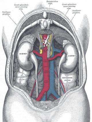
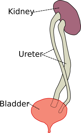
The human urinary system, also known as the urinary tract or renal system, consists of the kidneys, ureters, bladder, and the urethra. The purpose of the urinary system is to eliminate waste from the body, regulate blood volume and blood pressure, control levels of electrolytes and metabolites, and regulate blood pH. The urinary tract is the body's drainage system for the eventual removal of urine. The kidneys have an extensive blood supply via the renal arteries which leave the kidneys via the renal vein. Each kidney consists of functional units called nephrons. Following filtration of blood and further processing, wastes exit the kidney via the ureters, tubes made of smooth muscle fibres that propel urine towards the urinary bladder, where it is stored and subsequently expelled from the body by urination. The female and male urinary system are very similar, differing only in the length of the urethra.

<span class="mw-page-title-main">Cystoscopy</span> Medical procedure; endoscopy of the urinary bladder via the urethra

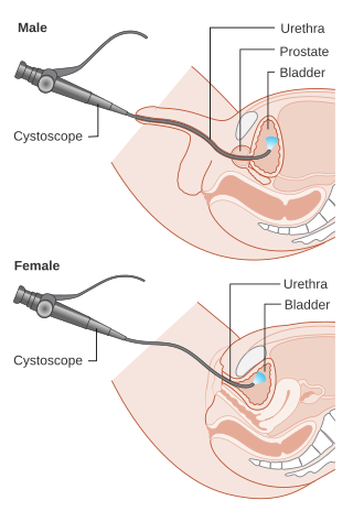
Cystoscopy is endoscopy of the urinary bladder via the urethra. It is carried out with a cystoscope.

The excretory system is a passive biological system that removes excess, unnecessary materials from the body fluids of an organism, so as to help maintain internal chemical homeostasis and prevent damage to the body. The dual function of excretory systems is the elimination of the waste products of metabolism and to drain the body of used up and broken down components in a liquid and gaseous state. In humans and other amniotes, most of these substances leave the body as urine and to some degree exhalation, mammals also expel them through sweating.

<span class="mw-page-title-main">Hydronephrosis</span> Medical condition

Hydronephrosis describes hydrostatic dilation of the renal pelvis and calyces as a result of obstruction to urine flow downstream. Alternatively, hydroureter describes the dilation of the ureter, and hydronephroureter describes the dilation of the entire upper urinary tract.

Horseshoe kidney, also known as ren arcuatus, renal fusion or super kidney, is a congenital disorder affecting about 1 in 500 people that is more common in men, often asymptomatic, and usually diagnosed incidentally. In this disorder, the patient's kidneys fuse to form a horseshoe-shape during development in the womb. The fused part is the isthmus of the horseshoe kidney. The abnormal anatomy can affect kidney drainage resulting in increased frequency of kidney stones and urinary tract infections as well as increase risk of certain renal cancers.

<span class="mw-page-title-main">Renal colic</span> Medical condition

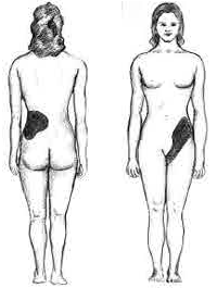
Renal colic, also known as ureteric colic, is a type of abdominal pain commonly caused by obstruction of ureter from dislodged kidney stones. The most frequent site of obstruction is the vesico-ureteric junction (VUJ), the narrowest point of the upper urinary tract. Acute obstruction and the resultant urinary stasis can distend the ureter (hydroureter) and cause a reflexive peristaltic smooth muscle spasm, which leads to a very intense visceral pain transmitted via the ureteric plexus.

Ureteroscopy is an examination of the upper urinary tract, usually performed with a ureteroscope that is passed through the urethra and the bladder, and then directly into the ureter. The procedure is useful in the diagnosis and treatment of disorders such as kidney stones and urothelial carcinoma of the upper urinary tract. Smaller stones in the bladder or lower ureter can be removed in one piece, while bigger ones are usually broken before removal during ureteroscopy.

<span class="mw-page-title-main">Vesicoureteral reflux</span> Medical condition

Vesicoureteral reflux (VUR), also known as vesicoureteric reflux, is a condition in which urine flows retrograde, or backward, from the bladder into one or both ureters and then to the renal calyx or kidneys. Urine normally travels in one direction from the kidneys to the bladder via the ureters, with a one-way valve at the vesicoureteral (ureteral-bladder) junction preventing backflow. The valve is formed by oblique tunneling of the distal ureter through the wall of the bladder, creating a short length of ureter (1–2 cm) that can be compressed as the bladder fills. Reflux occurs if the ureter enters the bladder without sufficient tunneling, i.e., too "end-on".

<span class="mw-page-title-main">Posterior urethral valve</span> Medical condition

Posterior urethral valve (PUV) disorder is an obstructive developmental anomaly in the urethra and genitourinary system of male newborns. A posterior urethral valve is an obstructing membrane in the posterior male urethra as a result of abnormal in utero development. It is the most common cause of bladder outlet obstruction in male newborns. The disorder varies in degree, with mild cases presenting late due to milder symptoms. More severe cases can have renal and respiratory failure from lung underdevelopment as result of low amniotic fluid volumes, requiring intensive care and close monitoring. It occurs in about one in 8,000 babies.

<span class="mw-page-title-main">Ureteral stent</span> Medical device

A ureteral stent, or ureteric stent, is a thin tube inserted into the ureter to prevent or treat obstruction of the urine flow from the kidney. The length of the stents used in adult patients varies between 24 and 30 cm. Additionally, stents come in differing diameters or gauges, to fit different size ureters. The stent is usually inserted with the aid of a cystoscope. One or both ends of the stent may be coiled to prevent it from moving out of place; this is called a JJ stent, double J stent or pig-tail stent.

Pyelogram is a form of imaging of the renal pelvis and ureter.

<span class="mw-page-title-main">Ureterostomy</span>

A ureterostomy is the creation of a stoma for a ureter or kidney.

<span class="mw-page-title-main">Ovarian vein syndrome</span> Medical condition

Ovarian vein syndrome is a rare condition in which a dilated ovarian vein compresses the ureter. This causes chronic or colicky abdominal pain, back pain and/or pelvic pain. The pain can worsen on lying down or between ovulation and menstruation. There can also be an increased tendency towards urinary tract infection or pyelonephritis. The right ovarian vein is most commonly involved, although the disease can be left-sided or affect both sides. It is currently classified as a form of pelvic congestion syndrome.

Urologic diseases or conditions include urinary tract infections, kidney stones, bladder control problems, and prostate problems, among others. Some urologic conditions do not affect a person for that long and some are lifetime conditions. Kidney diseases are normally investigated and treated by nephrologists, while the specialty of urology deals with problems in the other organs. Gynecologists may deal with problems of incontinence in women.

<span class="mw-page-title-main">Duplicated ureter</span>

Duplicated ureter or duplex collecting system is a congenital condition in which the ureteric bud, the embryological origin of the ureter, splits, resulting in two ureters draining a single kidney. It is the most common renal abnormality, occurring in approximately 1% of the population.

<span class="mw-page-title-main">Ureteral cancer</span> Medical condition

Ureteral cancer is cancer of the ureters, muscular tubes that propel urine from the kidneys to the urinary bladder. It is also known as ureter cancer, renal pelvic cancer, and rarely ureteric cancer or uretal cancer. Cancer in this location is rare. Ureteral cancer becomes more likely in older adults, usually ages 70–80, who have previously been diagnosed with bladder cancer.

<span class="mw-page-title-main">Renal ultrasonography</span> Examination of one or both kidneys using medical ultrasound

Renal ultrasonography is the examination of one or both kidneys using medical ultrasound.

Ureteric stricture (ureteral stricture) is the pathological narrowing of the ureter which may lead to serious complications such as kidney failure.

References

  1. 1 2 3 4 5 6 7 8 9 10 Arthur D. Smith (Editor), Glenn Preminger (Editor), Gopal H. Badlani (Editor), Louis R. Kavoussi (Editor) (2019). "38. Ureteral Anatomy". Smith's textbook of endourology (4th ed.). John Wiley & Sons Ltd. pp. 455–464. ISBN   9781119245193.{{cite book}}: |last1= has generic name (help)CS1 maint: multiple names: authors list (link)
  2. 1 2 3 4 5 6 7 8 9 10 11 12 13 14 15 16 17 18 19 20 21 22 23 24 25 26 Standring, Susan, ed. (2016). "Ureter". Gray's anatomy : the anatomical basis of clinical practice (41st ed.). Philadelphia. pp. 1251–1254. ISBN   9780702052309. OCLC   920806541.{{cite book}}: CS1 maint: location missing publisher (link)
  3. Standring, Susan, ed. (2016). Gray's anatomy : the anatomical basis of clinical practice (41st ed.). Philadelphia. p. 1259. ISBN   9780702052309. OCLC   920806541.{{cite book}}: CS1 maint: location missing publisher (link)
  4. Visveswaran, kasi (2009). Essentials of Nephrology, 2/e. BI Publications Pvt Ltd. p. 61. ISBN   978-81-7225-323-3.
  5. 1 2 3 Wein, Alan J. (2011). Campbell-Walsh Urology (10th ed.). Elsevier. p. 31.
  6. Lowe, Alan Stevens, James S. (2005). Human histology (3rd ed.). Philadelphia & Toronto: Elsevier Mosby. p. 324. ISBN   0-3230-3663-5.{{cite book}}: CS1 maint: multiple names: authors list (link)
  7. 1 2 3 Young, Barbara; O'Dowd, Geraldine; Woodford, Phillip (2013-11-04). Wheater's functional histology: a text and colour atlas (6th ed.). Philadelphia: Elsevier. p. 314. ISBN   9780702047473.
  8. 1 2 3 4 5 Sadley, TW (2019). "Urinary system". Langman's medical embryology (14th ed.). Philadelphia: Wolters Kluwer. pp. 256–266. ISBN   9781496383907.
  9. 1 2 3 Moore, Keith L.; Dalley, Arthur F.; Agur, A. M. R. (2013). Clinically Oriented Anatomy. Lippincott Williams & Wilkins. p. 300. ISBN   978-1-4511-1945-9.
  10. 1 2 3 Ralston, Stuart H.; Penman, Ian D.; Strachan, Mark W.; Hobson, Richard P. (eds.) (2018). "Urolithiasis". Davidson's principles and practice of medicine (23rd ed.). Elsevier. pp. 431–2. ISBN   978-0-7020-7028-0.{{cite book}}: |first4= has generic name (help)
  11. Ordon, Michael; Schuler, Trevor D.; Ghiculete, Daniela; Pace, Kenneth T.; Honey, R. John D'A. (March 2013). "Third Place: Stones Lodge at Three Sites of Anatomic Narrowing in the Ureter: Clinical Fact or Fiction?". Journal of Endourology. 27 (3): 270–276. doi:10.1089/end.2012.0201. PMID   22984899.
  12. 1 2 3 4 5 6 7 Tullus, Kjell (January 2015). "Vesicoureteric reflux in children" (PDF). The Lancet. 385 (9965): 371–379. doi:10.1016/S0140-6736(14)60383-4. PMID   25164069. S2CID   1733077.
  13. 1 2 3 4 5 6 7 8 Ralston, Stuart H.; Penman, Ian D.; Strachan, Mark W.; Hobson, Richard P. (eds.) (2018). "Diseases of the collecting system and ureters". Davidson's principles and practice of medicine (23rd ed.). Elsevier. pp. 433–4. ISBN   978-0-7020-7028-0.{{cite book}}: |first4= has generic name (help)
  14. 1 2 3 4 5 "Ureteral obstruction - Symptoms and causes". Mayo Clinic. 2020. Retrieved 6 July 2020.
  15. Faenza, A.; Nardo, Bruno; Catena, Fausto; Scolari, Maria P.; d'Arcangelo, Giovanni Liviano; Buscaroli, Andrea; Rossi, Cristina; Zompatori, Maurizio (1 October 1999). "Ureteral stenosis after kidney transplantation". Transplant International. 12 (5): 334–340. doi:10.1007/s001470050236. ISSN   1432-2277. S2CID   71928271.
  16. Sali, Gaurav Mohan; Joshi, Hrishikesh B (23 September 2019). "Ureteric stents: Overview of current clinical applications and economic implications". International Journal of Urology. 27 (1): 7–15. doi: 10.1111/iju.14119 . PMID   31549458.
  17. R, Martin; Baker, H (14 October 2019). "Nursing care and management of patients with a nephrostomy". Nursing Times.
  18. 1 2 3 4 5 6 7 8 Ralston, Stuart H.; Penman, Ian D.; Strachan, Mark W.; Hobson, Richard P. (eds.) (2018). "Urothelial tumours". Davidson's principles and practice of medicine (23rd ed.). Elsevier. pp. 435–6. ISBN   978-0-7020-7028-0.{{cite book}}: |first4= has generic name (help)
  19. Stein, D. M.; Santucci, R. A. (July 2015). "An update on urotrauma". Current Opinion in Urology. 25 (4): 323–30. doi:10.1097/MOU.0000000000000184. PMID   26049876. S2CID   26994715.
  20. 1 2 Burks, F. N.; Santucci, R. A. (June 2014). "Management of iatrogenic ureteral injury". Therapeutic Advances in Urology. 6 (3): 115–24. doi:10.1177/1756287214526767. PMC   4003841 . PMID   24883109.
  21. Santucci, Richard A. "Ureteral Trauma". Medscape. Retrieved 11 April 2012.
  22. 1 2 3 4 Ralston, Stuart H.; Penman, Ian D.; Strachan, Mark W.; Hobson, Richard P. (eds.) (2018). "Investigation of renal and urinary tract disease:Imaging". Davidson's principles and practice of medicine (23rd ed.). Elsevier. pp. 389–390. ISBN   978-0-7020-7028-0.{{cite book}}: |first4= has generic name (help)
  23. 1 2 "Ureteroscopy". National Kidney Foundation. 2020. Retrieved 2020-07-04.
  24. 1 2 3 4 Kardong, Kenneth (2019). "The urogenital system". Vertebrates : comparative anatomy, function, evolution (8th ed.). New York: McGraw-Hill. pp. 546–552. ISBN   9781260092042.
  25. Marvalee H. Wake (15 September 1992). Hyman's Comparative Vertebrate Anatomy. University of Chicago Press. p. 583. ISBN   978-0-226-87013-7 . Retrieved 6 May 2013.
  26. Linzey, Donald W. (2020-08-04). Vertebrate Biology: Systematics, Taxonomy, Natural History, and Conservation. JHU Press. ISBN   978-1-4214-3734-7.
  27. Withers, Philip C.; Cooper, Christine E.; Maloney, Shane K.; Bozinovic, Francisco; Neto, Ariovaldo P. Cruz (2016-11-10). Ecological and Environmental Physiology of Mammals. Oxford University Press. ISBN   978-0-19-109268-8.
  28. 1 2 3 4 Marx, Franz Josef; Karenberg, Axel (2010). "Uro-words making history: Ureter and urethra". The Prostate. 70 (9): 952–958. doi: 10.1002/pros.21129 . PMID   20166127. S2CID   32778667.
  29. 1 2 3 4 Tefekli, Ahmet; Cezayirli, Fatin (2013). "The History of Urinary Stones: In Parallel with Civilization". The Scientific World Journal. 2013: 423964. doi: 10.1155/2013/423964 . PMC   3856162 . PMID   24348156.
  30. Nahon, I; Waddington, G; Dorey, G; Adams, R (2011). "The history of urologic surgery: from reeds to robotics". Urologic Nursing. 31 (3): 173–80. doi:10.7257/1053-816X.2011.31.3.173. PMID   21805756.
  31. 1 2 3 4 5 6 Thomas, Adrian M. K.; Banerjee, Arpan K. (2013). "Contrast media and the renal tract". The History of Radiology. OUP Oxford. ISBN   978-0-19-166971-2.