SOD2

Last updated
SOD2
Protein SOD2 PDB 1ap5.png
Available structures
PDB Ortholog search: PDBe RCSB
Identifiers
Aliases SOD2 , IPOB, MNSOD, MVCD6, IPO-B, Mn-SOD, superoxide dismutase 2, mitochondrial, superoxide dismutase 2, GClnc1
External IDs OMIM: 147460 MGI: 98352 HomoloGene: 530 GeneCards: SOD2
Orthologs
SpeciesHumanMouse
Entrez
Ensembl
UniProt
RefSeq (mRNA)

NM_013671

RefSeq (protein)

NP_038699

Location (UCSC) Chr 6: 159.67 – 159.76 Mb Chr 17: 13.23 – 13.26 Mb
PubMed search [3] [4]
Wikidata
View/Edit Human View/Edit Mouse

Superoxide dismutase 2, mitochondrial (SOD2), also known as manganese-dependent superoxide dismutase (MnSOD), is an enzyme which in humans is encoded by the SOD2 gene on chromosome 6. [5] [6] A related pseudogene has been identified on chromosome 1. Alternative splicing of this gene results in multiple transcript variants. [5] This gene is a member of the iron/manganese superoxide dismutase family. It encodes a mitochondrial protein that forms a homotetramer and binds one manganese ion per subunit. This protein binds to the superoxide byproducts of oxidative phosphorylation and converts them to hydrogen peroxide and diatomic oxygen. Mutations in this gene have been associated with idiopathic cardiomyopathy (IDC), premature aging, sporadic motor neuron disease, and cancer. [5]

Structure

The SOD2 gene contains five exons interrupted by four introns, an uncharacteristic 5′-proximal promoter that possesses a GC-rich region in place of the TATA or CAAT, and an enhancer in the second intron. The proximal promoter region contains multiple binding sites for transcription factors, including specific-1 (Sp1), activator protein 2 (AP-2), and early growth response 1 (Egr-1). [6] This gene is a mitochondrial member of the iron/manganese superoxide dismutase family. [5] [7] It encodes a mitochondrial matrix protein that forms a homotetramer and binds one manganese ion per subunit. [5] [6] The manganese site forms a trigonal bipyramidal geometry with four ligands from the protein and a fifth solvent ligand. This solvent ligand is a hydroxide believed to serve as the electron acceptor of the enzyme. The active site cavity consists of a network of side chains of several residues associated by hydrogen bonding, extending from the aqueous ligand of the metal. Of note, the highly conserved residue Tyr34 plays a key role in the hydrogen-bonding network, as nitration of this residue inhibits the protein's catalytic ability. [8] This protein also possesses an N-terminal mitochondrial leader sequence which targets it to the mitochondrial matrix, where it converts mitochondrial-generated reactive oxygen species from the respiratory chain to H2. [6] Alternate transcriptional splice variants, encoding different isoforms, have been characterized. [5]

Function

As a member of the iron/manganese superoxide dismutase family, this protein transforms toxic superoxide, a byproduct of the mitochondrial electron transport chain, into hydrogen peroxide and diatomic oxygen. [5] This function allows SOD2 to clear mitochondrial reactive oxygen species (ROS) and, as a result, confer protection against cell death. [7] As a result, this protein plays an antiapoptotic role against oxidative stress, ionizing radiation, and inflammatory cytokines. [6]

The SOD2 proton-coupled electron transfer mechanism SOD2 Proton-Coupled Electron Transfer Mechanism.tif
The SOD2 proton-coupled electron transfer mechanism

Mechanism

SOD2 uses cyclic proton-coupled electron transfer reactions to convert superoxide (O2•-) into either oxygen (O2) or hydrogen peroxide (H2O2), depending on the oxidation state of the manganese metal and the protonation status of the active site.

Mn3+ + O2•- ↔ Mn2+ + O2

Mn2+ + O2•- + 2H+ ↔ Mn3+ + H2O2

The protons of the active site have been directly visualized and revealed that SOD2 utilizes a series of proton transfers among its active site residues per electron transfer step. [9] The findings demonstrate the use of unusual chemistry by the enzyme that include a glutamine that is cyclically deprotonated and protonated and amino acids with pKas that are significantly different from expected values. Low-barrier and short-strong hydrogen bonds are seen contributing to catalysis by promoting proton transfers and stabilizing intermediates in a fashion similar to those of some catalytic Asp-Ser-His triads. [10]


Clinical significance

The SOD2 enzyme is an important constituent in apoptotic signaling and oxidative stress, most notably as part of the mitochondrial death pathway and cardiac myocyte apoptosis signaling. [11] Programmed cell death is a distinct genetic and biochemical pathway essential to metazoans. An intact death pathway is required for successful embryonic development and the maintenance of normal tissue homeostasis. Apoptosis has proven to be tightly interwoven with other essential cell pathways. The identification of critical control points in the cell death pathway has yielded fundamental insights for basic biology, as well as provided rational targets for new therapeutics a normal embryologic processes, or during cell injury (such as ischemia-reperfusion injury during heart attacks and strokes) or during developments and processes in cancer, an apoptotic cell undergoes structural changes including cell shrinkage, plasma membrane blebbing, nuclear condensation, and fragmentation of the DNA and nucleus. This is followed by fragmentation into apoptotic bodies that are quickly removed by phagocytes, thereby preventing an inflammatory response. [12] It is a mode of cell death defined by characteristic morphological, biochemical and molecular changes. It was first described as a "shrinkage necrosis", and then this term was replaced by apoptosis to emphasize its role opposite mitosis in tissue kinetics. In later stages of apoptosis the entire cell becomes fragmented, forming a number of plasma membrane-bounded apoptotic bodies which contain nuclear and or cytoplasmic elements. The ultrastructural appearance of necrosis is quite different, the main features being mitochondrial swelling, plasma membrane breakdown and cellular disintegration. Apoptosis occurs in many physiological and pathological processes. It plays an important role during embryonal development as programmed cell death and accompanies a variety of normal involutional processes in which it serves as a mechanism to remove "unwanted" cells.

Cancer risk

Numerous studies have reported associations between SOD2 polymorphisms and cancer risk, but results have been inconsistent. An updated meta-analysis of such studies revealed that SOD2 polymorphisms are related to the development of non-Hodgkin lymphoma, lung cancer, and colorectal cancer. [13]

Role in oxidative stress

Most notably, SOD2 is pivotal in reactive oxygen species (ROS) release during oxidative stress by ischemia-reperfusion injury, specifically in the myocardium as part of a heart attack (also known as ischemic heart disease). Ischemic heart disease, which results from an occlusion of one of the major coronary arteries, is currently still the leading cause of morbidity and mortality in western society. [14] [15] During ischemia reperfusion, ROS release substantially contribute to the cell damage and death via a direct effect on the cell as well as via apoptotic signals. SOD2 is known to have a capacity to limit the detrimental effects of ROS. As such, SOD2 is important for its cardioprotective effects. [16] In addition, SOD2 has been implicated in cardioprotection against ischemia-reperfusion injury, such as during ischemic preconditioning of the heart. [17] Although a large burst of ROS is known to lead to cell damage, a moderate release of ROS from the mitochondria, which occurs during nonlethal short episodes of ischemia, can play a significant triggering role in the signal transduction pathways of ischemic preconditioning leading to reduction of cell damage. It has even observed that during this release of ROS, SOD2 plays an important role hereby regulating apoptotic signaling and cell death.

Due to its cytoprotective effects, overexpression of SOD2 has been linked to increased invasiveness of tumor metastasis. [7] Its role in controlling ROS levels also involves it in ageing, cancer, and neurodegenerative disease. [8] Mutations in this gene have been associated with idiopathic cardiomyopathy (IDC), sporadic motor neuron disease, and cancer. A common polymorphism associated with greater susceptibility to various pathologies is found in the mitochondrial leader targeting sequence (Val9Ala). [18] Mice lacking Sod2 die shortly after birth, indicating that unchecked levels of superoxide are incompatible with mammalian life. [19] However, mice 50% deficient in Sod2 have a normal lifespan and minimal phenotypic defects but do suffer increased DNA damage and increased incidence of cancer. [20] In Drosophila melanogaster , over-expression of Sod2 has been shown to increase maximum lifespan by 20% in one study, [21] and by as much as 37% in another study. [22]

Yeast studies

In wild-type budding yeast Saccharomyces cerevisiae nuclear DNA fragmentation increased 3-fold during cellular aging, whereas in the absence of SOD2 nuclear DNA fragmentation increased by 5-fold during aging. [23] Production of reactive oxygen species also increased with cellular age, but by a greater amount in SOD2 mutant cells than in wild-type cells. In the fission yeast Schizosaccharomyces pombe , SOD2 deficiency, drastically increased cellular aging and decreased cell viability in the stationary phase of the growth cycle. [24]

Role in invertebrates

SOD2's significant role in oxidative stress management makes it an essential component of the mitochondria. As a result, SOD2 similarly to SOD1 and SOD3 is highly conserved in vertebrates as well as in invertebrates. In the study Multiple measures of functionality exhibit progressive decline in a parallel, stochastic fashion in Drosophilla Sod2 mutants. [25] In SOD2 mutants there was a cascade of deterioration within the organ systems. These deterioration were not linear in that one organ's system would fail then the other, rather on the contrary the deterioration were parallel, meaning that various systems would be affected at any given time. The build up of ROS's in the flies did play a substantial role in affecting the organ system s of the flies in such a way, that though not all observed flies suffered permanent damage, the damages that were observed were like those associated with old age in mature fruit flies. [20] The tissues that are affected in light of defective SOD2 in invertebrates are the muscles, heart, and brain. ROS's effect on these tissue results in not only loss of cellular function in most cases, but a substantial loss in longevity. [21] Though SOD2's role in oxidative stress management is one that has been accepted for both vertebrates and invertebrates, its necessity has been questioned by a study that was conducted on Caenorhabditis elegans (C. elegans). The correlation between the lack of defective SOD2 and loss of longevity and function is generally understood, however it was discovered that the removal of some of the five members of the SOD family including SOD2 resulted in the increase in longevity in mutant C. elegans compared to the wild type. [26]

Animal studies

When animals are exercised at a relatively high work rate, exercise training promotes an increase in myocardial MnSOD activity. Increased MnSOD activity is required to achieve optimal training-induced protection against both ischemia/reperfusion(IR)-induced cardiac arrhythmias and infarction Using an antisense oligonucleotide against MnSOD to prevent ExTr-induced increases in myocardial MnSOD activity, it was demonstrated that an increase in myocardial MnSOD activity is required to provide training-induced protection against IR-induced myocardial infarction. [27] Using a MnSOD gene silencing approach, reported that prevention of the ExTr-induced increase in myocardial MnSOD resulted in a loss of training-induced protection against IR-mediated arrhythmias. [28]

In a mouse model, mitochondrial oxidative stress caused by SOD2 deficiency promoted cellular senescence and aging phenotypes in the skin including an increase in DNA double-strand breaks [29] (see DNA damage theory of aging). Loss of epidermal SOD2 in mice induced cellular senescence, which irreversibly arrested proliferation of a fraction of keratinocytes. [30] In older mice SOD2 deficiency delayed wound closure and reduced epidermal thickness.

Mutant mice with a connective tissue specific lack of SOD2 had a reduced lifespan and a premature onset of aging-related phenotypes such as weight loss, skin atrophy, kyphosis (curvature of the spine), osteoporosis, and muscle degeneration. [31]

SOD2 over-expression was found to extend lifespan in mice. [32]

Interactions

The SOD2 gene has been shown to bind:

The SOD2 protein has been shown to interact with HIV-1 Tat and HIV-1 Vif. [33]

Related Research Articles

<span class="mw-page-title-main">Apoptosis</span> Programmed cell death in multicellular organisms

Apoptosis is a form of programmed cell death that occurs in multicellular organisms and in some eukaryotic, single-celled microorganisms such as yeast. Biochemical events lead to characteristic cell changes (morphology) and death. These changes include blebbing, cell shrinkage, nuclear fragmentation, chromatin condensation, DNA fragmentation, and mRNA decay. The average adult human loses between 50 and 70 billion cells each day due to apoptosis. For an average human child between eight and fourteen years old, each day the approximate lost is 20 to 30 billion cells.

<span class="mw-page-title-main">Superoxide dismutase</span> Class of enzymes

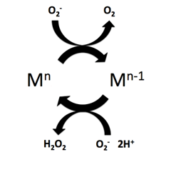
Superoxide dismutase (SOD, EC 1.15.1.1) is an enzyme that alternately catalyzes the dismutation (or partitioning) of the superoxide (O
2
) radical into ordinary molecular oxygen (O2) and hydrogen peroxide (H
2
O
2
). Superoxide is produced as a by-product of oxygen metabolism and, if not regulated, causes many types of cell damage. Hydrogen peroxide is also damaging and is degraded by other enzymes such as catalase. Thus, SOD is an important antioxidant defense in nearly all living cells exposed to oxygen. One exception is Lactobacillus plantarum and related lactobacilli, which use a different mechanism to prevent damage from reactive O
2
.

The free radical theory of aging states that organisms age because cells accumulate free radical damage over time. A free radical is any atom or molecule that has a single unpaired electron in an outer shell. While a few free radicals such as melanin are not chemically reactive, most biologically relevant free radicals are highly reactive. For most biological structures, free radical damage is closely associated with oxidative damage. Antioxidants are reducing agents, and limit oxidative damage to biological structures by passivating them from free radicals.

<span class="mw-page-title-main">Reactive oxygen species</span> Highly reactive molecules formed from diatomic oxygen (O₂)

In chemistry and biology, reactive oxygen species (ROS) are highly reactive chemicals formed from diatomic oxygen (O2), water, and hydrogen peroxide. Some prominent ROS are hydroperoxide (O2H), superoxide (O2-), hydroxyl radical (OH.), and singlet oxygen. ROS are pervasive because they are readily produced from O2, which is abundant. ROS are important in many ways, both beneficial and otherwise. ROS function as signals, that turn on and off biological functions. They are intermediates in the redox behavior of O2, which is central to fuel cells. ROS are central to the photodegradation of organic pollutants in the atmosphere. Most often however, ROS are discussed in a biological context, ranging from their effects on aging and their role in causing dangerous genetic mutations.

<span class="mw-page-title-main">SOD1</span> Protein-coding gene in the species Homo sapiens

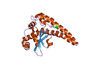
Superoxide dismutase [Cu-Zn] also known as superoxide dismutase 1 or hSod1 is an enzyme that in humans is encoded by the SOD1 gene, located on chromosome 21. SOD1 is one of three human superoxide dismutases. It is implicated in apoptosis, familial amyotrophic lateral sclerosis and Parkinson's disease.

<span class="mw-page-title-main">GPX1</span> Protein-coding gene in the species Homo sapiens

Glutathione peroxidase 1, also known as GPx1, is an enzyme that in humans is encoded by the GPX1 gene on chromosome 3. This gene encodes a member of the glutathione peroxidase family. Glutathione peroxidase functions in the detoxification of hydrogen peroxide, and is one of the most important antioxidant enzymes in humans.

Glisodin is the registered trademark of a nutritional supplement based on two constituents:

<span class="mw-page-title-main">Brain mitochondrial carrier protein 1</span> Protein-coding gene in the species Homo sapiens

Brain mitochondrial carrier protein 1 is a protein that in humans is encoded by the SLC25A14 gene.

<span class="mw-page-title-main">ENDOG</span> Protein-coding gene in the species Homo sapiens

Endonuclease G, mitochondrial is an enzyme that in humans is encoded by the ENDOG gene. This protein primarily participates in caspase-independent apoptosis via DNA degradation when translocating from the mitochondrion to nucleus under oxidative stress. As a result, EndoG has been implicated in cancer, aging, and neurodegenerative diseases such as Parkinson’s disease (PD). Regulation of its expression levels thus holds potential to treat or ameliorate those conditions.

<span class="mw-page-title-main">VDAC2</span> Protein-coding gene in the species Homo sapiens

Voltage-dependent anion-selective channel protein 2 is a protein that in humans is encoded by the VDAC2 gene on chromosome 10. This protein is a voltage-dependent anion channel and shares high structural homology with the other VDAC isoforms. VDACs are generally involved in the regulation of cell metabolism, mitochondrial apoptosis, and spermatogenesis. Additionally, VDAC2 participates in cardiac contractions and pulmonary circulation, which implicate it in cardiopulmonary diseases. VDAC2 also mediates immune response to infectious bursal disease (IBD).

<span class="mw-page-title-main">Rottlerin</span> Chemical compound

Rottlerin (mallotoxin) is a polyphenol natural product isolated from the Asian tree Mallotus philippensis. Rottlerin displays a complex spectrum of pharmacology.

<span class="mw-page-title-main">Mitochondrial ROS</span> Reactive oxygen species produced by mitochondria

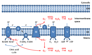
Mitochondrial ROS are reactive oxygen species (ROS) that are produced by mitochondria. Generation of mitochondrial ROS mainly takes place at the electron transport chain located on the inner mitochondrial membrane during the process of oxidative phosphorylation. Leakage of electrons at complex I and complex III from electron transport chains leads to partial reduction of oxygen to form superoxide. Subsequently, superoxide is quickly dismutated to hydrogen peroxide by two dismutases including superoxide dismutase 2 (SOD2) in mitochondrial matrix and superoxide dismutase 1 (SOD1) in mitochondrial intermembrane space. Collectively, both superoxide and hydrogen peroxide generated in this process are considered as mitochondrial ROS.

<span class="mw-page-title-main">ADP/ATP translocase 2</span> Protein-coding gene in humans

ADP/ATP translocase 2 is a protein that in humans is encoded by the SLC25A5 gene on the X chromosome.

Oxidation response is stimulated by a disturbance in the balance between the production of reactive oxygen species and antioxidant responses, known as oxidative stress. Active species of oxygen naturally occur in aerobic cells and have both intracellular and extracellular sources. These species, if not controlled, damage all components of the cell, including proteins, lipids and DNA. Hence cells need to maintain a strong defense against the damage. The following table gives an idea of the antioxidant defense system in bacterial system.

Non-Homologous Isofunctional Enzymes (NISE) are two evolutionarily unrelated enzymes that catalyze the same chemical reaction. Enzymes that catalyze the same reaction are sometimes referred to as analogous as opposed to homologous, however it is more appropriate to name them as Non-homologous Isofunctional Enzymes, hence the acronym (NISE). These enzymes all serve the same end function but do so in different organisms without detectable similarity in primary and possibly tertiary structures.

Diallyl trisulfide (DATS), also known as Allitridin, is an organosulfur compound with the formula S(SCH2CH=CH2)2. It is one of several compounds produced by hydrolysis of allicin, including diallyl disulfide and diallyl tetrasulfide; DATS is one of the most potent.

<span class="mw-page-title-main">Superoxide dismutase mimetics</span> Synthetic compounds

Superoxide dismutase (SOD) mimetics are synthetic compounds that mimic the native superoxide dismutase enzyme. SOD mimetics effectively convert the superoxide anion, a reactive oxygen species, into hydrogen peroxide, which is further converted into water by catalase. Reactive oxygen species are natural byproducts of cellular respiration and cause oxidative stress and cell damage, which has been linked to causing cancers, neurodegeneration, age-related declines in health, and inflammatory diseases. SOD mimetics are a prime interest in therapeutic treatment of oxidative stress because of their smaller size, longer half-life, and similarity in function to the native enzyme.

<span class="mw-page-title-main">Mitochondrial theory of ageing</span> Theory of ageing

The mitochondrial theory of ageing has two varieties: free radical and non-free radical. The first is one of the variants of the free radical theory of ageing. It was formulated by J. Miquel and colleagues in 1980 and was developed in the works of Linnane and coworkers (1989). The second was proposed by A. N. Lobachev in 1978.

Kidney ischemia is a disease with a high morbidity and mortality rate. Blood vessels shrink and undergo apoptosis which results in poor blood flow in the kidneys. More complications happen when failure of the kidney functions result in toxicity in various parts of the body which may cause septic shock, hypovolemia, and a need for surgery. What causes kidney ischemia is not entirely known, but several pathophysiology relating to this disease have been elucidated. Possible causes of kidney ischemia include the activation of IL-17C and hypoxia due to surgery or transplant. Several signs and symptoms include injury to the microvascular endothelium, apoptosis of kidney cells due to overstress in the endoplasmic reticulum, dysfunctions of the mitochondria, autophagy, inflammation of the kidneys, and maladaptive repair.

<span class="mw-page-title-main">Iron superoxide dismutase</span> Enzyme that catalyses reduction of superoxides

Iron superoxide dismutase (FeSOD) is a metalloenzyme that belongs to the superoxide dismutases family of enzymes. Like other superoxide dismutases, it catalyses the dismutation of superoxides into diatomic oxygen and hydrogen peroxide. Found primarily in prokaryotes such as Escherichia coli and present in all strict anaerobes, examples of FeSOD have also been isolated from eukaryotes, such as Vigna unguiculata.

References

  1. 1 2 3 GRCh38: Ensembl release 89: ENSG00000112096 - Ensembl, May 2017
  2. 1 2 3 GRCm38: Ensembl release 89: ENSMUSG00000006818 - Ensembl, May 2017
  3. "Human PubMed Reference:". National Center for Biotechnology Information, U.S. National Library of Medicine.
  4. "Mouse PubMed Reference:". National Center for Biotechnology Information, U.S. National Library of Medicine.
  5. 1 2 3 4 5 6 7 "Entrez Gene: SOD2 superoxide dismutase 2, mitochondrial".
  6. 1 2 3 4 5 6 7 8 9 10 11 12 13 Becuwe P, Ennen M, Klotz R, Barbieux C, Grandemange S (Dec 2014). "Manganese superoxide dismutase in breast cancer: from molecular mechanisms of gene regulation to biological and clinical significance". Free Radical Biology & Medicine. 77: 139–151. doi:10.1016/j.freeradbiomed.2014.08.026. PMID   25224035.
  7. 1 2 3 Pias EK, Ekshyyan OY, Rhoads CA, Fuseler J, Harrison L, Aw TY (Apr 2003). "Differential effects of superoxide dismutase isoform expression on hydroperoxide-induced apoptosis in PC-12 cells". The Journal of Biological Chemistry. 278 (15): 13294–301. doi: 10.1074/jbc.M208670200 . PMID   12551919.
  8. 1 2 Perry JJ, Hearn AS, Cabelli DE, Nick HS, Tainer JA, Silverman DN (Apr 2009). "Contribution of human manganese superoxide dismutase tyrosine 34 to structure and catalysis". Biochemistry. 48 (15): 3417–24. doi:10.1021/bi8023288. PMC   2756076 . PMID   19265433.
  9. 1 2 Azadmanesh J, Lutz WE, Coates L, Weiss KL, Borgstahl GE (April 2021). "Direct detection of coupled proton and electron transfers in human manganese superoxide dismutase". Nature Communications. 12 (1): 2079. Bibcode:2021NatCo..12.2079A. doi: 10.1038/s41467-021-22290-1 . PMC   8024262 . PMID   33824320.
  10. Agback P, Agback T (July 2018). "Direct evidence of a low barrier hydrogen bond in the catalytic triad of a Serine protease". Scientific Reports. 8 (1): 10078. Bibcode:2018NatSR...810078A. doi:10.1038/s41598-018-28441-7. PMC   6031666 . PMID   29973622.
  11. Danial NN, Korsmeyer SJ (January 2004). "Cell death: critical control points". Cell. 116 (2): 205–19. doi: 10.1016/s0092-8674(04)00046-7 . PMID   14744432. S2CID   10764012.
  12. Kerr JF, Wyllie AH, Currie AR (Aug 1972). "Apoptosis: a basic biological phenomenon with wide-ranging implications in tissue kinetics". British Journal of Cancer. 26 (4): 239–57. doi:10.1038/bjc.1972.33. PMC   2008650 . PMID   4561027.
  13. Kang SW (2015). "Superoxide dismutase 2 gene and cancer risk: evidence from an updated meta-analysis". Int J Clin Exp Med. 8 (9): 14647–55. PMC   4658836 . PMID   26628947.
  14. Murray CJ, Lopez AD (May 1997). "Alternative projections of mortality and disability by cause 1990-2020: Global Burden of Disease Study". Lancet. 349 (9064): 1498–504. doi:10.1016/S0140-6736(96)07492-2. PMID   9167458. S2CID   10556268.
  15. Braunwald E, Kloner RA (Nov 1985). "Myocardial reperfusion: a double-edged sword?". The Journal of Clinical Investigation. 76 (5): 1713–9. doi:10.1172/JCI112160. PMC   424191 . PMID   4056048.
  16. Maslov LN, Naryzhnaia NV, Podoksenov IuK, Prokudina ES, Gorbunov AS, Zhang I, Peĭ ZhM (Jan 2015). "[Reactive oxygen species are triggers and mediators of an increase in cardiac tolerance to impact of ischemia-reperfusion]". Rossiĭskii Fiziologicheskiĭ Zhurnal Imeni I.M. Sechenova / Rossiĭskaia Akademiia Nauk. 101 (1): 3–24. PMID   25868322.
  17. Liem DA, Honda HM, Zhang J, Woo D, Ping P (Dec 2007). "Past and present course of cardioprotection against ischemia-reperfusion injury". Journal of Applied Physiology. 103 (6): 2129–36. doi:10.1152/japplphysiol.00383.2007. PMID   17673563. S2CID   24815784.
  18. Muller FL, Lustgarten MS, Jang Y, Richardson A, Van Remmen H (Aug 2007). "Trends in oxidative aging theories". Free Radical Biology & Medicine. 43 (4): 477–503. doi:10.1016/j.freeradbiomed.2007.03.034. PMID   17640558.
  19. Li Y, Huang TT, Carlson EJ, Melov S, Ursell PC, Olson JL, Noble LJ, Yoshimura MP, Berger C, Chan PH, Wallace DC, Epstein CJ (Dec 1995). "Dilated cardiomyopathy and neonatal lethality in mutant mice lacking manganese superoxide dismutase". Nature Genetics. 11 (4): 376–81. doi:10.1038/ng1295-376. PMID   7493016. S2CID   10900822.
  20. 1 2 Van Remmen H, Ikeno Y, Hamilton M, Pahlavani M, Wolf N, Thorpe SR, Alderson NL, Baynes JW, Epstein CJ, Huang TT, Nelson J, Strong R, Richardson A (Dec 2003). "Life-long reduction in MnSOD activity results in increased DNA damage and higher incidence of cancer but does not accelerate aging". Physiological Genomics. 16 (1): 29–37. doi:10.1152/physiolgenomics.00122.2003. PMID   14679299.
  21. 1 2 Curtis C, Landis GN, Folk D, Wehr NB, Hoe N, Waskar M, Abdueva D, Skvortsov D, Ford D, Luu A, Badrinath A, Levine RL, Bradley TJ, Tavaré S, Tower J (2007). "Transcriptional profiling of MnSOD-mediated lifespan extension in Drosophila reveals a species-general network of aging and metabolic genes". Genome Biology. 8 (12): R262. doi: 10.1186/gb-2007-8-12-r262 . PMC   2246264 . PMID   18067683.
  22. Sun J, Folk D, Bradley TJ, Tower J (June 2002). "Induced overexpression of mitochondrial Mn-superoxide dismutase extends the life span of adult Drosophila melanogaster". Genetics. 161 (2): 661–72. doi:10.1093/genetics/161.2.661. PMC   1462135 . PMID   12072463.
  23. Muid KA, Karakaya HÇ, Koc A (February 2014). "Absence of superoxide dismutase activity causes nuclear DNA fragmentation during the aging process". Biochem. Biophys. Res. Commun. 444 (2): 260–3. doi:10.1016/j.bbrc.2014.01.056. hdl: 11147/5542 . PMID   24462872.
  24. Ogata T, Senoo T, Kawano S, Ikeda S (January 2016). "Mitochondrial superoxide dismutase deficiency accelerates chronological aging in the fission yeast Schizosaccharomyces pombe". Cell Biol. Int. 40 (1): 100–6. doi: 10.1002/cbin.10556 . PMID   26507459. S2CID   205563521.
  25. Piazza N, Hayes M, Martin I, Duttaroy A, Grotewiel M, Wessells R (2009). "Multiple measures of functionality exhibit progressive decline in a parallel, stochastic fashion in Drosophila Sod2 null mutants". Biogerontology. 10 (5): 637–48. doi:10.1007/s10522-008-9210-2. PMC   2800787 . PMID   19148770.
  26. Van Raamsdonk JM, Hekimi S (February 2009). "Deletion of the mitochondrial superoxide dismutase sod-2 extends lifespan in Caenorhabditis elegans". PLOS Genetics. 5 (2): e1000361. doi: 10.1371/journal.pgen.1000361 . PMC   2628729 . PMID   19197346.
  27. Yamashita N, Hoshida S, Otsu K, Asahi M, Kuzuya T, Hori M (1999). "Exercise provides direct biphasic cardioprotection via manganese superoxide dismutase activation". The Journal of Experimental Medicine. 189 (11): 1699–706. doi:10.1084/jem.189.11.1699. PMC   2193084 . PMID   10359573.
  28. Hamilton KL, Quindry JC, French JP, Staib J, Hughes J, Mehta JL, Powers SK (2004). "MnSOD antisense treatment and exercise-induced protection against arrhythmias". Free Radical Biology & Medicine. 37 (9): 1360–8. doi:10.1016/j.freeradbiomed.2004.07.025. PMID   15454275.
  29. Velarde MC, Flynn JM, Day NU, Melov S, Campisi J (January 2012). "Mitochondrial oxidative stress caused by Sod2 deficiency promotes cellular senescence and aging phenotypes in the skin". Aging (Albany NY). 4 (1): 3–12. doi:10.18632/aging.100423. PMC   3292901 . PMID   22278880.
  30. Velarde MC, Demaria M, Melov S, Campisi J (August 2015). "Pleiotropic age-dependent effects of mitochondrial dysfunction on epidermal stem cells". Proc. Natl. Acad. Sci. U.S.A. 112 (33): 10407–12. Bibcode:2015PNAS..11210407V. doi: 10.1073/pnas.1505675112 . PMC   4547253 . PMID   26240345.
  31. Treiber N, Maity P, Singh K, Kohn M, Keist AF, Ferchiu F, Sante L, Frese S, Bloch W, Kreppel F, Kochanek S, Sindrilaru A, Iben S, Högel J, Ohnmacht M, Claes LE, Ignatius A, Chung JH, Lee MJ, Kamenisch Y, Berneburg M, Nikolaus T, Braunstein K, Sperfeld AD, Ludolph AC, Briviba K, Wlaschek M, Florin L, Angel P, Scharffetter-Kochanek K (April 2011). "Accelerated aging phenotype in mice with conditional deficiency for mitochondrial superoxide dismutase in the connective tissue". Aging Cell. 10 (2): 239–54. doi:10.1111/j.1474-9726.2010.00658.x. PMID   21108731. S2CID   46458295.
  32. Hu D, Cao P, Thiels E, Chu CT, Wu GY, Oury TD, Klann E (March 2007). "Hippocampal long-term potentiation, memory, and longevity in mice that overexpress mitochondrial superoxide dismutase". Neurobiol Learn Mem. 87 (3): 372–84. doi:10.1016/j.nlm.2006.10.003. PMC   1847321 . PMID   17129739.
  33. Woollard SM, Bhargavan B, Yu F, Kanmogne GD (Jun 2014). "Differential effects of Tat proteins derived from HIV-1 subtypes B and recombinant CRF02_AG on human brain microvascular endothelial cells: implications for blood-brain barrier dysfunction". Journal of Cerebral Blood Flow and Metabolism. 34 (6): 1047–59. doi:10.1038/jcbfm.2014.54. PMC   4050250 . PMID   24667918.

Further reading