Schistosoma bovis

Last updated

Schistosoma bovis
Scientific classification OOjs UI icon edit-ltr.svg
Domain: Eukaryota
Kingdom: Animalia
Phylum: Platyhelminthes
Class: Trematoda
Order: Diplostomida
Family: Schistosomatidae
Genus: Schistosoma
Species:
S. bovis
Binomial name
Schistosoma bovis
Sonsino, 1876

Schistosoma bovis is a two-host blood fluke, that causes intestinal schistosomiasis in ruminants in North Africa, Mediterranean Europe and the Middle East. S. bovis is mostly transmitted by Bulinus freshwater snail species. It is one of nine haematobium group species and exists in the same geographical areas as Schistosoma haematobium , with which it can hybridise. S. bovis-haematobium hybrids can infect humans, and have been reported in Senegal since 2009, and a 2013 outbreak in Corsica.

Contents

Taxonomy and identification

Schistosoma bovis is a digenetic, two-host blood fluke. It was discovered by Italian parasitologist Prospero Sonsino at Zagazig meat market in Egypt in 1876 from a bull. [1] It is generally similar to other schistosomes, but Sonsino knew that it was larger and its eggs were different from those of the human species (first and only known schistosome at the time), Schistosoma haematobium, discovered by a German physician Theodor Bilharz in 1852, [2] [3] then known as Bilharzia haematobium or Distomum haematobium. Sonsino gave the name Bilharzia bovis [4] following the genus classification introduced by another German physician Heinrich Meckel von Hemsbach in 1856 for the species described by Bilharz. [5] [6] The eggs of S. bovis are not only larger than those of other schistosomes, but also have thicker spine with an elongated shape in the form a spindle. [7] [8] The eggs are therefore the best and simplest identification key from other species. [9] [10]

Sonsino later revised the name as Bilharzia crassa in 1877, and then as Gynaecophorus crassa in 1992. Though the genus name remained confusing, Louis-Joseph Alcide Railliet (in 1893) and Raphaël Blanchard (in 1895) revived and maintained the species name, bovis. [4] The valid genus name Schistosoma was accepted by the International Commission on Zoological Nomenclature (ICZN) in 1954, [11] following the name created by David Friedrich Weinland in 1858. [12] Thus, the original Bilharzia bovis became Schistosoma bovis. [4]

Hybrids between S. bovis and the human schistosome, Schistosoma haematobium were first described in 2009 in Northern Senegalese children, and to a lesser degree hybrids between S. bovis and cattle schistosome S. curassoni found only in cattle. [13] S. bovis-haematobium hybrids were also found during the 2013 outbreak traced to the Cavu river on Corsica. [14] As a hybrid increases the host range of the parent species, they affect transmission, and as is known from other schistosome hybrid pairings, morbidity and drug susceptibility, so they are epidemiologically important. [13]

Life cycle

Schistosoma life cycle Schistosoma life cycle.svg
Schistosoma life cycle

Schistosoma bovis infects two hosts, namely ruminants (cattle, goats, sheep, horses and camels) and freshwater snails ( Bulinus sp. and Planorbarius sp.). [15] :392 Experimental infections have been proven in Planorbarius metidjensis snails, which are native to Northwestern Africa and the Iberian peninsula.[ citation needed ]

In water, its free swimming infective larval cercariae can burrow into the skin of its definite host, the ruminant, upon contact. The cercariae enter the host's blood stream, and travel to the liver to mature into adult flukes. Adult flukes can coat themselves with host antigen thus avoiding detection by the host immune system. After a period of about three weeks the young flukes migrate to the mesenteric veins of the gut to copulate. The female fluke lays eggs, which migrate into the lumen of the gut and leave the host upon defecation. In fresh water, the eggs hatch, forming free swimming miracidia. [16] [17] [18]

Miracidia penetrate into the intermediate host, the freshwater snails [19] of the Bulinus spp., (e.g. B. globosus , B. forskalii , B. nyassanus and B. truncatus ), except in Spain, [15] :20 Portugal and Morocco, where Planorbarius metidjensis can transmit. [20] Inside the snail, the miracidium sheds its epithelium, and develops into a mother sporocyst. After two weeks the mother begins forming daughter sporocysts. One month – or more with cooler ambient temperatures – after a miracidium has penetrated into the snail, hundreds to thousands of cercariae of the same sex begin to be released through special areas of the sporocyst wall. [15] :30 The cercariae cycle from the top of the water to the bottom in search of a host. They can enter the host epithelium within minutes. [21] [15] :34

Geographical distribution

S. bovis infects snails in Africa north of the equator, Europe (Sardinia, Corsica, Spain) and the Middle East as far as Iraq. [15] :20S. bovis-haematobium hybrids have been reported first in Senegal in the early 1990s, [22] and then an outbreak in 2013 in Corsica. [23]

Diagnosis

Laboratory

The diagnosis of schistosomiasis can be made by microscopically examining the feces for the egg. The S. bovis egg is terminally spiked, spindle shaped, and the largest in size compared to other Schistosoma eggs at 202 μm length and 72μm width. [15] :396 In chronic infections, or if eggs are difficult to find, an intradermal injection of Schistosome antigen to form a wheal can determine infection. Alternatively diagnosis can be made by complement fixation tests. [19]

As of 2012 commercial serological tests have included ELISA and an indirect immunofluorescence test, hampered by a low sensitivity ranging from 21% to 71%. [24] Exposure to any Schistosoma eggs or cercariae can cause false positive serological test results for individual Schistosoma species, unless highly specific antigens are used. [15] :402

Various polymerase chain reaction (PCR) assays to differentiate S. bovis from other schistosomes in urine and naturally infected snails for surveillance purposes have been described since 2010. [25]

Pathology

The ova are initially deposited in the muscularis propria of the gut which leads to ulceration of the overlaying tissue. Infections are characterized by pronounced acute inflammation, blood and reactive epithelial changes. Granulomas and multinucleated giant cells may be seen.[ citation needed ]

Immunopathology

The immune system responds to eggs in the liver causing hypersensitivity; an immune response is necessary to prevent damage to hepatocytes. The hosts' antibodies bind to the tegument of the schistosome but not for long since the tegument is shed every few hours. The schistosome can also take on host proteins. Schistomiasis can be divided into three phases; Within the haematobium group S. bovis and S. curassoni appear to be closely related: (1) the migratory phase lasting from penetration to maturity, (2) the acute phase which occurs when the schistosomes begin producing eggs, and (3) the chronic phase which occurs mainly in endemic areas. [19]

Gene expression

S. bovis is one of the few non-bacterial species with a known moonlighting protein. Glyceraldehyde-3-phosphate dehydrogenase (GAPD) is also a common MP in bacteria. [26]

Treatment

Historically, antimonials and trichlorphon were tested against visceral schistosome infection in cattle. [27] :245–248,265–273 Antimony affects phosphofructokinase activity in Schistosoma, hycanthone intercalates Schistosoma DNA and the organophosphorus metabolite dichlorvos inhibits acetylcholinesterase, "but progressively less so in S. bovis". [15] :44

Since the 1980s the drug of choice is praziquantel, a quinolone derivative which disrupts membranes, leading to calcium influx. It clears eggs from stool, and affects adult but not immature worms. [15] :44–45 Damaged and dying flukes can be trapped in the liver and cause fatal portal vein thrombosis. [27]

Disease prevention

The main cause of schistosomiasis is the dumping of human and animal waste into water supplies. Hygienic disposal of waste would be sufficient to eliminate the disease. [19]

Related Research Articles

<span class="mw-page-title-main">Schistosomiasis</span> Human disease caused by parasitic worms called schistosomes

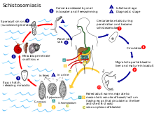
Schistosomiasis, also known as snail fever, bilharzia, and Katayama fever, is a disease caused by parasitic flatworms called schistosomes. The urinary tract or the intestines may be infected. Symptoms include abdominal pain, diarrhea, bloody stool, or blood in the urine. Those who have been infected for a long time may experience liver damage, kidney failure, infertility, or bladder cancer. In children, it may cause poor growth and learning difficulty.

<span class="mw-page-title-main">Trematoda</span> Class of parasitic flatworms

Trematoda is a class of flatworms known as flukes or trematodes. They are obligate internal parasites with a complex life cycle requiring at least two hosts. The intermediate host, in which asexual reproduction occurs, is usually a snail. The definitive host, where the flukes sexually reproduce, is a vertebrate. Infection by trematodes can cause disease in all five traditional vertebrate classes: mammals, birds, amphibians, reptiles, and fish.

<i>Schistosoma</i> Genus of flukes

Schistosoma is a genus of trematodes, commonly known as blood flukes. They are parasitic flatworms responsible for a highly significant group of infections in humans termed schistosomiasis, which is considered by the World Health Organization as the second-most socioeconomically devastating parasitic disease, with hundreds of millions infected worldwide.

Schistosoma japonicum is an important parasite and one of the major infectious agents of schistosomiasis. This parasite has a very wide host range, infecting at least 31 species of wild mammals, including nine carnivores, 16 rodents, one primate (human), two insectivores and three artiodactyls and therefore it can be considered a true zoonosis. Travelers should be well-aware of where this parasite might be a problem and how to prevent the infection. S. japonicum occurs in the Far East, such as China, the Philippines, Indonesia and Southeast Asia.

<i>Schistosoma mansoni</i> Species of fluke

Schistosoma mansoni is a water-borne parasite of humans, and belongs to the group of blood flukes (Schistosoma). The adult lives in the blood vessels near the human intestine. It causes intestinal schistosomiasis. Clinical symptoms are caused by the eggs. As the leading cause of schistosomiasis in the world, it is the most prevalent parasite in humans. It is classified as a neglected tropical disease. As of 2021, the World Health Organization reports that 251.4 million people have schistosomiasis and most of it is due to S. mansoni. It is found in Africa, the Middle East, the Caribbean, Brazil, Venezuela and Suriname.

<i>Schistosoma intercalatum</i> Species of fluke

Schistosoma intercalatum is a parasitic worm found in parts of western and central Africa. There are two strains: the Lower Guinea strain and the Zaire strain. S. intercalatum is one of the major agents of the rectal form of schistosomiasis, also called bilharzia. It is a trematode, and being part of the genus Schistosoma, it is commonly referred to as a blood-fluke since the adult resides in blood vessels.

<i>Schistosoma haematobium</i> Species of fluke

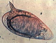
Schistosoma haematobium is a species of digenetic trematode, belonging to a group (genus) of blood flukes (Schistosoma). It is found in Africa and the Middle East. It is the major agent of schistosomiasis, the most prevalent parasitic infection in humans. It is the only blood fluke that infects the urinary tract, causing urinary schistosomiasis, and is the leading cause of bladder cancer. The diseases are caused by the eggs.

<span class="mw-page-title-main">Theodor Bilharz</span> German physician

Theodor Maximilian Bilharz was a German physician who made pioneering discoveries in the field of parasitology. His contributions led to the foundation of tropical medicine. He is best remembered as the discoverer of the blood fluke Schistosoma haematobium, the causative parasite of bloody urine (haematuria) known since ancient times in Egypt. The parasite, as the cause of bladder cancer, is declared by the International Agency for Research on Cancer as Group 1 carcinogen. The infection is known by an eponymous term bilharzia or bilharziasis, as well as by schistosomiasis.

<i>Trematocranus placodon</i> Species of fish

Trematocranus placodon is a species of cichlid fish endemic to Lake Malawi, Lake Malombe and the upper reaches of the Shire River in Africa. It is mainly a shallow-water species that prefers to occupy areas with patches of Vallisneria, but it can occur as deep as 31 m (102 ft). It can reach a total length of up to 25 cm (9.8 in).

Schistosoma indicum is a species of digenetic trematode in the family Schistosomatidae. The parasite is widespread in domestic animals in India and other Asian countries.

<span class="mw-page-title-main">Schistosomiasis vaccine</span>

A Schistosomiasis vaccine is a vaccine against Schistosomiasis, a parasitic disease caused by several species of fluke of the genus Schistosoma. No effective vaccine for the disease exists yet. Schistosomiasis affects over 200 million people worldwide, mainly in rural agricultural and peri-urban areas of the third world, and approximately 10% suffer severe health complications from the infection. While chemotherapeutic drugs, such as praziquantel, oxamniquine and metrifonate both no longer on the market, are currently considered safe and effective for the treatment of schistosomiasis, reinfection occurs frequently following drug treatment, thus a vaccine is sought to provide long-term treatment. Additionally, experimental vaccination efforts have been successful in animal models of schistosomiasis.

Schistosoma mekongi is a species of trematodes, also known as flukes. It is one of the five major schistosomes that account for all human infections, the other four being S. haematobium, S. mansoni, S. japonicum, and S. intercalatum. This trematode causes schistosomiasis in humans.

<i>Indoplanorbis</i> Genus of gastropods

Indoplanorbis is a genus of air-breathing freshwater snail. Its only member species is Indoplanorbis exustus, an aquatic pulmonate gastropod mollusk in the family Planorbidae, the ram's horn snails. The species is widely distributed across the tropics. It serves as an important intermediate host for several trematode parasites. The invasive nature and ecological tolerance of Indoplanorbis exustus add to its importance in veterinary and medical science.

<i>Schistosoma spindale</i> Species of fluke

Schistosoma spindale is a species of digenetic trematode in the family Schistosomatidae. It causes intestinal schistosomiasis in the ruminants.

<i>Bulinus forskalii</i> Species of gastropod

Bulinus forskalii is a species of tropical freshwater snail with a sinistral shell, an aquatic gastropod mollusk in the family Bulinidae, the ramshorn snails and their allies.

Bulinus nasutus is a species of tropical freshwater snail with a sinistral shell, an aquatic gastropod mollusk in the family Planorbidae, the ramshorn snails and their allies.

<span class="mw-page-title-main">Bivitellobilharzia nairi</span> Species of fluke

Bivitellobilharzia nairi is a species of trematodes, part of the family Schistosomatidae. This is a fairly new identified endoparasite that was found in 1945 by Mudaliar and Ramanujachari, who first recorded the parasite in India. Researchers collected fecal samples of the Indian rhinoceros and were startled to find B. nairi eggs.

<span class="mw-page-title-main">Cavu</span> River in Corsica, France

The Cavu or rivière de Cavu, is a short river in the Corse-du-Sud department of Corsica which discharges into the Tyrrhenian Sea, and the Mediterranean Sea. In 2014 the Cavu became the first place of re-emerging schistosomiasis in Europe. As of 2016 120 people have become infected after bathing in it.

Carcinogenic parasites are parasitic organisms that depend on other organisms for their survival, and cause cancer in such hosts. Three species of flukes (trematodes) are medically-proven carcinogenic parasites, namely the urinary blood fluke, the Southeast Asian liver fluke and the Chinese liver fluke. S. haematobium is prevalent in Africa and the Middle East, and is the leading cause of bladder cancer. O. viverrini and C. sinensis are both found in eastern and southeastern Asia, and are responsible for cholangiocarcinoma. The International Agency for Research on Cancer declared them in 2009 as a Group 1 biological carcinogens in humans.

<i>Planorbarius metidjensis</i> Species of gastropod

Planorbarius metidjensis is a freshwater lung snail.

References

  1. Pitchford RJ (1965). "Differences in the egg morphology and certain biological characteristics of some African and Middle Eastern schistosomes, genus Schistosoma, with terminal-spined eggs". Bulletin of the World Health Organization. 32 (1): 105–120. PMC   2555201 . PMID   14292062.
  2. Southgate V, Knowles R (1975). "Observations on Schistosoma bovis Sonsino, 1876". Journal of Natural History. 9 (3): 273–314. Bibcode:1975JNatH...9..273S. doi:10.1080/00222937500770201. ISSN   0022-2933.
  3. Khalil M (1924). "On the Morphology of Schistosoma bovis". Journal of Helminthology. 2 (2): 81–86. doi:10.1017/S0022149X00003059. ISSN   1475-2697. S2CID   85079589.
  4. 1 2 3 Styles CW (1896). "The inspection of meats for animal parasites". Bulletin of the U.S. Department of Agriculture. Vol. 10. U.S. Government Printing Office. p. 60.
  5. Meckel von Hemsbach H (2018) [1856]. Mikrogeologie: Ueber die Concremente im thierischen Organismus (Reprint 2018 ed.). Berlin: De Gruyter. p. 114. ISBN   978-3-11-171606-0. OCLC   1029825577.
  6. Lalchhandama K (2017). "The making of oncology: Helminthology as the cornerstone". Science Vision. 17 (2): 78–93. doi: 10.33493/scivis.17.02.04 . S2CID   86495376.
  7. Schwetz J (1956). "Some new Comparative Investigations on Three Physopsis Borne Schistosomes: Schistosoma Haematobium, S. Bovis and S. Intercalatum 1". The American Journal of Tropical Medicine and Hygiene. 5 (6): 1071–1085. doi:10.4269/ajtmh.1956.5.1071. ISSN   0002-9637. PMID   13381883.
  8. Touassem R (1987). "Egg polymorphism of Schistosoma bovis". Veterinary Parasitology. 23 (3–4): 185–191. doi:10.1016/0304-4017(87)90004-5. PMID   3564347.
  9. Alves W (1949). "The Eggs of Schistosoma bovis , S. mattheei and S. haematobium". Journal of Helminthology. 23 (3–4): 127–134. doi:10.1017/S0022149X00032466. PMID   15402061. S2CID   41238161.
  10. Soentjens P, Cnops L, Huyse T, Yansouni C, De Vos D, Bottieau E, Clerinx J, Van Esbroeck M (2016). "Diagnosis and Clinical Management of Schistosoma haematobium – Schistosoma bovis Hybrid Infection in a Cluster of Travelers Returning From Mali". Clinical Infectious Diseases. 63 (12): 1626–1629. doi: 10.1093/cid/ciw493 . ISSN   1058-4838. PMID   27941144.
  11. Hemming F (1954). Opinions and Declarations Rendered by the International Commission on Zoological Nomenclature (Volume 4 Part 16). Vol. 4. London: International Trust for Zoological Nomenclature. pp. 177–200.
  12. Weinland DF (1858). Human Cestoides: An Essay on the Tapeworms of Man. Cambridge (UK): Metcalf and Company. p. 87.
  13. 1 2 Huyse T, Webster BL, Geldof S, Stothard JR, Diaw OT, Polman K, Rollinson D (2009). "Bidirectional introgressive hybridization between a cattle and human schistosome species". PLOS Pathogens. 5 (9): e1000571. doi: 10.1371/journal.ppat.1000571 . PMC   2731855 . PMID   19730700.
  14. Jérôme Boissier, Sébastien Grech-Angelini, Bonnie L Webster, et al. (2016). "Outbreak of urogenital schistosomiasis in Corsica (France): an epidemiological case study" (PDF). The Lancet Infectious Diseases. 16 (8): 971–979. doi:10.1016/S1473-3099(16)00175-4. PMID   27197551. S2CID   3725312.
  15. 1 2 3 4 5 6 7 8 9 Adel A. F. Mahmoud (2001). Schistosomiasis. Imperial College Press. p. 524. ISBN   978-1-86094-146-7 . Retrieved 31 July 2016.
  16. Agnew AM, Murare HM, Lucas SB, Doenhoff MJ (1989). "Schistosoma bovis as an immunological analogue of S. haematobium". Parasite Immunology. 11 (4): 329–340. doi:10.1111/j.1365-3024.1989.tb00671.x. ISSN   0141-9838. PMID   2506507. S2CID   45120768.
  17. Bushara HO, Omer OH, Malik KH, Taylor MG (1994). "The effect of multiple transfers of immune serum on maturingSchistosoma bovis infections in calves". Parasitology Research. 80 (3): 198–202. doi:10.1007/BF00932674. ISSN   1432-1955. PMID   8036232. S2CID   21345815.
  18. de la Torre-Escudero E, Pérez-Sánchez R, Manzano-Román R, Oleaga A (2017). "Schistosoma bovis -host interplay: Proteomics for knowing and acting". Molecular and Biochemical Parasitology. 215: 30–39. doi:10.1016/j.molbiopara.2016.07.009. PMID   27485556.
  19. 1 2 3 4 Black, J. (2005). Microbiology: Principles and Explorations. Wiley, New York.
  20. Mouahid A, Théron A (Dec 1987). "Schistosoma bovis: variability of cercarial production as related to the snail hosts: Bulinus truncatus, B. wright and Planorbarius metidjensis". Int J Parasitol. 17 (8): 1431–4. doi:10.1016/0020-7519(87)90078-6. PMID   3440697.
  21. Roberts, L., Janovy, J. (1996). Foundations of Parasitology (5th ed.). Wm. C. Brown Publishers, Dubuque, Iowa.
  22. Webster BL, Diaw OT, Seye MM, Webster JP, Rollinson D (2013). "Introgressive hybridization of Schistosoma haematobium group species in Senegal: species barrier break down between ruminant and human schistosomes". PLOS Neglected Tropical Diseases. 7 (4): e2110. doi: 10.1371/journal.pntd.0002110 . ISSN   1935-2735. PMC   3617179 . PMID   23593513.
  23. Boissier J, Grech-Angelini S, Webster BL, Allienne JF, Huyse T, Mas-Coma S, Toulza E, Barré-Cardi H, Rollinson D, Kincaid-Smith J, Oleaga A (2016). "Outbreak of urogenital schistosomiasis in Corsica (France): an epidemiological case study". The Lancet. Infectious Diseases. 16 (8): 971–979. doi: 10.1016/S1473-3099(16)00175-4 . ISSN   1474-4457. PMID   27197551.
  24. Kinkel HF, Dittrich S, Bäumer B, Weitzel T (2012). "Evaluation of eight serological tests for diagnosis of imported schistosomiasis". Clin Vaccine Immunol. 19 (6): 948–53. doi:10.1128/CVI.05680-11. PMC   3370443 . PMID   22441394.
  25. Akinwale OP, Hock TT, Chia-Kwung F, Zheng Q, Haimo S, Ezeh C, Gyang PV (2014). "Differentiating Schistosoma haematobium from Schistosoma magrebowiei and other closely related schistosomes by polymerase chain reaction amplification of a species specific mitochondrial gene". Tropical Parasitology. 4 (1): 38–42. doi: 10.4103/2229-5070.129163 . PMC   3992801 . PMID   24754026.
  26. Henderson B, Martin A (2011). "Bacterial Moonlighting Proteins and Bacterial Virulence". In Dobrindt U, Hacker JH, Svanborg C (eds.). Between Pathogenicity and Commensalism. Current Topics in Microbiology and Immunology. Vol. 358. Berlin, Heidelberg: Springer Berlin Heidelberg. pp. 155–213. doi:10.1007/82_2011_188. ISBN   978-3-642-36559-1. ISSN   0070-217X. PMID   22143554.
  27. 1 2 Reinecke, RK (1983). "Phylum plathelminthes". Veterinary helminthology. Durban, SouthAfrica: Butterworths.