
The medulla oblongata or simply medulla is a long stem-like structure which makes up the lower part of the brainstem. It is anterior and partially inferior to the cerebellum. It is a cone-shaped neuronal mass responsible for autonomic (involuntary) functions, ranging from vomiting to sneezing. The medulla contains the cardiac, respiratory, vomiting and vasomotor centers, and therefore deals with the autonomic functions of breathing, heart rate and blood pressure as well as the sleep–wake cycle.

The pons is part of the brainstem that in humans and other mammals, lies inferior to the midbrain, superior to the medulla oblongata and anterior to the cerebellum.
The brainstem is the stalk-like part of the brain that interconnects the cerebrum and diencephalon with the spinal cord. In the human brain, the brainstem is composed of the midbrain, the pons, and the medulla oblongata. The midbrain is continuous with the thalamus of the diencephalon through the tentorial notch.

In neuroanatomy, the trigeminal nerve (lit. triplet nerve), also known as the fifth cranial nerve, cranial nerve V, or simply CN V, is a cranial nerve responsible for sensation in the face and motor functions such as biting and chewing; it is the most complex of the cranial nerves. Its name (trigeminal, from Latin tri- 'three', and -geminus 'twin') derives from each of the two nerves (one on each side of the pons) having three major branches: the ophthalmic nerve (V1), the maxillary nerve (V2), and the mandibular nerve (V3). The ophthalmic and maxillary nerves are purely sensory, whereas the mandibular nerve supplies motor as well as sensory (or "cutaneous") functions. Adding to the complexity of this nerve is that autonomic nerve fibers as well as special sensory fibers (taste) are contained within it.

In neuroanatomy, a neural pathway is the connection formed by axons that project from neurons to make synapses onto neurons in another location, to enable neurotransmission. Neurons are connected by a single axon, or by a bundle of axons known as a nerve tract, or fasciculus. Shorter neural pathways are found within grey matter in the brain, whereas longer projections, made up of myelinated axons, constitute white matter.

The pyramidal tracts include both the corticobulbar tract and the corticospinal tract. These are aggregations of efferent nerve fibers from the upper motor neurons that travel from the cerebral cortex and terminate either in the brainstem (corticobulbar) or spinal cord (corticospinal) and are involved in the control of motor functions of the body.

The spinothalamic tract is a part of the anterolateral system or the ventrolateral system, a sensory pathway to the thalamus. From the ventral posterolateral nucleus in the thalamus, sensory information is relayed upward to the somatosensory cortex of the postcentral gyrus.

The dorsal column–medial lemniscus pathway (DCML) is a sensory pathway of the central nervous system that conveys sensations of fine touch, vibration, two-point discrimination, and proprioception from the skin and joints. It transmits information from the body to the primary somatosensory cortex in the postcentral gyrus of the parietal lobe of the brain. The pathway receives information from sensory receptors throughout the body, and carries this in nerve tracts in the white matter of the dorsal column of the spinal cord to the medulla, where it is continued in the medial lemniscus, on to the thalamus and relayed from there through the internal capsule and transmitted to the somatosensory cortex. The name dorsal-column medial lemniscus comes from the two structures that carry the sensory information: the dorsal columns of the spinal cord, and the medial lemniscus in the brainstem.

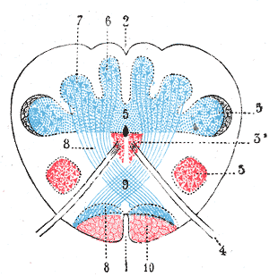
In neuroanatomy, the medial lemniscus, also known as Reil's band or Reil's ribbon, is a large ascending bundle of heavily myelinated axons that decussate (cross) in the brainstem, specifically in the medulla oblongata. The medial lemniscus is formed by the crossings of the internal arcuate fibers. The internal arcuate fibers are composed of axons of nucleus gracilis and nucleus cuneatus. The axons of the nucleus gracilis and nucleus cuneatus in the medial lemniscus have cell bodies that lie contralaterally.

The inferior olivary nucleus (ION) is a structure found in the medulla oblongata underneath the superior olivary nucleus. In vertebrates, the ION is known to coordinate signals from the spinal cord to the cerebellum to regulate motor coordination and learning. These connections have been shown to be tightly associated, as degeneration of either the cerebellum or the ION results in degeneration of the other.
Stereognosis is the ability to perceive and recognize the form of an object in the absence of visual and auditory information, by using tactile information to provide cues from texture, size, spatial properties, and temperature, etc. In humans, this sense, along with tactile spatial acuity, vibration perception, texture discrimination and proprioception, is mediated by the dorsal column-medial lemniscus pathway of the central nervous system. Stereognosis tests determine whether or not the parietal lobe of the brain is intact. Typically, these tests involved having the patient identify common objects placed in their hand without any visual cues. Stereognosis is a higher cerebral associative cortical function.

The spinocerebellar tract is a nerve tract originating in the spinal cord and terminating in the same side (ipsilateral) of the cerebellum.

The upper part of the posterior district of the medulla oblongata is occupied by the inferior cerebellar peduncle, a thick rope-like strand situated between the lower part of the fourth ventricle and the roots of the glossopharyngeal and vagus nerves.

Cerebellar peduncles connect the cerebellum to the brain stem. There are six cerebellar peduncles in total, three on each side:

The posterior thoracic nucleus, is a group of interneurons found in the medial part of lamina VII, also known as the intermediate zone, of the spinal cord. It is mainly located from the cervical vertebra C7 to lumbar L3–L4 levels and is an important structure for proprioception of the lower limb.
The lateral corticospinal tract is the largest part of the corticospinal tract. It extends throughout the entire length of the spinal cord, and on transverse section appears as an oval area in front of the posterior column and medial to the posterior spinocerebellar tract.

In neuroanatomy, the internal arcuate fibers or internal arcuate tract are the axons of second-order sensory neurons that compose the gracile and cuneate nuclei of the medulla oblongata. These second-order neurons begin in the gracile and cuneate nuclei in the medulla. They receive input from first-order sensory neurons, which provide sensation to many areas of the body and have cell bodies in the dorsal root ganglia of the dorsal root of the spinal nerves. Upon decussation from one side of the medulla to the other, also known as the sensory decussation, they are then called the medial lemniscus.
In neuroanatomy, the dorsal column nuclei are a pair of nuclei in the dorsal columns in the brainstem. The name refers collectively to the cuneate nucleus and gracile nucleus, which are situated at the lower end of the medulla oblongata. Both nuclei contain second-order neurons of the dorsal column–medial lemniscus pathway, which convey fine touch and proprioceptive information from the body to the brain. The dorsal column nuclei project to the thalamus.

The spinal cord is a long, thin, tubular structure made up of nervous tissue that extends from the medulla oblongata in the brainstem to the lumbar region of the vertebral column (backbone) of vertebrate animals. The center of the spinal cord is hollow and contains a structure called central canal, which contains cerebrospinal fluid. The spinal cord is also covered by meninges and enclosed by the neural arches. Together, the brain and spinal cord make up the central nervous system.
