
The inferior vena cava is a large vein that carries the deoxygenated blood from the lower and middle body into the right atrium of the heart. It is formed by the joining of the right and the left common iliac veins, usually at the level of the fifth lumbar vertebra.

The rectus abdominis muscle, also known as the "abdominal muscle" or simply the "abs", is a pair of segmented skeletal muscle on the ventral aspect of a person's abdomen. The paired muscle is separated at the midline by a band of dense connective tissue called the linea alba, and the connective tissue defining each lateral margin of the rectus abdominus is the linea semilunaris. The muscle extends from the pubic symphysis, pubic crest and pubic tubercle inferiorly, to the xiphoid process and costal cartilages of the 5th–7th ribs superiorly.

The celiacartery, also known as the celiac trunk or truncus coeliacus, is the first major branch of the abdominal aorta. It is about 1.25 cm in length. Branching from the aorta at thoracic vertebra 12 (T12) in humans, it is one of three anterior/ midline branches of the abdominal aorta.

In human anatomy, the internal thoracic artery (ITA), also known as the internal mammary artery, is an artery that supplies the anterior chest wall and the breasts. It is a paired artery, with one running along each side of the sternum, to continue after its bifurcation as the superior epigastric and musculophrenic arteries.
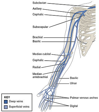
The ulnar veins are venae comitantes of the ulnar artery. They drain the superficial venous palmar arch. They arise in the hand and terminate by uniting with the radial veins to form the brachial veins. They mostly drain the medial aspect of the forearm. They receive the venae comitantes of the anterior and posterior interosseous arteries near the elbow, as well as a large branch from the median cubital vein. The ulnar veins are larger than the radial veins.

In human anatomy, the inguinal triangle is a region of the abdominal wall. It is also known by the eponym Hesselbach's triangle, after Franz Kaspar Hesselbach.

In human anatomy, the inferior epigastric artery is an artery that arises from the external iliac artery. It is accompanied by the inferior epigastric vein; inferiorly, these two inferior epigastric vessels together travel within the lateral umbilical fold The inferior epigastric artery then traverses the arcuate line of rectus sheath to enter the rectus sheath, then anastomoses with the superior epigastric artery within the rectus sheath.

In human anatomy, inferior epigastric vein are 1-2 veins accompanying the inferior epigastric artery. They drain into the external iliac vein just proximal to the inguinal ligament.
In human anatomy, the superior epigastric artery is a terminal branch of the internal thoracic artery that provides arterial supply to the abdominal wall, and upper rectus abdominis muscle. It enters the rectus sheath to descend upon the inner surface of the rectus abdominis muscle. It ends by anastomosing with the inferior epigastric artery.

In human anatomy, the internal thoracic vein is the vein that drains the chest wall and breasts.

The inferior ophthalmic vein is a vein of the orbit that - together with the superior ophthalmic vein - represents the principal drainage system of the orbit. It begins from a venous network in the front of the orbit, then passes backwards through the lower orbit. It drains several structures of the orbit. It may end by splitting into two branches, one draining into the pterygoid venous plexus and the other ultimately into the cavernous sinus.
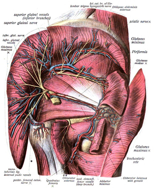
The inferior gluteal veins are venae comitantes of the inferior gluteal artery. They commence in the superior/proximal posterior thigh. They enter the pelvis through the lower part of the greater sciatic foramen. They converge to form a single vessel before emptying into the distal portion of the internal iliac vein.

The lateral circumflex femoral artery is an artery in the upper thigh. It is usually a branch of the profunda femoris artery, and produces three branches. It is mostly distributed to the muscles of the lateral thigh, supplying arterial blood to muscles of the knee extensor group.

The lateral umbilical fold is an elevation of the peritoneum lining the inner/posterior surface of the lower anterior abdominal wall formed by the underlying inferior epigastric artery and inferior epigastric vein which the peritoneum covers. Superiorly, the lateral umbilical fold ends where the vessels reach and enter the rectus sheath at the arcuate line of rectus sheath; in spite of the name, the lateral umbilical folds do not extend as far superiorly as the umbilicus. Inferiorly, it extends to just medial to the deep inguinal ring.

The arcuate line of rectus sheath is a line of demarcation corresponding to the free inferior margin of the posterior layer of the rectus sheath inferior to which only the anterior layer of the rectus sheath is present and the rectus abdominis muscle is therefore in direct contact with the transversalis fascia. The arcuate line is concave inferior-wards.

The deep circumflex iliac artery is an artery in the pelvis that travels along the iliac crest of the pelvic bone.

The lumbar veins are four pairs of veins running along the inside of the posterior abdominal wall, and drain venous blood from parts of the abdominal wall. Each lumbar vein accompanies a single lumbar artery. The lower two pairs of lumbar veins all drain directly into the inferior vena cava, whereas the fate of the upper two pairs is more variable.

The following outline is provided as an overview of and topical guide to human anatomy:
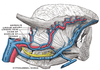
The dorsal lingual veins are some of the lingual veins. They provide venous drainage to the dorsum of the tongue, and the sides of the tongue. Between the hyoglossus and genioglossus, dorsal lingual veins unite with those lingual veins that are venae comitantes of the lingual artery; these consolidated lingual veins then empty into the internal jugular vein proximal to the greater cornu of hyoid bone.
Free-flap breast reconstruction is a type of autologous-tissue breast reconstruction applied after mastectomy for breast cancer, without the emplacement of a breast implant prosthesis. As a type of plastic surgery, the free-flap procedure for breast reconstruction employs tissues, harvested from another part of the woman's body, to create a vascularised flap, which is equipped with its own blood vessels. Breast-reconstruction mammoplasty can sometimes be realised with the application of a pedicled flap of tissue that has been harvested from the latissimus dorsi muscle, which is the broadest muscle of the back, to which the pedicle (“foot”) of the tissue flap remains attached until it successfully grafts to the recipient site, the mastectomy wound. Moreover, if the volume of breast-tissue excised was of relatively small mass, breast augmentation procedures, such as autologous-fat grafting, also can be applied to reconstruct the breast lost to mastectomy.
