Tom Brown (engineer)

Last updated

Tom Brown
Tom Brown, engineer.jpg
Tom Brown. Inventor of medical ultrasound. Taken in 1960's.
Born(1933-04-10)10 April 1933
Died(2019-12-13)13 December 2019
NationalityScottish, British
Known forDeveloping obstetric ultrasound
Scientific career
FieldsIndustrial design, Medical Ultrasound
InstitutionsKelvin & Hughes Ltd, University of Glasgow, Honeywell

Thomas Graham Brown (10 April 1933 in Glasgow – 13 December 2019) was a Scottish engineer who was most notable for collaborating in the design of the first medical ultrasound machine along with the obstetrician and designer Ian Donald, a physician at the University of Glasgow and industrial designer and obstetrician John MacVicar. [1] [2]

Contents

Life

In 1944, Brown enrolled at Allan Glen's School in Glasgow. [3] [4] In April 1951, after completing school and making an exploratory visit to the company to meet the chief engineer, he joined Kelvin & Hughes Ltd at the time a Glasgow manufacturer of scientific instruments as a technical apprentice. [1] Two years into his five year apprenticeship, he started working for Alex Rankin and to specialise in non-destructive testing. [4]

Career

In 1956 Brown was promoted to research and development engineer at Kelvin & Hughes Ltd. [1]

Western Infirmary

It was in late 1956 when Brown first met Ian Donald. Brown, although relatively young at twenty-three, had previously worked on an automatic flaw detector for testing of industrial products. [5] It was while working in the Western Infirmary installing a bulb in an operating theatre that Brown found out that Donald was experimenting with the flaw detector. [6] Brown immediately looked up Donald in the Infirmary directory, phoned him and arranged a meeting. [6] When they met, Brown noticed that the Mark IIb was not manufactured by Kelvin & Hughes but instead had been manufactured under contract. He also noticed that the machine had been converted from using a double probe, one to produce pulses and one to receive the pulses, to a single probe. [6] Not wanting to insult Donald by explaining why the machine was not working correctly, Brown offered to try and source another machine from somewhere. [6] Brown phoned Alex Rankin, the man who collaborated with Brown on the automatic flaw detector, for help. [6] Rankin offered to gift the latest Mk IV Flaw Detector which was subsequently forwarded to Glasgow Central station from the Barkingside Labs location of Kelvin & Hughes, for delivery to Brown. [6]

B-mode scanner

Brown approached deputy chairman Bill Slater who sent Brown to see Bill Halliday, the company's chief research scientist for an opinion on building the machine. [7] After Brown delivered his spiel to Halliday it was several months before Brown received a reply in the form of a memo which stated that £500 had been allocated by Smiths for the development and that Brown was able to spend half a day per week working with Donald. [5]

The new B-mode scanner was also known by the name Bed-Table Scanner and was built out of an amalgamation of medical and industrial parts. Brown managed to scrounge an older Mark IV flaw detector in Glasgow along with a 6-inch electrostatically-deflected Cathode-ray tube taken from the company stores in Glasgow. From the companies Barkingside R&D department, Brown found a experimental weld-testing machine. Both these machines were cannibalised for parts. To measure the position of the transducer, Brown selected an 'X-Y' orthogonal measuring frame system. This was measured in place by a sine/cosine potentiometer that was used to calculate the position of the transducer from the angle of its rotations. This was an exceedingly expensive piece of electronic equipment and was more than their £500 budget. However, Brown managed to scrounge a damaged component and repair it. The machine was built on top an old hospital bed and made extensive use of Meccano chains and sprockets. [5] By late 1957 the first contact B-mode scanner was constructed and in clinical use by that year. [5]

The design was patented by Kelvin & Hughes in 1957 with Brown being named the inventor with commercial rights assigned to the company. [4] In a landmark paper in June 1958, published by Donald, McVicar and Brown in The Lancet, they discussed the development of the A-mode scanner and decisions that led up to the B-mode scanner. [8] Although the images described in the paper were very crude, they were the first successful application of obstetric ultrasound.

In 1961 Kelvin Hughes merged with Smiths Industries. [5] In 1963, Brown became director of the medical ultrasonics department in Glasgow after Alex Rankin died. [4] In 1964 the Glasgow operation of Kelvin Hughes was the subject of a takeover bid by the aviation division of Smiths Industries. [4] with the factory at Hillington being eventually closed in 1966, when Smiths pulled out of Scotland. [5] The design the group created was gradually evolved by them before it was transferred to Smith Industrials of England where it was improved by Brown, to become a commercial product known as the Diasonograph. [4]

Later career

In 1965, Brown was appointed to a post of chief engineer at Honeywell with a move to Hemel Hempstead. [4] At Honeywell he worked on the design of open-heart surgery and coronary care machines, as well as prefabricated operating theatres. [4] In 1967, Brown left Honeywell to work at Nuclear Enterprises in Edinburgh, the business that bought the medical ultrasound unit from Kelvin & Hughes in 1966. [4] As Nuclear Enterprises did not buy the patent rights for the ultrasound machine designs, they instead went to a firm in the United States. [9] So to get around his own patents, Brown decided to develop a 3-D ultrasound machine and to formally study the problem. In 1970 Brown became a research fellow to study medical physics and three-dimensional imaging at the University of Edinburgh. [10] In 1973 Brown was appointed as a team leader on the development of multiplanar 3D scanners at Sonicaid in Livingston, West Lothian. [1] [4] Brown developed a contact scanner that could produce three-dimensional stereoscopic virtual image of body tissue. [10] The new machine known as the Multiplanar Scanner was finally developed by 1976 and shown at an American Institute of Ultrasound in Medicine meeting in the same year and put into production in 1977. [4] However, sales to UK and overseas hospitals were poor and the machine was finally withdrawn in 1979 [4] and the Sonicaid project in Livingston closed. [10] Brown's foresight in the design of the Multiplanar Scanner machine was admirable and it was a step in the right direction but at the time computing resources were meagre, being insufficient to achieve the desired results. [4]

Brown was unable to find any further work in the medical instrumentation industry and decided to move back to working in the oil and gas industry where he worked until 1998. [10] After he retired in 1999, he worked part time as a quality manager at the radiological protection centre in St George's Hospital in Tooting, London. [1] [4] In 2002, he moved back to Scotland to finally retire. [1] In 2005, Brown founded a small firm, NoStrain to recognise and help Sonographers who suffered from musculoskeletal disorders. [1] He died on 13 December 2019. [11]

Marriage

In 1958, Brown married Geira née Stevens and had three daughters and six grandchildren. [11]

Awards and honours

In 1982, Brown along with Donald were elected as the first honorary life members of the British Medical Ultrasound Society. [12] In 1996 he was awarded the Ian Donald Gold medal Award for Technical Merit. [13] In 2007, Brown was awarded an honorary fellowship ad eundem of the Royal College of Obstetricians and Gynaecologists. [14] In 2014 Brown was inducted into the Scottish Engineering Hall of Fame [1] and awarded an honorary fellowship of the Institution of Engineers and Shipbuilders in Scotland. [15]

Related Research Articles

<span class="mw-page-title-main">Medical ultrasound</span> Diagnostic and therapeutic technique

Medical ultrasound includes diagnostic techniques using ultrasound, as well as therapeutic applications of ultrasound. In diagnosis, it is used to create an image of internal body structures such as tendons, muscles, joints, blood vessels, and internal organs, to measure some characteristics or to generate an informative audible sound. The usage of ultrasound to produce visual images for medicine is called medical ultrasonography or simply sonography, or echography. The practice of examining pregnant women using ultrasound is called obstetric ultrasonography, and was an early development of clinical ultrasonography. The machine used is called an ultrasound machine, a sonograph or an echograph. The visual image formed using this technique is called an ultrasonogram, a sonogram or an echogram.

<span class="mw-page-title-main">Medical imaging</span> Technique and process of creating visual representations of the interior of a body

Medical imaging is the technique and process of imaging the interior of a body for clinical analysis and medical intervention, as well as visual representation of the function of some organs or tissues (physiology). Medical imaging seeks to reveal internal structures hidden by the skin and bones, as well as to diagnose and treat disease. Medical imaging also establishes a database of normal anatomy and physiology to make it possible to identify abnormalities. Although imaging of removed organs and tissues can be performed for medical reasons, such procedures are usually considered part of pathology instead of medical imaging.

<span class="mw-page-title-main">Govan</span> Area of Glasgow, Scotland

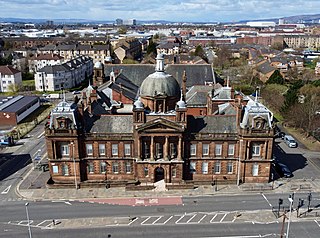
Govan is a district, parish, and former burgh now part of south-west Glasgow, Scotland. It is situated 2.5 miles (4.0 km) west of Glasgow city centre, on the south bank of the River Clyde, opposite the mouth of the River Kelvin and the district of Partick. Historically it was part of the County of Lanark.

<span class="mw-page-title-main">Obstetric ultrasonography</span> Use of medical ultrasonography in pregnancy

Obstetric ultrasonography, or prenatal ultrasound, is the use of medical ultrasonography in pregnancy, in which sound waves are used to create real-time visual images of the developing embryo or fetus in the uterus (womb). The procedure is a standard part of prenatal care in many countries, as it can provide a variety of information about the health of the mother, the timing and progress of the pregnancy, and the health and development of the embryo or fetus.

Sonicaid Ltd was a medical electronics company headquartered in West Sussex best known for its range of Doppler fetal monitors. The company also developed early ultrasound scanners. The word "Sonicaid" is in generic use for Doppler fetal monitors. Sonicaid is now a registered trademark of Huntleigh Healthcare.

In obstetrics, gestational age is a measure of the age of a pregnancy taken from the beginning of the woman's last menstrual period (LMP), or the corresponding age of the gestation as estimated by a more accurate method, if available. Such methods include adding 14 days to a known duration since fertilization, or by obstetric ultrasonography. The popularity of using this measure of pregnancy is largely due to convenience: menstruation is usually noticed, while there is generally no convenient way to discern when fertilization or implantation occurred.

<span class="mw-page-title-main">Ian Donald</span>

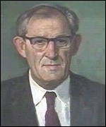
Ian Donald was an English physician who pioneered the diagnostic use of ultrasound in obstetrics, enabling the visual discovery of abnormalities during pregnancy. Donald was born in Cornwall, England, to a Scottish family of physicians. He was educated in Scotland and South Africa before studying medicine at the University of London in 1930, and became the third generation of doctors in his family. At the start of World War II, Donald was drafted into the Royal Air Force as a medical officer, where he developed an interest in radar and sonar. In 1952, at St Thomas' Hospital, he used what he learned in the RAF to build a respirator for newborn babies with respiratory problems.

<span class="mw-page-title-main">University of Glasgow Medical School</span> Medical school of the University of Glasgow, Scotland

<span class="mw-page-title-main">Dugald Baird</span> British medical professor

Sir Dugald Baird FRCOG was a British medical doctor and a professor of obstetrics and gynaecology. Baird was most notable and influential in calling for the liberalising of abortion. In his delivery of the Sandoz lecture in November 1961, titled the Fifth Freedom, he advocated for freedom from the tyranny of fertility.

<span class="mw-page-title-main">Allan Glen's School</span> Secondary school in Scotland

Allan Glen's School was, for most of its existence, a local authority, selective secondary school for boys in Glasgow, Scotland, charging nominal fees for tuition.

<span class="mw-page-title-main">Kypros Nicolaides</span> Greek-Cypriot fetal medicine specialist

Kyprianos "Kypros" Nicolaides is a Greek Cypriot physician of British citizenship, Professor of Fetal Medicine at King's College Hospital, London. He is one of the pioneers of fetal medicine and his discoveries have revolutionised the field. He was elected to the US National Academy of Medicine in 2020 for 'improving the care of pregnant women worldwide with pioneering rigorous and creative approaches, and making seminal contributions to prenatal diagnosis and every major obstetrical disorder'. This is considered to be one of the highest honours in the fields of health and medicine and recognises individuals who have demonstrated outstanding professional achievement and commitment to service.

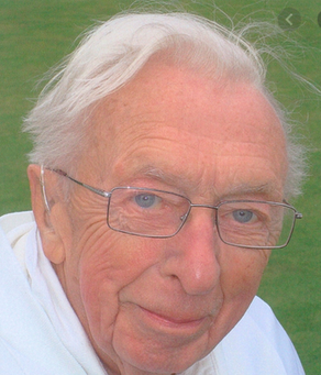
Stuart Campbell DSc FRCPEd FRCOG FACOG, was born in Glasgow, Scotland, and graduated from the medical school of Glasgow University. During his training he worked with Ian Donald, who had published some of the first papers on the use of ultrasound in obstetrics.

<span class="mw-page-title-main">Riverside Museum</span> Transport museum in Glasgow, Scotland.

The Riverside Museum is a museum in Glasgow, designed by Zaha Hadid Architects, housed in a building at Pointhouse Quay in the Glasgow Harbour regeneration district of Glasgow, Scotland. The building opened in June 2011, winning the 2013 European Museum of the Year Award. It houses many exhibits of national and international importance. The Govan-Partick Bridge will provide a pedestrian link from the museum across the Clyde to Govan. It is set to be completed in 2024.

<span class="mw-page-title-main">Institution of Engineers and Shipbuilders in Scotland</span> Scottish professional body

The Institution of Engineers and Shipbuilders in Scotland (IESIS) is a multi-disciplinary professional body and learned society, founded in Scotland, for professional engineers in all disciplines and for those associated with or taking an interest in their work. Its main activities are an annual series of evening talks on engineering, open to all, and a range of school events aimed at encouraging young people to consider engineering careers.

The Scottish Engineering Hall of Fame honours "those engineers from, or closely associated with, Scotland who have achieved, or deserve to achieve, greatness", as selected by an independent panel representing Scottish engineering institutions, academies, museums and archiving organisations.

<span class="mw-page-title-main">Dugald Cameron</span>

Dugald Cameron, OBE, FCSD, FRSA is a Scottish artist and industrial designer.

Professor Donald Macleod FRCSEd, FFAEM (Hon), FFSEM (Hon), FISM was a Scottish former rugby union player and a former President of the Scottish Rugby Union. A retired surgeon, he was the Scotland national rugby union team doctor for many years.

<span class="mw-page-title-main">Charles Richard Whitfield</span> Northern Irish obstetrician and gynaecologist

Charles Richard Whitfield FRCOG, FRCP(G) was a Northern Irish obstetrician and gynaecologist who was a pioneer of maternal-fetal (perinatal) medicine. His primary interest was in fetal medicine, a branch of obstetrics and gynaecology that focuses on the assessment of the development, growth and health of the baby in the womb. He was also an early proponent of subspecialisation within the fields of obstetrics and gynaecology, a practice that is common today.

<span class="mw-page-title-main">John MacVicar</span> British physician

John MacVicar was a British physician who was most notable for pioneering the diagnostic use of ultrasound in obstetrics as well as later, being a clinical educator. MacVicar was part of a team along with physician Ian Donald and engineer Tom Brown, who developed the worlds first obstetric ultrasound machine in 1963. Using the new technique of ultrasound, MacVicar's research transformed the treatment of gynaecological conditions in pregnant women, through the use of clinical trials.

References

  1. 1 2 3 4 5 6 7 8 "Thomas Graham Brown". The Scottish Engineering Hall of Fame. The Institution of Engineers and Shipbuilders in Scotland. Retrieved 26 August 2019.
  2. "Imaging the fetus – the history of obstetric ultrasound". University of Glasgow. Glasgow University News. 26 April 2013. Retrieved 27 August 2019.
  3. McArdle, Helen (21 December 2019). "Scotland's ultrasound pioneer Tom Brown dies". Herald & Times Group. Glasgow Herald. Retrieved 18 December 2020.
  4. 1 2 3 4 5 6 7 8 9 10 11 12 13 14 Adrian M. K. Thomas; Arpan K. Banerjee; Uwe Busch (5 December 2005). Classic Papers in Modern Diagnostic Radiology. Springer Science & Business Media. p. 214. ISBN   978-3-540-26988-5 . Retrieved 27 August 2019.
  5. 1 2 3 4 5 6 Nicholson, Deborah (2003). Secrets of success:the development of obstetric ultrasound in Scotland, 1963-1990 (PDF) (Phd Thesis). University of Glasgow. Retrieved 18 July 2019.
  6. 1 2 3 4 5 6 Brown, Tom (Unpublished article) Development of ultrasonic scanning techniques in Scotland 1956-1979, 1994
  7. "Looking at the Unborn: Historical Aspects of Obstetric Ultrasound" (PDF). The History of Modern Biomedicine Research Group. Wellcome Institute for the History of Medicine: Wellcome Trust. 10 March 1998. p. 11,38. Retrieved 6 August 2019.
  8. Donald, Ian; Macvicar, J; Brown, T.G (June 1958). "Investigation of Abdominal Masses by Pulsed Ultrasound". The Lancet. 271 (7032): 1188–1195. doi:10.1016/S0140-6736(58)91905-6. PMID   13550965.
  9. "Looking at the Unborn: Historical Aspects of Obstetric Ultrasound" (PDF). The History of Modern Biomedicine Research Group. Wellcome Institute for the History of Medicine: Wellcome Trust. 10 March 1998. p. 32. Retrieved 26 October 2019.
  10. 1 2 3 4 "Looking at the Unborn: Historical Aspects of Obstetric Ultrasound" (PDF). The History of Modern Biomedicine Research Group. Wellcome Institute for the History of Medicine: Wellcome Trust. 10 March 1998. p. 5. Retrieved 26 October 2019.
  11. 1 2 "Obituary: Thomas Graham Brown, engineer who was a pioneer in the invention and development of ultrasound imaging". JPIMedia Ltd. The Scotsman. 20 December 2019. Retrieved 20 December 2019.
  12. Tansey, E M; Christie, D A, eds. (10 March 1998). Looking at the Unborn - Historical Aspects of Obstetric Ultrasound (PDF). Vol. 55. London: Wellcome Institute for the History of Medicine. p. 5. Retrieved 27 August 2019.
  13. "Ian Donald Award for Technical Merit". International Society of Ultrasound in Obstetrics and Gynecology. Awards. Retrieved 28 September 2019.
  14. "Fellows ad eundem of the RCOG". Royal College of Obstetricians and Gynaecologists. 2007. Retrieved 27 August 2019.
  15. "Honorary Members and Fellows". The Institution of Engineers & Shipbuilders. Retrieved 27 August 2019.