Trabeculae of spleen

Last updated
Trabeculae of spleen
Gray1189.png
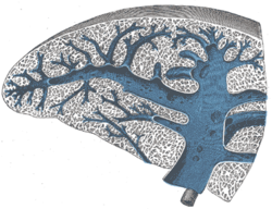
Transverse section of the spleen, showing the trabecular tissue and the splenic vein and its tributaries
Details
Identifiers
Latin trabeculae splenicae
FMA 16027
Anatomical terminology

The fibroelastic coat of the spleen invests the organ, and at the hilum is reflected inward upon the vessels in the form of sheaths. From these sheaths, as well as from the inner surface of the fibroelastic coat, numerous small fibrous bands, the trabeculae of the spleen (or splenic trabeculae), emerge from all directions; these uniting, constitute the frame-work of the spleen.

The spleen therefore consists of a number of small spaces or areolae, formed by the trabeculae; in these areolae is contained the splenic pulp.

See also

Related Research Articles

<span class="mw-page-title-main">Spleen</span> Organ recycling old red blood cells and also housing lymphocytes

The spleen is an organ found in almost all vertebrates. Similar in structure to a large lymph node, it acts primarily as a blood filter. The word spleen comes from Ancient Greek σπλήν (splḗn).

<span class="mw-page-title-main">Portal vein</span> Short thick vein formed by the union of the superior mesenteric vein and the splenic vein

The portal vein or hepatic portal vein (HPV) is a blood vessel that carries blood from the gastrointestinal tract, gallbladder, pancreas and spleen to the liver. This blood contains nutrients and toxins extracted from digested contents. Approximately 75% of total liver blood flow is through the portal vein, with the remainder coming from the hepatic artery proper. The blood leaves the liver to the heart in the hepatic veins.

<span class="mw-page-title-main">Splenic artery</span> Artery which supplies blood to the spleen

In human anatomy, the splenic artery or lienal artery, an older term, is the blood vessel that supplies oxygenated blood to the spleen. It branches from the celiac artery, and follows a course superior to the pancreas. It is known for its tortuous path to the spleen.

<span class="mw-page-title-main">Splenic vein</span> Vein that drains blood from the spleen, stomach and pancreas

In human anatomy, the splenic vein is a blood vessel that drains blood from the spleen, the stomach fundus and part of the pancreas. It is part of the hepatic portal system.

<span class="mw-page-title-main">Reticular fiber</span> Type of connective tissue in animals

Reticular fibers, reticular fibres or reticulin is a type of fiber in connective tissue composed of type III collagen secreted by reticular cells. They are mainly composed reticulin protein and form a network or mesh. Reticular fibers crosslink to form a fine meshwork (reticulin). This network acts as a supporting mesh in soft tissues such as liver, bone marrow, and the tissues and organs of the lymphatic system.

<span class="mw-page-title-main">Splenic injury</span> Injury to the spleen

A splenic injury, which includes a ruptured spleen, is any injury to the spleen. The rupture of a normal spleen can be caused by trauma, such as a traffic collision.

<span class="mw-page-title-main">Left gastroepiploic artery</span>

The left gastroepiploic artery, the largest branch of the splenic artery, runs from left to right about a finger's breadth or more from the greater curvature of the stomach, between the layers of the greater omentum, and anastomoses with the right gastroepiploic.

<span class="mw-page-title-main">Colic flexures</span> Two abrupt bends in the large intestine

In the anatomy of the human digestive tract, there are two colic flexures, or curvatures in the transverse colon. The right colic flexure is also known as the hepatic flexure, and the left colic flexure is also known as the splenic flexure. Note that "right" refers to the patient's anatomical right, which may be depicted on the left of a diagram.

<span class="mw-page-title-main">Transverse colon</span> Longest section of the large intestine

In human anatomy, the transverse colon is the longest and most movable part of the colon.

<span class="mw-page-title-main">Greater omentum</span> Fat sheath under abdominal wall

The greater omentum is a large apron-like fold of visceral peritoneum that hangs down from the stomach. It extends from the greater curvature of the stomach, passing in front of the small intestines and doubles back to ascend to the transverse colon before reaching to the posterior abdominal wall. The greater omentum is larger than the lesser omentum, which hangs down from the liver to the lesser curvature. The common anatomical term "epiploic" derives from "epiploon", from the Greek epipleein, meaning to float or sail on, since the greater omentum appears to float on the surface of the intestines. It is the first structure observed when the abdominal cavity is opened anteriorly.

<span class="mw-page-title-main">White pulp</span> Type of tissue in the spleen

White pulp is a histological designation for regions of the spleen, that encompasses approximately 25% of splenic tissue. White pulp consists entirely of lymphoid tissue.

<span class="mw-page-title-main">Red pulp</span> Type of tissue in the spleen

The red pulp of the spleen is composed of connective tissue known also as the cords of Billroth and many splenic sinusoids that are engorged with blood, giving it a red color. Its primary function is to filter the blood of antigens, microorganisms, and defective or worn-out red blood cells.

<span class="mw-page-title-main">Splenic plexus</span>

The splenic plexus is formed by branches from the celiac plexus, the left celiac ganglion, and from the right vagus nerve.

<span class="mw-page-title-main">Tunica albuginea (penis)</span> Anatomical structure of the penis

The tunica albuginea is the fibrous envelope that extends the length of the corpora cavernosa penis and corpus spongiosum penis. It is a bi-layered structure that includes an outer longitudinal layer and an inner circular layer.

The fibrous envelope of the corpus cavernosum urethrae is thinner, whiter in color, and more elastic than that of the corpora cavernosa penis. It is called the trabeculae of corpus spongiosum of penis.

<span class="mw-page-title-main">Accessory spleen</span> Small nodule found apart from the main body of the spleen

An accessory spleen is a small nodule of splenic tissue found apart from the main body of the spleen. Accessory spleens are found in approximately 10 percent of the population and are typically around 1 centimetre in diameter. They may resemble a lymph node or a small spleen. They form either by the result of developmental anomalies or trauma. They are medically significant in that they may result in interpretation errors in diagnostic imaging or continued symptoms after therapeutic splenectomy. Polysplenia is the presence of multiple accessory spleens rather than one normal spleen.

<span class="mw-page-title-main">Trabecular arteries</span>

The trabecular arteries are the name of the branches of the splenic artery after it passes into the trabeculae of the spleen, where it branches. When these arteries then reach the white pulp, and become covered with periarteriolar lymphoid sheaths, the name changes again to central arteries. Branches of the central arteries are given to the red pulp, and these are called penicillar arteries).

<span class="mw-page-title-main">Trabecular veins</span> Veins inside the spleen

The trabecular veins are the largest veins inside the spleen. They drain the blood collected in the sinuses of the pulp.

Schweigger-Seidel sheath is a phagocytic sleeve that is part of a sheathed arteriole of the spleen, and is sometimes referred to as a splenic ellipsoid. It is a spindle-shaped thickening in the walls of the second part of the arterial branches forming the penicilli in the spleen. It is named after German physiologist Franz Schweigger-Seidel (1834-1871).

<span class="mw-page-title-main">Splenic lymph node</span>

The splenic lymph nodes are found at the splenic hilum and in relation to the tail of the pancreas.

References

PD-icon.svgThis article incorporates text in the public domain from page 1283 of the 20th edition of Gray's Anatomy (1918)