Lingual tonsils

Last updated
Lingual tonsils
Illu04 tongue.jpg
Tongue
Details
System Immune system (lymphatic system)
Identifiers
Latin tonsilla lingualis
TA98 A05.1.04.022
TA2 2830
FMA 54836
Anatomical terminology

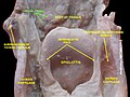
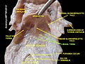
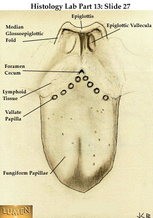
The lingual tonsils are a collection of lymphatic tissue located in the lamina propria of the root of the tongue. [1] This lymphatic tissue consists of the lymphatic nodules rich in cells of the immune system (immunocytes). [2] The immunocytes initiate the immune response when the lingual tonsils get in contact with invading microorganisms (pathogenic bacteria, viruses or parasites). [2] [3] [4]

Contents

Structure

Microanatomy

Lingual tonsils are covered externally by stratified squamous epithelium (nonkeratinized) that invaginates inward forming tonsillar crypts. Beneath the epithelium is a layer of lymphoid nodules containing lymphocytes. Mucous glands located at the root of the tongue are drained through several ducts into the crypts of the lingual tonsils. [2] [3] Secretions of these mucous glands keep the crypts clean and free of any debris.

Blood supply

Lingual tonsils are located on posterior aspect of tongue which is supplied through: [1]

Nerve supply

Lingual tonsils are innervated by tonsillar nerves from the tonsilar plexus, formed by the glossopharyngeal and vagus nerves. [1]

Function

Tonsils and the throat-lingual tonsils labelled Blausen 0861 Tonsils&Throat Anatomy2.png
Tonsils and the throat–lingual tonsils labelled

Like other lymphatic tissues, the function of lingual tonsils is to prevent infections. These tonsils contain B and T lymphocytes which get activated when harmful bacteria and viruses come in contact with tonsils. B lymphocytes kill pathogens by producing antibodies against them, while T lymphocytes directly kill them releasing cytotoxic substances or indirectly by stimulating other cells of the immune system. [2] [3] [4]

Clinical significance

Cancer

Squamous cell carcinoma is a type of neoplasm that can affect lingual tonsils. [5]

Sleep apnea

Enlarged or hypertrophic lingual tonsils have the potential to cause or exacerbate sleep apnea. [6]

Additional images

Related Research Articles

<span class="mw-page-title-main">Lymphatic system</span> Organ system in vertebrates complementary to the circulatory system

The lymphatic system, or lymphoid system, is an organ system in vertebrates that is part of the immune system, and complementary to the circulatory system. It consists of a large network of lymphatic vessels, lymph nodes, lymphoid organs, lymphatic tissue and lymph. Lymph is a clear fluid carried by the lymphatic vessels back to the heart for re-circulation. The Latin word for lymph, lympha, refers to the deity of fresh water, "Lympha".

<span class="mw-page-title-main">Lymph node</span> Organ of the lymphatic system

A lymph node, or lymph gland, is a kidney-shaped organ of the lymphatic system and the adaptive immune system. A large number of lymph nodes are linked throughout the body by the lymphatic vessels. They are major sites of lymphocytes that include B and T cells. Lymph nodes are important for the proper functioning of the immune system, acting as filters for foreign particles including cancer cells, but have no detoxification function.

<span class="mw-page-title-main">Ileum</span> Final section of the small intestine

The ileum is the final section of the small intestine in most higher vertebrates, including mammals, reptiles, and birds. In fish, the divisions of the small intestine are not as clear and the terms posterior intestine or distal intestine may be used instead of ileum. Its main function is to absorb vitamin B12, bile salts, and whatever products of digestion that were not absorbed by the jejunum.

<span class="mw-page-title-main">Epithelium</span> Tissue lining the surfaces of organs in animals

Epithelium or epithelial tissue is a thin, continuous, protective layer of compactly packed cells with little extracellular matrix. Epithelial tissues line the outer surfaces of organs and blood vessels throughout the body, as well as the inner surfaces of cavities in many internal organs. An example is the epidermis, the outermost layer of the skin. Epithelial tissue is one of the four basic types of animal tissue, along with connective tissue, muscle tissue and nervous tissue. These tissues also lack blood or lymph supply. The tissue is supplied by nerves.

<span class="mw-page-title-main">Palatine tonsil</span> Lymphoid organs at the back of the throat on both sides

Palatine tonsils, commonly called the tonsils and occasionally called the faucial tonsils, are tonsils located on the left and right sides at the back of the throat, which can often be seen as flesh-colored, pinkish lumps. Tonsils only present as "white lumps" if they are inflamed or infected with symptoms of exudates and severe swelling.

<span class="mw-page-title-main">Lamina propria</span> Thin connective layer forming part of the mucous membranes

The lamina propria is a thin layer of connective tissue that forms part of the moist linings known as mucous membranes or mucosae, which line various tubes in the body, such as the respiratory tract, the gastrointestinal tract, and the urogenital tract.

<span class="mw-page-title-main">Adenoid</span> Type of tonsil

In anatomy, the pharyngeal tonsil, also known as the nasopharyngeal tonsil or adenoid, is the superior-most of the tonsils. It is a mass of lymphatic tissue located behind the nasal cavity, in the roof of the nasopharynx, where the nose blends into the throat. In children, it normally forms a soft mound in the roof and back wall of the nasopharynx, just above and behind the uvula.

<span class="mw-page-title-main">Submandibular gland</span> Human salivary gland

The paired submandibular glands are major salivary glands located beneath the floor of the mouth. In adult humans, they each weigh about 15 grams and contribute some 60–67% of unstimulated saliva secretion; on stimulation their contribution decreases in proportion as parotid gland secretion rises to 50%. The average length of the normal adult human submandibular salivary gland is approximately 27 mm, while the average width is approximately 14.3 mm.

Gut-associated lymphoid tissue (GALT) is a component of the mucosa-associated lymphoid tissue (MALT) which works in the immune system to protect the body from invasion in the gut.

The mucosa-associated lymphoid tissue (MALT), also called mucosa-associated lymphatic tissue, is a diffuse system of small concentrations of lymphoid tissue found in various submucosal membrane sites of the body, such as the gastrointestinal tract, nasopharynx, thyroid, breast, lung, salivary glands, eye, and skin. MALT is populated by lymphocytes such as T cells and B cells, as well as plasma cells, dendritic cells and macrophages, each of which is well situated to encounter antigens passing through the mucosal epithelium. In the case of intestinal MALT, M cells are also present, which sample antigen from the lumen and deliver it to the lymphoid tissue. MALT constitute about 50% of the lymphoid tissue in human body. Immune responses that occur at mucous membranes are studied by mucosal immunology.

<span class="mw-page-title-main">Keratosis pharyngis</span> Medical condition

Keratosis Pharyngis is a medical condition where keratin grows on the surface of the pharynx, that is the part of the throat at the back of the mouth. Keratin is a protein that normally occurs as the main component of hair and nails. It is characterized by the presence of whitish-yellow dots on the pharyngeal wall, tonsils or lingual tonsils. They are firmly adherent and cannot be wiped off. The surrounding region does not show any sign or inflammation or any other symptoms that make affect the rest of the body.

<span class="mw-page-title-main">Head and neck anatomy</span>

This article describes the anatomy of the head and neck of the human body, including the brain, bones, muscles, blood vessels, nerves, glands, nose, mouth, teeth, tongue, and throat.

<span class="mw-page-title-main">Waldeyer's tonsillar ring</span> A ringed arrangement of lymphoid tissue in the pharynx

Waldeyer's tonsillar ring is a ringed arrangement of lymphoid organs in the pharynx. Waldeyer's ring surrounds the naso- and oropharynx, with some of its tonsillar tissue located above and some below the soft palate.

<span class="mw-page-title-main">Tonsil</span> Lymphoid organs in the mouth and throat

The tonsils are a set of lymphoid organs facing into the aerodigestive tract, which is known as Waldeyer's tonsillar ring and consists of the adenoid tonsil, two tubal tonsils, two palatine tonsils, and the lingual tonsils. These organs play an important role in the immune system.

<span class="mw-page-title-main">Mantle zone</span>

The mantle zone of a lymphatic nodule is an outer ring of small lymphocytes surrounding a germinal center.

<span class="mw-page-title-main">Pharynx</span> Part of the throat that is behind the mouth and nasal cavity

The pharynx is the part of the throat behind the mouth and nasal cavity, and above the esophagus and trachea. It is found in vertebrates and invertebrates, though its structure varies across species. The pharynx carries food to the esophagus and air to the larynx. The flap of cartilage called the epiglottis stops food from entering the larynx.

<span class="mw-page-title-main">Tonsillar crypts</span> Deep indentations in human tonsils

The human palatine tonsils (PT) are covered by stratified squamous epithelium that extends into deep and partly branched tonsillar crypts, of which there are about 10 to 30. The crypts greatly increase the contact surface between environmental influences and lymphoid tissue. In an average adult palatine tonsil the estimated epithelial surface area of the crypts is 295 cm2, in addition to the 45 cm2 of epithelium covering the oropharyngeal surface.

<span class="mw-page-title-main">Lingual papillae</span> Structure giving the tongue its characteristic rough texture

Lingual papillae are small structures on the upper surface of the tongue that give it its characteristic rough texture. The four types of papillae on the human tongue have different structures and are accordingly classified as circumvallate, fungiform, filiform, and foliate. All except the filiform papillae are associated with taste buds.

Carcinoma of the tonsil is a type of squamous cell carcinoma. The tonsil is the most common site of squamous cell carcinoma in the oropharynx. It comprises 23.1% of all malignancies of the oropharynx. The tumors frequently present at advanced stages, and around 70% of patients present with metastasis to the cervical lymph nodes. . The most reported complaints include sore throat, otalgia or dysphagia. Some patients may complain of feeling the presence of a lump in the throat. Approximately 20% patients present with a node in the neck as the only symptom.

<span class="mw-page-title-main">Anatomical terms of microanatomy</span> Anatomical terminology is used to describe microanatomical (or histological) structures

Anatomical terminology is used to describe microanatomical structures. This helps describe precisely the structure, layout and position of an object, and minimises ambiguity. An internationally accepted lexicon is Terminologia Histologica.

References

PD-icon.svgThis article incorporates text in the public domain from page 1138 of the 20th edition of Gray's Anatomy (1918)

  1. 1 2 3 Moore, Keith L. (2013-02-13). Clinically oriented anatomy. Dalley, Arthur F., II,, Agur, A. M. R. (Seventh ed.). Philadelphia. ISBN   978-1451119459. OCLC   813301028.{{cite book}}: CS1 maint: location missing publisher (link)
  2. 1 2 3 4 Mescher, Anthony L. (22 February 2013). Junqueira's basic histology : text and atlas. Junqueira, Luiz Carlos Uchôa, 1920- (Thirteenth ed.). New York. ISBN   9780071807203. OCLC   854567882.{{cite book}}: CS1 maint: location missing publisher (link)
  3. 1 2 3 Ross, Michael H. (2011). Histology : a text and atlas : with correlated cell and molecular biology. Pawlina, Wojciech. (6th ed.). Philadelphia: Wolters Kluwer/Lippincott Williams & Wilkins Health. ISBN   9780781772006. OCLC   548651322.
  4. 1 2 Abbas, Abul K. (2012). Cellular and molecular immunology. Lichtman, Andrew H., Pillai, Shiv. (7th ed.). Philadelphia: Elsevier/Saunders. ISBN   9781437715286. OCLC   698580696.
  5. Theodoraki, M-N.; Veit, J. A.; Hoffmann, T. K.; Greve, J. (2017-06-26). "Synchronous bilateral tonsil carcinoma: case presentation and review of the literature". Infectious Agents and Cancer. 12: 38. doi: 10.1186/s13027-017-0146-5 . ISSN   1750-9378. PMC   5485684 . PMID   28670333.
  6. Badełek-izdebska, Małgorzata; Dębska, Małgorzata; Zawadzka-Głos, Lidia (2015-12-23). "Lingual Tonsil Hypertrophy as a Cause of Obstructive Sleep Apnea Syndrome – Case Report". New Medicine. 19 (4): 130–132. doi: 10.5604/14270994.1191789 . ISSN   1427-0994.